
📖 Step 2 — Learning Material
🔹 1️⃣ Introduction
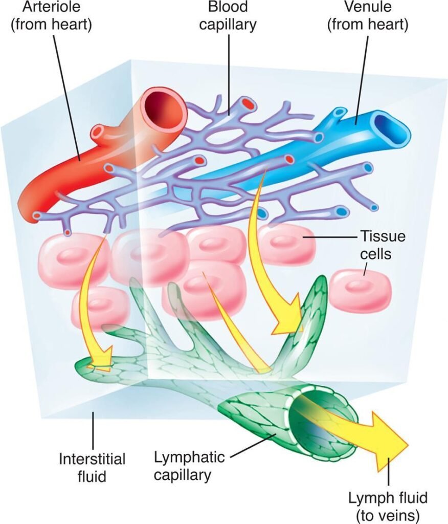
The cardiovascular system continuously delivers oxygen and nutrients to tissues while removing metabolic waste products. Adequate blood flow is essential for maintaining normal cellular function and organ survival. Capillaries serve as the major exchange vessels where movement of fluids, gases, nutrients, and waste products occurs between blood and tissues. The lymphatic system helps return excess interstitial fluid back to the circulation and prevents tissue swelling.
Regulation of blood flow is a dynamic physiological process involving pressure gradients, vascular resistance, vessel diameter, local metabolic activity, neural control, and hormonal influences. Disturbances in capillary exchange or lymphatic drainage can lead to edema, which is commonly seen clinically as ankle swelling, pulmonary edema, or tissue congestion.
Understanding these mechanisms is important for explaining exercise physiology, shock, heart failure, inflammation, and many vascular disorders.
🔹 2️⃣ Foundation Concepts
Key Definitions
- Blood Flow: Volume of blood passing through a vessel per unit time.
- Perfusion: Blood supply reaching tissues.
- Resistance: Opposition to blood flow within vessels.
- Basal Tone: Partial continuous contraction of vascular smooth muscle.
- Capillary Exchange: Movement of fluid and solutes between blood and interstitial fluid.
- Starling Forces: Forces controlling fluid movement across capillary walls.
- Hydrostatic Pressure: Pressure exerted by fluid on vessel walls.
- Oncotic Pressure: Osmotic pressure generated mainly by plasma proteins.
- Lymphatics: Vessels returning excess interstitial fluid to circulation.
- Edema: Excess accumulation of fluid in interstitial tissues.
- Autoregulation: Ability of tissues to maintain relatively constant blood flow despite pressure changes.
Essential Terminology
- Vasodilation
- Vasoconstriction
- Capillary permeability
- Interstitial fluid
- Venous return
- Filtration
- Reabsorption
- Tissue perfusion
- Local metabolic control
🔹 3️⃣ Core Learning — Curriculum Coverage
Hemodynamics: Pressure, Flow and Resistance
🧠 CORE
- Blood flows from high pressure to low pressure.
- Flow depends on pressure gradient and resistance.
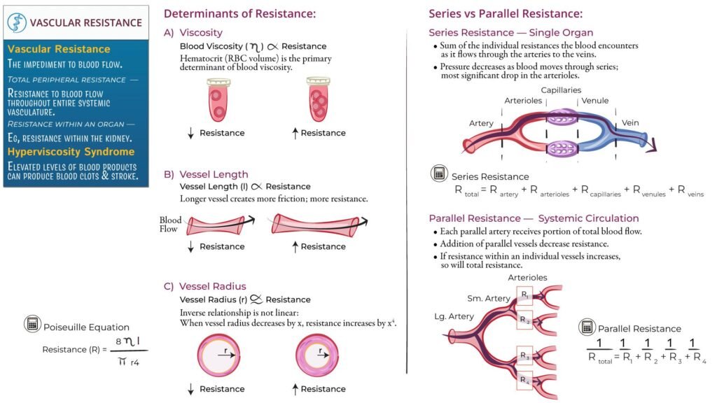
- Arterioles are the major resistance vessels.
- Vessel radius is the most important determinant of resistance.
- Blood viscosity and vessel length also affect resistance.
- Ohm’s law explains relationship between pressure, flow, and resistance.
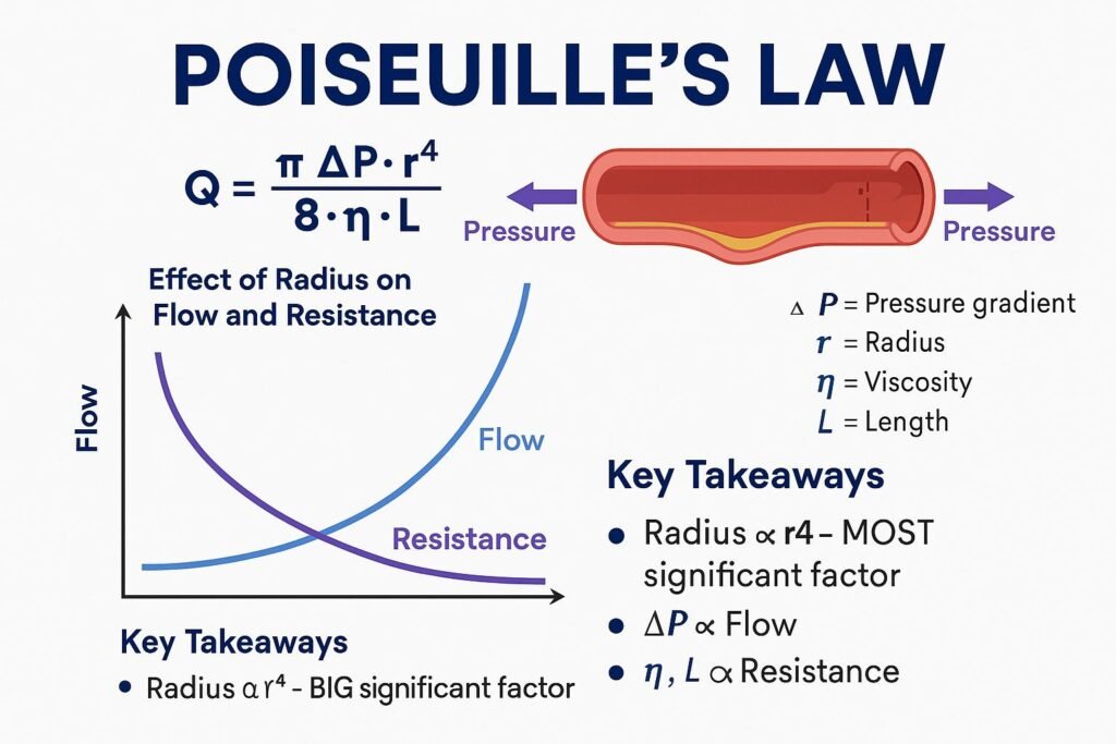
- Poiseuille’s law explains effect of vessel radius on blood flow.
- Small changes in vessel diameter produce major changes in flow.
- Basal vascular tone maintains partial vasoconstriction at rest.
🔬 CONCEPT EXPLAINED
Blood flow follows a pressure gradient similar to movement of current in an electrical circuit.
According to Ohm’s law:
Q=ΔP/R
Where:
- Q = Blood flow
- ΔP = Pressure difference
- R = Resistance
Thus:
- Increased pressure gradient → increased flow
- Increased resistance → decreased flow
Resistance mainly depends on vessel radius. Poiseuille’s law states:
Q=πΔPr4/ 8ηl
Where:
- r = vessel radius
- η = viscosity
- l = vessel length
The fourth-power relationship means:
- Slight vasodilation greatly increases flow
- Slight vasoconstriction markedly decreases flow
Stepwise Mechanism of Increased Blood Flow
- Tissue metabolism increases
- Local metabolites accumulate
- Arteriolar smooth muscle relaxes
- Vessel diameter increases
- Resistance decreases
- Blood flow increases
- Oxygen delivery improves
Basal Tone
Blood vessels normally remain partially constricted due to continuous low-level smooth muscle contraction called basal tone. This allows vessels to:
- Dilate when more blood is needed
- Constrict when flow must decrease
Without basal tone, vascular regulation would become ineffective.
⚠️ CLINICAL IMPORTANCE
- Hypertension increases vascular resistance.
- Atherosclerosis narrows vessel lumen and reduces blood flow.
- Shock causes inadequate tissue perfusion.
- Excessive vasoconstriction may lead to tissue ischemia.
Regulation of Blood Flow and Vascular Tone
🧠 CORE
- Blood flow is regulated locally and systemically.
- Local metabolic control matches blood flow to tissue demand.
- Vasodilators increase tissue perfusion.
- Vasoconstrictors reduce blood flow.
- Autoregulation maintains stable tissue perfusion.
- Endothelium actively regulates vascular tone.
- Basal tone allows rapid vascular adjustment.
🔬 CONCEPT EXPLAINED
Local Metabolic Vasodilator Hypothesis
Active tissues produce metabolites that relax vascular smooth muscle and increase local blood flow.
Important Local Vasodilators
- CO₂
- H⁺ ions
- Adenosine
- Potassium ions
- Nitric oxide
- Lactate
Sequential Mechanism
- Tissue activity increases
- Oxygen consumption rises
- Metabolites accumulate
- Arteriolar smooth muscle relaxes
- Vasodilation occurs
- Resistance decreases
- Blood flow increases
Physiological Vasoconstrictors
- Sympathetic stimulation
- Norepinephrine
- Endothelin
- Angiotensin II
Physiological Vasodilators
- Nitric oxide
- Prostacyclin
- Histamine
- Bradykinin
Autoregulation
Organs like brain, kidney, and heart maintain relatively constant blood flow despite changes in blood pressure.
Mechanism:
- Blood pressure increases
- Vessel wall stretches
- Smooth muscle contracts reflexly
- Vessel diameter decreases
- Flow normalizes
This is called the myogenic mechanism.
⚠️ CLINICAL IMPORTANCE
- Impaired autoregulation may cause cerebral ischemia.
- Excess vasoconstriction contributes to hypertension.
- Septic shock causes widespread vasodilation and hypotension.
- Endothelial dysfunction contributes to vascular disease.
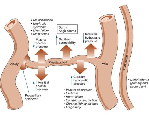
Capillary Exchange and Starling Forces
🧠 CORE
- Capillaries are the main exchange vessels.
- Exchange occurs by diffusion, filtration, and reabsorption.
- Capillary walls are thin for efficient exchange.
- Starling forces determine fluid movement.
- Hydrostatic pressure pushes fluid outward.
- Oncotic pressure pulls fluid inward.
- Balance between these forces prevents edema.
🔬 CONCEPT EXPLAINED
Histology Correlation
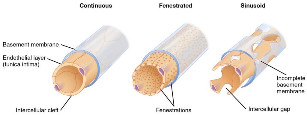
Microscopic Appearance of Capillaries
- Lined by simple squamous endothelial cells
- Thin basement membrane present
- Minimal wall thickness for exchange
- Pericytes may surround capillaries
Types of Capillaries
Continuous Capillaries
- Tight endothelial junctions
- Least permeable
- Found in brain and muscle
Fenestrated Capillaries
- Small pores present
- Increased permeability
- Found in kidneys and endocrine glands
Sinusoidal Capillaries
- Large gaps between cells
- Highly permeable
- Found in liver and bone marrow
Structure → Function Relationship
- Thin endothelial lining allows rapid diffusion
- Fenestrations increase filtration
- Sinusoids allow passage of proteins and cells
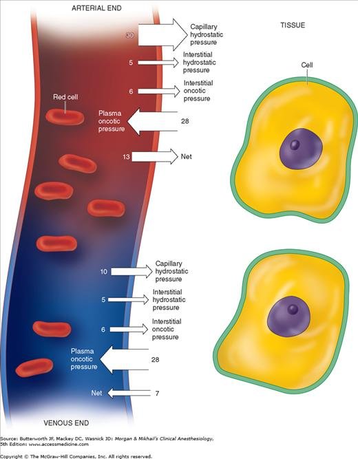
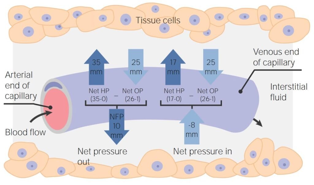
Starling Forces
Fluid movement depends on balance between:
- Capillary hydrostatic pressure
- Plasma oncotic pressure
- Interstitial hydrostatic pressure
- Interstitial oncotic pressure
Sequential Mechanism of Filtration
- Blood enters arterial end of capillary
- Capillary hydrostatic pressure is high
- Fluid moves outward into interstitial space
- Nutrients reach tissues
Sequential Mechanism of Reabsorption
- Blood reaches venous end
- Hydrostatic pressure falls
- Plasma oncotic pressure predominates
- Fluid returns into capillary
⚠️ CLINICAL IMPORTANCE
Causes of Edema
Increased Capillary Hydrostatic Pressure
- Heart failure
- Venous obstruction
Reduced Plasma Oncotic Pressure
- Liver disease
- Nephrotic syndrome
- Malnutrition
Increased Capillary Permeability
- Inflammation
- Burns
- Allergic reactions
Lymphatic Obstruction
- Filariasis
- Tumors
- Surgery
Lymphatic System and Interstitial Fluid Balance
🧠 CORE
- Lymphatics return excess tissue fluid to circulation.
- Lymph flow prevents edema formation.
- Lymphatics absorb proteins from interstitial fluid.
- One-way valves maintain forward flow.
- Skeletal muscle activity promotes lymph movement.
🔬 CONCEPT EXPLAINED
Histology Correlation
Microscopic Appearance of Lymphatic Capillaries
- Thin endothelial lining
- Irregular lumen
- Incomplete basement membrane
- Overlapping endothelial cells form valve-like openings
Structure → Function Relationship
- Thin walls allow uptake of excess fluid
- Valve-like openings prevent backflow
- Highly permeable structure absorbs proteins and large particles
Mechanism of Lymph Formation and Flow
- Excess interstitial fluid accumulates
- Interstitial pressure rises
- Endothelial flaps open
- Fluid enters lymphatics
- Skeletal muscle contraction compresses vessels
- Valves prevent backward flow
- Lymph returns to venous circulation
Effect of Interstitial Fluid Pressure
- Increased interstitial pressure increases lymph flow.
- Negative interstitial pressure reduces lymph entry.
⚠️ CLINICAL IMPORTANCE
- Lymphatic obstruction causes lymphedema.
- Cancer surgery may impair lymph drainage.
- Filarial infection may produce elephantiasis.
- Persistent edema impairs tissue healing.
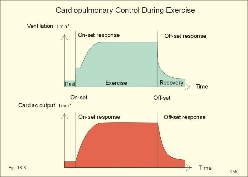
Blood Flow During Exercise
🧠 CORE
- Exercise increases cardiac output.
- Active muscles receive increased blood flow.
- Blood is redistributed according to tissue demand.
- Sympathetic activity increases during exercise.
- Local metabolic vasodilation overrides sympathetic vasoconstriction in active muscles.
🔬 CONCEPT EXPLAINED
Sequential Circulatory Changes During Exercise
- Motor cortex initiates movement
- Sympathetic nervous system activates
- Heart rate increases
- Stroke volume increases
- Cardiac output rises
- Active muscles produce metabolites
- Local vasodilation occurs
- Blood flow to skeletal muscle increases
Organ Blood Flow Changes
Skeletal Muscle
- Markedly increased flow
Heart
- Coronary blood flow increases
Brain
- Flow remains relatively constant
Skin
- Flow increases for heat loss
Liver and GIT
- Flow decreases temporarily
⚠️ CLINICAL IMPORTANCE
- Poor coronary circulation may cause angina during exercise.
- Peripheral vascular disease reduces exercise tolerance.
- Heart failure limits increase in cardiac output.
⚙️ 4️⃣ Functional Flow
| Structure | Function | Outcome |
|---|---|---|
| Arterioles | Control resistance | Regulation of tissue perfusion |
| Capillary endothelium | Exchange surface | Nutrient and fluid movement |
| Plasma proteins | Generate oncotic pressure | Fluid reabsorption |
| Lymphatic vessels | Return excess fluid | Prevention of edema |
| Vascular smooth muscle | Vasoconstriction/vasodilation | Blood flow regulation |
🩺 5️⃣ Clinical Correlation
Congestive Heart Failure
Increased venous pressure raises capillary hydrostatic pressure causing ankle edema and pulmonary edema.
Hypoproteinemia
Reduced plasma proteins decrease oncotic pressure leading to generalized edema.
Inflammation
Increased capillary permeability allows protein leakage into tissues producing swelling.
Lymphedema
Obstruction of lymphatics causes accumulation of protein-rich interstitial fluid.
📌 6️⃣ Summary Points
- Blood flow is directly proportional to pressure gradient.
- Vessel radius is the most important determinant of resistance.
- Arterioles are the major resistance vessels.
- Basal tone allows rapid vascular regulation.
- Capillaries are the primary exchange vessels.
- Hydrostatic pressure favors filtration.
- Plasma oncotic pressure favors reabsorption.
- Lymphatics prevent accumulation of interstitial fluid.
- Edema occurs when filtration exceeds drainage.
- Active tissues receive increased blood flow due to local metabolites.
- Fenestrated capillaries are highly permeable.
- Exercise redistributes blood flow according to metabolic demand.