
📖 Step 2 — Learning Material
🔹 1️⃣ Introduction
Blood vessels form the transportation network of the body and are responsible for carrying blood between the heart and tissues. They include arteries, veins, capillaries, and lymphatic vessels. Structurally, vessel walls are specially designed according to their functions such as pressure regulation, nutrient exchange, and blood return to the heart.
During embryonic development, blood vessels arise early to establish circulation necessary for fetal survival and organ development. Abnormal development of vessels can lead to clinically important congenital disorders such as coarctation of the aorta.
Histologically, arteries and veins can be differentiated by the thickness of their walls, arrangement of smooth muscle, elastic tissue, and lumen size. Understanding vessel histology is essential for interpreting vascular diseases, hypertension, aneurysm formation, edema, and circulatory disorders.
Lymphatic vessels play an important role in maintaining tissue fluid balance and immune defense by draining excess interstitial fluid back into circulation.
🔹 2️⃣ Foundation Concepts
Key Definitions
- Blood vessels: Tubular structures that transport blood throughout the body
- Artery: Vessel carrying blood away from the heart
- Vein: Vessel carrying blood toward the heart
- Capillary: Microscopic vessel for exchange between blood and tissues
- Lymphatic vessel: Thin-walled channel carrying lymph
- Tunica intima: Innermost vessel layer lined by endothelium
- Tunica media: Middle muscular layer
- Tunica adventitia: Outer connective tissue layer
- Elastic artery: Artery containing abundant elastic fibers
- Muscular artery: Artery containing more smooth muscle
- Coarctation of aorta: Congenital narrowing of the aorta
Essential Terminology
- Endothelium
- Elastic lamina
- Smooth muscle
- Vasoconstriction
- Vasodilation
- Vasa vasorum
- Anastomosis
- Portal circulation
- Sinusoid
- Lymph
Basic Overview
- Arteries withstand high pressure from the heart
- Veins act as capacitance vessels and contain valves
- Capillaries allow exchange of gases and nutrients
- Lymphatics return excess tissue fluid to circulation
- Vessel walls are organized into three layers
- Embryonic vessels develop from mesoderm
- Developmental defects may produce congenital vascular abnormalities
🔹 3️⃣ Core Learning — Curriculum Coverage
Histological Organization of Blood Vessels
🧠 CORE
- Most vessels contain three layers:
- Tunica intima
- Tunica media
- Tunica adventitia
- Endothelium lines all blood vessels
- Arteries have thick muscular walls
- Veins have thinner walls and wider lumens
- Elastic fibers provide recoil
- Smooth muscle regulates vessel diameter
- Connective tissue provides support
- Large vessels contain vasa vasorum
🔬 CONCEPT EXPLAINED
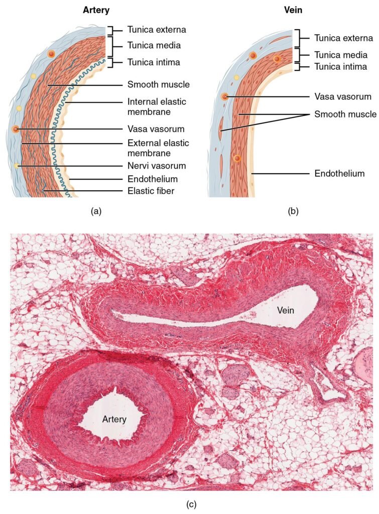
The structure of blood vessels is directly related to their function. Blood leaving the heart travels under high pressure; therefore, arteries require thick walls rich in elastic tissue and smooth muscle. Elastic tissue allows vessels to stretch during systole and recoil during diastole, helping maintain continuous blood flow.
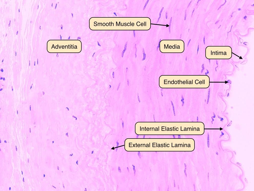
The tunica intima consists of endothelium resting on a basement membrane. Endothelial cells provide a smooth surface that reduces friction and regulate clotting, inflammation, and vascular tone.
The tunica media contains concentric layers of smooth muscle and elastic fibers. This layer is thickest in arteries because arteries regulate blood pressure and blood distribution through vasoconstriction and vasodilation.
The tunica adventitia contains collagen fibers, nerves, and small vessels called vasa vasorum. It anchors vessels to surrounding tissues and nourishes large vessel walls.
Veins experience much lower pressure than arteries; therefore, they contain thinner walls and larger lumens. Their reduced elastic tissue reflects lower hemodynamic stress.
⚠️ CLINICAL IMPORTANCE
- Damage to endothelium contributes to atherosclerosis
- Loss of elastic tissue may cause aneurysm formation
- Thickened arterial walls increase peripheral resistance and hypertension
- Weak venous walls predispose to varicose veins
- Impaired venous return may cause edema
Microscopic Structure of Arteries
🧠 CORE
- Arteries carry blood away from the heart
- Thick tunica media is characteristic
- Elastic arteries contain abundant elastic lamellae
- Muscular arteries contain more smooth muscle
- Small arteries regulate tissue perfusion
- Arterioles are resistance vessels
- Internal elastic lamina is prominent in muscular arteries
- Arteries maintain blood pressure
🔬 CONCEPT EXPLAINED
Elastic Arteries
Examples include:
- Aorta
- Pulmonary trunk
These vessels receive blood directly from the heart. Their tunica media contains multiple elastic lamellae that allow expansion during ventricular systole and recoil during diastole. This recoil maintains continuous blood flow even when the heart relaxes.
Muscular Arteries
These distribute blood to organs. Their tunica media contains many layers of smooth muscle. Smooth muscle contraction changes vessel diameter and regulates blood supply according to tissue demand.
Arterioles
Arterioles contain only a few layers of smooth muscle but play a major role in controlling peripheral resistance and systemic blood pressure.
Structure → Function Relationship:
- Thick media → withstands pressure
- Elastic fibers → maintain pulsatile flow
- Smooth muscle → controls vascular tone
⚠️ CLINICAL IMPORTANCE
- Arteriolar constriction contributes to hypertension
- Atherosclerosis commonly affects muscular arteries
- Loss of elasticity in aging increases systolic blood pressure
- Chronic hypertension causes thickening of arterial walls
Microscopic Structure of Veins
🧠 CORE
- Veins return blood to the heart
- Thin tunica media
- Large irregular lumen
- Thick adventitia
- Valves prevent backflow
- Low-pressure vessels
- Veins act as blood reservoirs
🔬 CONCEPT EXPLAINED
Veins operate under low pressure; therefore, they do not require thick muscular walls. Their walls are thinner and more collapsible compared to arteries.
The tunica adventitia is usually the thickest layer and contains collagen fibers for support. Large veins may contain longitudinal smooth muscle bundles.
Venous valves are folds of tunica intima that prevent backflow of blood, especially in limbs where blood must move against gravity.
Large lumens reduce resistance to blood flow and allow veins to store a large volume of blood.
Histological Differentiation Under Light Microscope:
| Feature | Artery | Vein |
|---|---|---|
| Wall thickness | Thick | Thin |
| Lumen | Small, round | Large, irregular |
| Tunica media | Thick | Thin |
| Elastic tissue | Prominent | Less |
| Valves | Absent | Present |
⚠️ CLINICAL IMPORTANCE
- Valve failure leads to varicose veins
- Venous stasis predisposes to thrombosis
- Increased venous pressure causes edema
- Dilated veins may occur in portal hypertension
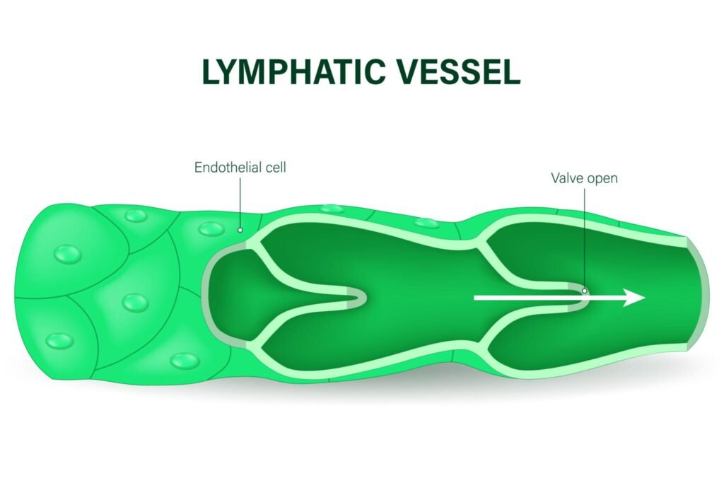
Histology of Lymphatic Vessels
🧠 CORE
- Thin-walled vessels
- Lined by endothelium
- Carry lymph instead of blood
- Contain valves
- Lack thick muscular layer
- Drain excess interstitial fluid
- Participate in immune defense
🔬 CONCEPT EXPLAINED
Lymphatic vessels begin as blind-ended capillaries in tissues. Their walls are extremely thin and highly permeable, allowing entry of proteins, fluid, bacteria, and immune cells.
Unlike blood capillaries, lymphatics have incomplete basement membranes and loosely arranged endothelial cells, facilitating fluid uptake.
Larger lymphatic vessels contain valves to maintain one-way lymph flow toward veins.
Functionally, lymphatics prevent tissue fluid accumulation and return leaked plasma proteins to circulation.
⚠️ CLINICAL IMPORTANCE
- Obstruction of lymphatics causes lymphedema
- Filariasis can block lymphatic drainage
- Cancer spreads through lymphatic channels
- Impaired lymph drainage causes tissue swelling
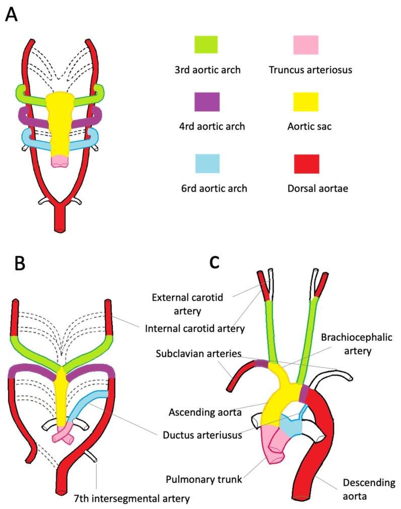
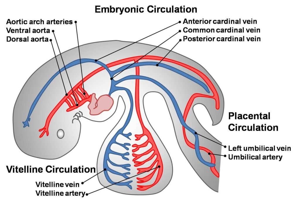
Development of the Arterial System
🧠 CORE
- Arterial system develops from mesoderm
- Blood islands form primitive vessels
- Aortic arches contribute to major arteries
- Dorsal aortae fuse to form descending aorta
- Remodeling produces adult arterial pattern
- Umbilical arteries connect fetus to placenta
🔬 CONCEPT EXPLAINED
The vascular system begins developing during the third week of embryogenesis from mesodermal cells called angioblasts. These cells form blood islands, which develop into primitive blood vessels.
The embryonic arterial system initially consists of paired dorsal aortae connected to the heart by six pairs of aortic arches.
Important derivatives:
| Aortic Arch | Major Derivative |
|---|---|
| 1st | Maxillary artery |
| 2nd | Stapedial artery |
| 3rd | Common carotid artery |
| 4th | Part of aortic arch/subclavian |
| 6th | Pulmonary arteries and ductus arteriosus |
The dorsal aortae later fuse to form the descending aorta.
Development → Adult Link:
Embryonic arch remodeling creates the mature arterial circulation supplying head, neck, thorax, and limbs.
⚠️ CLINICAL IMPORTANCE
- Abnormal arch development may cause vascular rings
- Defects can compress trachea or esophagus
- Interrupted aortic arch is life-threatening
- Persistent ductus arteriosus affects circulation
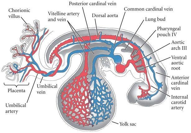
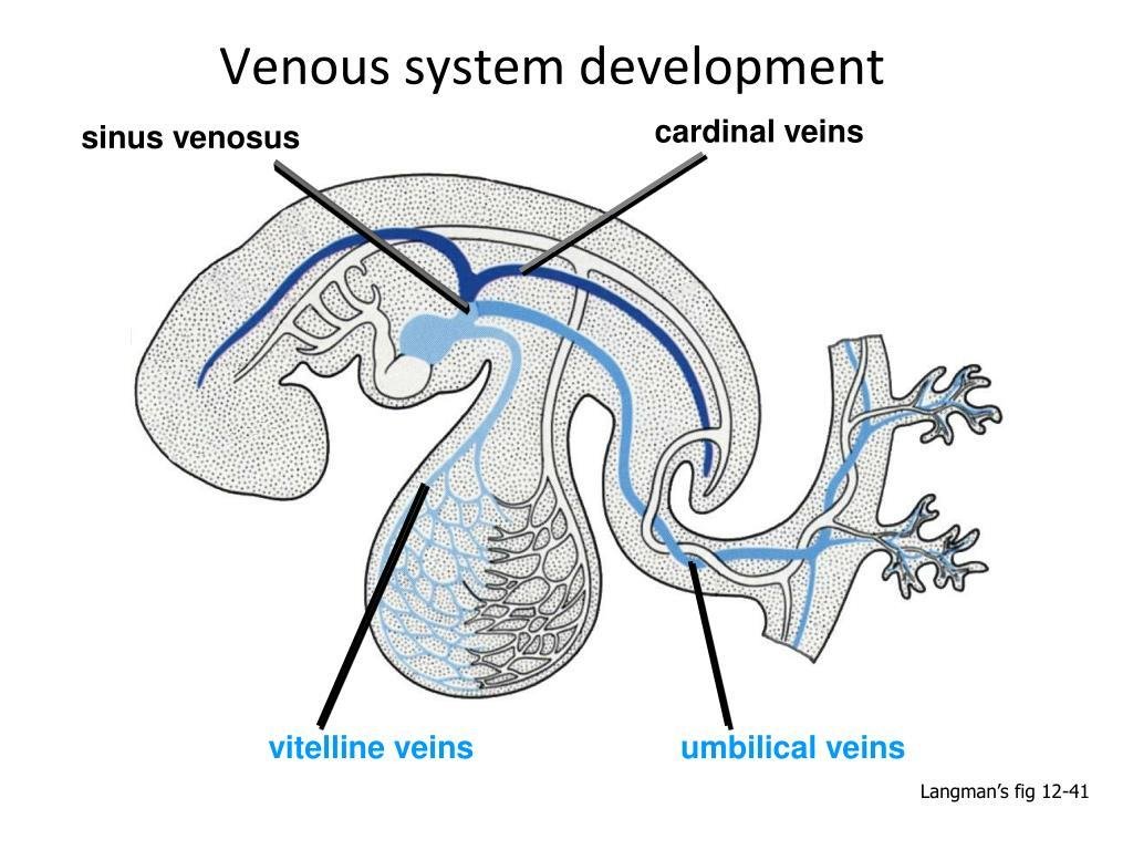
Development of the Venous System
🧠 CORE
- Venous system develops from three paired veins:
- Vitelline veins
- Umbilical veins
- Cardinal veins
- Extensive remodeling occurs
- Right-sided channels predominate
- Venous asymmetry develops gradually
- Sinusoids form in liver
🔬 CONCEPT EXPLAINED
The embryonic venous system initially consists of symmetrical paired veins draining into the sinus venosus.
Vitelline Veins
- Drain yolk sac
- Form portal vein and hepatic sinusoids
Umbilical Veins
- Carry oxygenated blood from placenta
- Left umbilical vein persists
Cardinal Veins
- Drain embryonic body
- Form superior vena cava and systemic veins
As development proceeds, many veins regress while others enlarge, producing the asymmetric adult venous pattern.
⚠️ CLINICAL IMPORTANCE
- Persistent left superior vena cava may occur
- Abnormal venous return can impair circulation
- Portal hypertension relates to portal venous system abnormalities
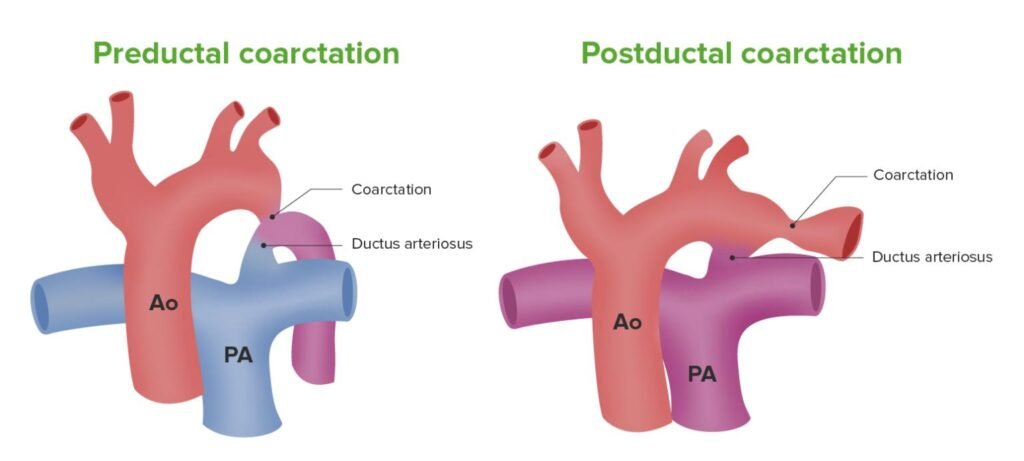
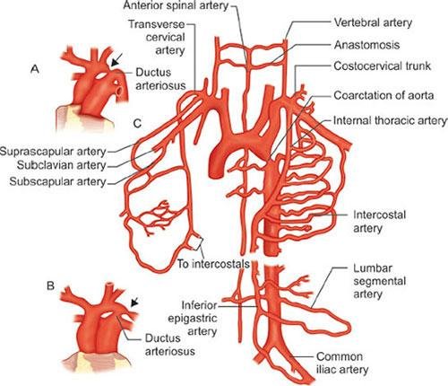
Congenital Abnormality — Coarctation of Aorta
🧠 CORE
- Congenital narrowing of aorta
- Usually near ductus arteriosus
- Causes upper body hypertension
- Reduces lower limb perfusion
- May be preductal or postductal
- Collateral circulation develops
🔬 CONCEPT EXPLAINED
Coarctation of the aorta usually occurs near the insertion of the ductus arteriosus. Narrowing obstructs blood flow from the left ventricle to the lower body.
Preductal Type
- Narrowing proximal to ductus arteriosus
- Severe in infancy
- Lower body supplied through ductus arteriosus
Postductal Type
- Narrowing distal to ductus arteriosus
- Collateral circulation develops through intercostal arteries
Why Symptoms Occur:
- Increased resistance proximal to narrowing raises blood pressure in upper limbs
- Reduced distal flow causes weak femoral pulses and lower limb ischemia
⚠️ CLINICAL IMPORTANCE
- Hypertension in upper limbs
- Weak lower limb pulses
- Rib notching on X-ray due to enlarged collateral arteries
- Left ventricular hypertrophy may develop
- Severe untreated cases may cause heart failure
⚙️ 4️⃣ Functional Flow
| Structure | Function | Outcome |
|---|---|---|
| Thick arterial media | Withstands pressure | Maintains blood flow |
| Elastic lamellae | Recoil during diastole | Continuous circulation |
| Smooth muscle | Vasoconstriction/vasodilation | BP regulation |
| Venous valves | Prevent backflow | Efficient venous return |
| Thin lymphatic wall | Fluid absorption | Prevents edema |
| Endothelium | Smooth blood flow | Reduces thrombosis |
🩺 5️⃣ Clinical Correlation
Hypertension
Thickened arterial walls and increased smooth muscle contraction elevate peripheral resistance, increasing blood pressure.
Varicose Veins
Failure of venous valves causes blood pooling and vein dilation, especially in lower limbs.
Atherosclerosis
Endothelial injury promotes lipid deposition within arterial walls leading to narrowing and ischemia.
Lymphedema
Obstruction of lymphatic drainage causes accumulation of tissue fluid and chronic swelling.
Coarctation of Aorta
Congenital narrowing of aorta causes upper limb hypertension with weak femoral pulses.
📌 6️⃣ Summary Points
- Blood vessels contain three layers: intima, media, and adventitia
- Arteries have thick tunica media and smaller lumen
- Veins have thinner walls and valves
- Elastic arteries maintain continuous blood flow
- Arterioles are major resistance vessels
- Lymphatics drain excess tissue fluid
- Endothelium regulates vascular function
- Aortic arches form major arteries
- Cardinal veins contribute to systemic venous system
- Coarctation of aorta causes upper limb hypertension
- Rib notching suggests collateral circulation in coarctation
- Vessel structure is directly related to hemodynamic function