
📖 Step 2 — Learning Material
🔹 1️⃣ Introduction
The cardiac conduction system is a specialized network of modified cardiac muscle cells that generates and spreads electrical impulses throughout the heart.
It is located mainly in the right atrium, atrioventricular region, interventricular septum, and ventricular walls.
This system allows the heart to beat rhythmically without requiring direct nervous stimulation.
The SA node normally acts as the natural pacemaker of the heart.
Proper conduction ensures coordinated atrial contraction followed by ventricular contraction.
Clinically, disturbances in impulse generation or conduction can cause palpitations, arrhythmias, heart block, and sudden cardiac symptoms.
🔹 2️⃣ Foundation Concepts
Key Definitions
- Automaticity: Ability of cardiac cells to generate impulses spontaneously.
- Rhythmicity: Ability to generate impulses at regular intervals.
- Conductivity: Ability to transmit electrical impulses from one cardiac cell to another.
- Pacemaker: Tissue that initiates the heartbeat.
- Normal pacemaker: SA node.
- Ectopic pacemaker: Abnormal pacemaker outside the SA node.
- Functional syncytium: Cardiac muscle behaves as one unit due to gap junctions.
- Gap junctions: Low-resistance channels between cardiac muscle cells allowing rapid ion movement.
- AV nodal delay: Short delay of impulse at AV node before passing to ventricles.
🔹 3️⃣ Core Learning — Curriculum Coverage
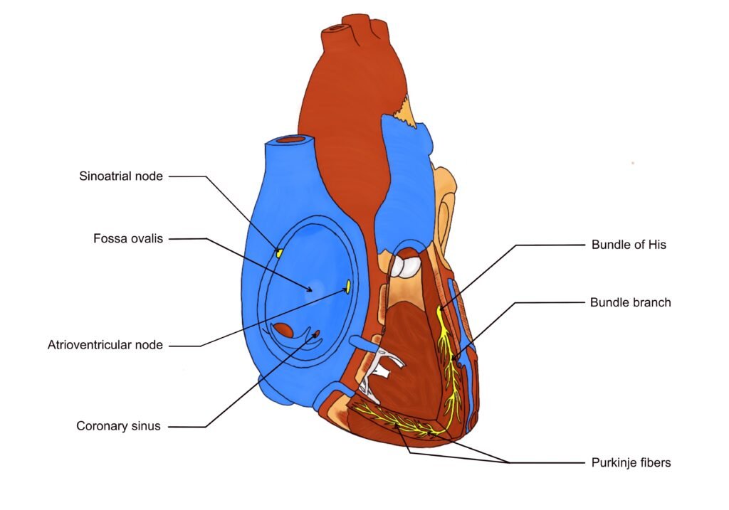
A. Specialized Conduction System of the Heart
🧠 CORE
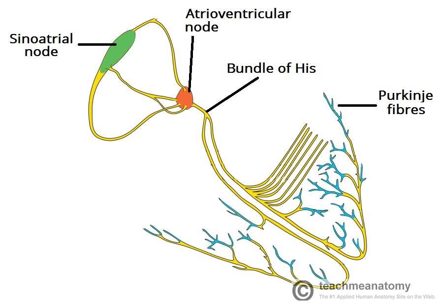
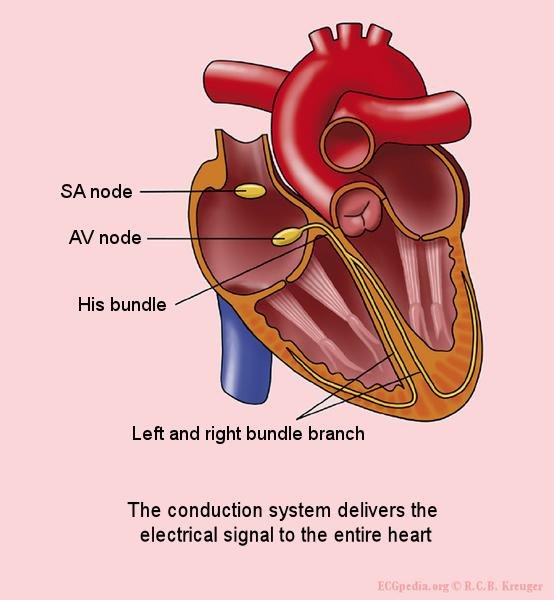
- The cardiac conduction system consists of:
- SA node
- AV node
- Bundle of His
- Right and left bundle branches
- Purkinje fibers
- These are modified cardiac muscle cells, not nerves.
- They generate and conduct impulses.
- The system coordinates atrial and ventricular contraction.
- SA node initiates normal rhythm.
- AV node delays impulse transmission.
- Purkinje fibers rapidly distribute impulses to ventricles.
- Conduction follows a fixed sequence from atria to ventricles.
🔬 CONCEPT EXPLAINED
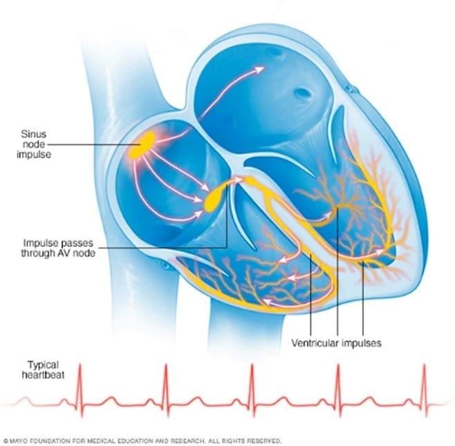
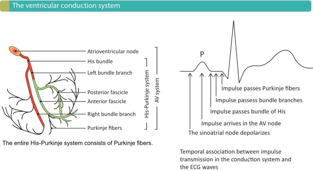
The heart has its own intrinsic electrical system. The impulse begins in the SA node, spreads across both atria, reaches the AV node, passes through the Bundle of His, travels down the right and left bundle branches, and finally spreads through Purkinje fibers to ventricular muscle.
This arrangement exists because atria must contract first to fill the ventricles, and ventricles must contract afterward to pump blood into the pulmonary trunk and aorta.
The conduction system is made of specialized cardiac muscle cells. These cells are adapted for impulse generation and rapid conduction rather than strong contraction.
⚠️ CLINICAL IMPORTANCE
Failure of this system may cause:
- Irregular heartbeat
- Palpitations
- Arrhythmias
- AV block
- Poor ventricular filling
- Reduced cardiac output
B. SA Node — Normal Pacemaker of the Heart
🧠 CORE
- SA node is the normal pacemaker of the heart.
- It is located in the right atrium near the opening of the superior vena cava.
- It has the highest spontaneous firing rate.
- It generates regular impulses.
- It initiates atrial depolarization.
- Its activity determines normal heart rate.
- It is influenced by sympathetic and parasympathetic nerves.
- It produces sinus rhythm.
🔬 CONCEPT EXPLAINED
The SA node acts as the natural pacemaker because its cells depolarize faster than other parts of the conduction system. This means it reaches threshold first and starts the heartbeat before other potential pacemaker tissues can fire.
Its location in the right atrium allows the impulse to spread quickly across both atria. This causes atrial contraction and helps push blood into the ventricles before ventricular systole begins.
⚠️ CLINICAL IMPORTANCE
If the SA node fails or becomes slow, another part of the conduction system may take over. This is called an ectopic pacemaker. It may produce abnormal rhythm and palpitations.
C. AV Node and AV Nodal Delay
🧠 CORE
- AV node is located in the lower part of the right atrium.
- It receives impulses from the atria.
- It delays the impulse before ventricular conduction.
- This delay allows ventricular filling.
- It protects ventricles from excessively rapid atrial impulses.
- It connects atrial rhythm to ventricular rhythm.
- After AV node, impulse enters Bundle of His.
🔬 CONCEPT EXPLAINED
The AV node is important because atria and ventricles should not contract at the same time. A brief delay at the AV node allows the atria to complete contraction and empty blood into the ventricles.
After this delay, the impulse passes to the ventricles through the Bundle of His and Purkinje system. This timing improves ventricular filling and increases the efficiency of cardiac pumping.
⚠️ CLINICAL IMPORTANCE
Damage to the AV node may cause heart block. In heart block, impulses from atria may not properly reach the ventricles. This can cause slow heart rate, dizziness, weakness, or palpitations.
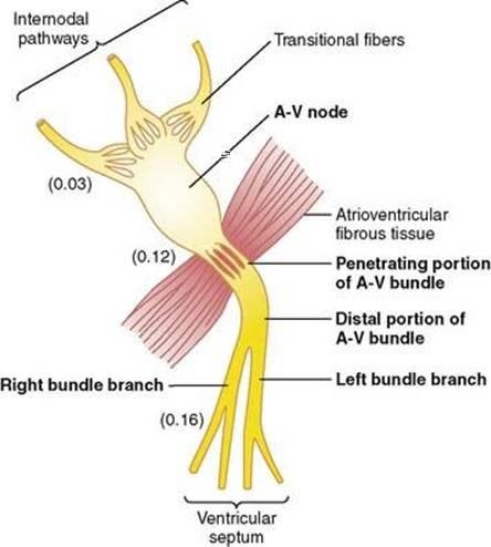
D. Bundle of His, Bundle Branches, and Purkinje Fibers
🧠 CORE
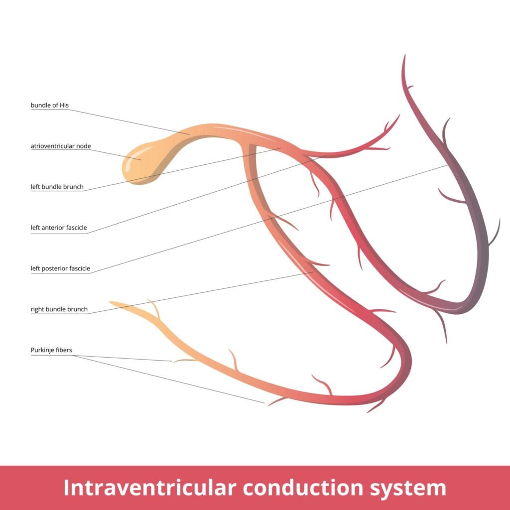
- Bundle of His is the only normal electrical connection between atria and ventricles.
- It passes through the fibrous skeleton of the heart.
- It divides into right and left bundle branches.
- Bundle branches travel along the interventricular septum.
- Purkinje fibers spread into ventricular walls.
- Purkinje fibers conduct impulses very rapidly.
- Rapid conduction allows coordinated ventricular contraction.
- Ventricular contraction begins near the apex and moves upward.
🔬 CONCEPT EXPLAINED
The fibrous skeleton electrically separates atria from ventricles. Therefore, the Bundle of His is necessary to transmit impulses from AV node into the ventricles.
The impulse then moves through right and left bundle branches and rapidly spreads through Purkinje fibers. This causes both ventricles to contract almost together. The pattern of spread helps blood move upward from the ventricular apex toward the outflow tracts.
⚠️ CLINICAL IMPORTANCE
Blockage in bundle branches can delay ventricular activation. This may reduce pumping efficiency and produce abnormal ECG findings.
E. Automaticity, Rhythmicity, Conductivity, and Ectopic Pacemakers
🧠 CORE
- Automaticity allows spontaneous impulse generation.
- Rhythmicity allows regular impulse formation.
- Conductivity allows impulse spread.
- SA node has the fastest natural rhythm.
- AV node, Bundle of His, and Purkinje fibers can act as backup pacemakers.
- Ectopic pacemaker arises outside the SA node.
- Causes include SA node failure, ischemia, abnormal electrolytes, or excessive sympathetic stimulation.
- Ectopic impulses may cause premature beats or arrhythmias.
🔬 CONCEPT EXPLAINED
Different parts of the conduction system can generate impulses, but the SA node normally dominates because it fires fastest. If the SA node becomes weak or another area becomes abnormally irritable, an ectopic pacemaker may appear.
Ectopic pacemakers disturb normal sequence of depolarization. This can make the heartbeat irregular or inefficient.
⚠️ CLINICAL IMPORTANCE
Ectopic pacemakers are important causes of:
- Palpitations
- Premature beats
- Tachyarrhythmias
- Irregular pulse
- Reduced cardiac efficiency
F. Sympathetic and Parasympathetic Innervation of the Heart
🧠 CORE
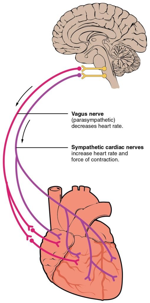
- Sympathetic nerves increase heart rate.
- Sympathetic stimulation increases conduction velocity.
- Sympathetic stimulation increases force of contraction.
- Parasympathetic supply comes mainly through the vagus nerve.
- Parasympathetic stimulation decreases heart rate.
- Parasympathetic stimulation slows AV conduction.
- SA node and AV node are strongly affected by autonomic nerves.
- Autonomic nerves modify heart activity but do not normally initiate heartbeat.
🔬 CONCEPT EXPLAINED
The heart beats intrinsically because of the SA node, but autonomic nerves adjust its rate according to body needs.
During exercise, stress, or fear, sympathetic stimulation increases SA node firing and speeds AV conduction. This increases cardiac output.
During rest, parasympathetic vagal activity slows the SA node and delays AV nodal conduction. This conserves energy and reduces heart rate.
⚠️ CLINICAL IMPORTANCE
Excess sympathetic activity may cause tachycardia and palpitations. Excess vagal activity may cause bradycardia or AV conduction slowing.
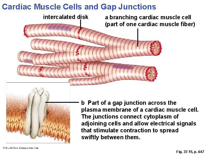
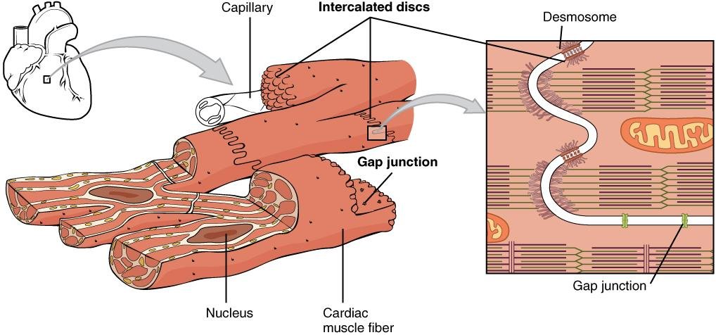
G. Gap Junctions and Functional Syncytium
🧠 CORE
- Cardiac muscle cells are connected by intercalated discs.
- Gap junctions are present in intercalated discs.
- Gap junctions allow ions to pass between cells.
- This permits rapid spread of depolarization.
- Atria behave as one functional syncytium.
- Ventricles behave as another functional syncytium.
- Fibrous skeleton separates atrial and ventricular syncytia.
- Functional syncytium allows coordinated contraction.
🔬 CONCEPT EXPLAINED
Cardiac muscle cells are separate cells anatomically, but functionally they behave like a connected unit. Gap junctions allow electrical current to pass from cell to cell.
This means that once one cardiac cell is depolarized, nearby cells also depolarize. As a result, atria contract together and ventricles contract together.
The fibrous skeleton prevents direct spread of impulses from atria to ventricles, forcing the impulse to pass through the AV node. This preserves proper timing.
⚠️ CLINICAL IMPORTANCE
Disturbed electrical coupling can impair coordinated contraction and contribute to arrhythmias.
⚙️ 4️⃣ Functional Flow
Structure → Function → Outcome
- SA node structure: Specialized pacemaker cells
→ Function: Spontaneous impulse generation
→ Outcome: Normal sinus rhythm - AV node slow conduction
→ Function: Delays impulse
→ Outcome: Allows ventricular filling - Bundle branches and Purkinje fibers
→ Function: Rapid ventricular conduction
→ Outcome: Coordinated ventricular contraction - Gap junctions
→ Function: Electrical connection between cardiac cells
→ Outcome: Functional syncytium - Sympathetic innervation
→ Function: Increases rate and conduction
→ Outcome: Increased cardiac output - Parasympathetic innervation
→ Function: Slows SA node and AV node
→ Outcome: Reduced heart rate
🩺 5️⃣ Clinical Correlation
Palpitations
Palpitations are the awareness of heartbeat. They may occur when impulse generation becomes abnormal or conduction becomes irregular.
Arrhythmias
Arrhythmias occur when the rhythm of the heart becomes abnormal. They may result from:
- Abnormal SA node activity
- Ectopic pacemaker
- AV nodal conduction defect
- Bundle branch block
- Excess sympathetic stimulation
- Electrolyte imbalance
Heart Block
Heart block occurs when impulse transmission from atria to ventricles is delayed or interrupted, commonly at the AV node or Bundle of His.
Ectopic Beats
Ectopic beats arise from abnormal pacemaker sites outside the SA node. They may cause premature or irregular heartbeats.
📌 6️⃣ Summary Points
- The SA node is the normal pacemaker of the heart.
- The conduction system is made of modified cardiac muscle cells.
- Normal impulse pathway is SA node → AV node → Bundle of His → bundle branches → Purkinje fibers.
- AV nodal delay allows proper ventricular filling.
- Purkinje fibers ensure rapid and coordinated ventricular contraction.
- Automaticity means spontaneous impulse generation.
- Ectopic pacemaker means abnormal pacemaker outside the SA node.
- Sympathetic stimulation increases heart rate and conduction.
- Parasympathetic stimulation decreases heart rate and slows AV conduction.
- Gap junctions allow cardiac muscle to act as a functional syncytium.
- Conduction defects commonly present as palpitations or arrhythmias.