
📖 Step 2 — Learning Material
🔹 1️⃣ Introduction
The cardiac cycle is the sequence of mechanical and electrical events that occur during one heartbeat. It includes coordinated contraction and relaxation of the atria and ventricles to maintain continuous blood circulation throughout the body. The heart valves ensure one-way blood flow and prevent backflow during pumping. Heart sounds are produced by valve closure and provide important clinical information regarding cardiac function.
This topic is central to understanding cardiovascular physiology because it integrates electrical activity, pressure changes, blood flow, valve mechanics, and heart sounds into one functional process. Knowledge of the cardiac cycle helps students understand ECG interpretation, murmurs, valvular diseases, and cardiac dysfunction.
Clinically, abnormalities in valve function can produce stenosis or regurgitation leading to breathlessness, fatigue, heart failure, and characteristic murmurs. Understanding normal physiology allows students to interpret pathological findings logically.
🔹 2️⃣ Foundation Concepts
Key Definitions
- Cardiac Cycle: Sequence of events occurring from one heartbeat to the next.
- Systole: Period of cardiac muscle contraction.
- Diastole: Period of cardiac muscle relaxation and chamber filling.
- Stroke Volume: Amount of blood ejected by one ventricle in one beat.
- End-Diastolic Volume (EDV): Volume present in ventricle before contraction.
- End-Systolic Volume (ESV): Blood remaining after ventricular contraction.
- Heart Sounds: Sounds produced mainly by valve closure and blood flow vibrations.
- Murmur: Abnormal heart sound due to turbulent blood flow.
- Stenosis: Narrowing of a valve opening.
- Regurgitation: Backflow of blood due to incomplete valve closure.
Essential Terminology
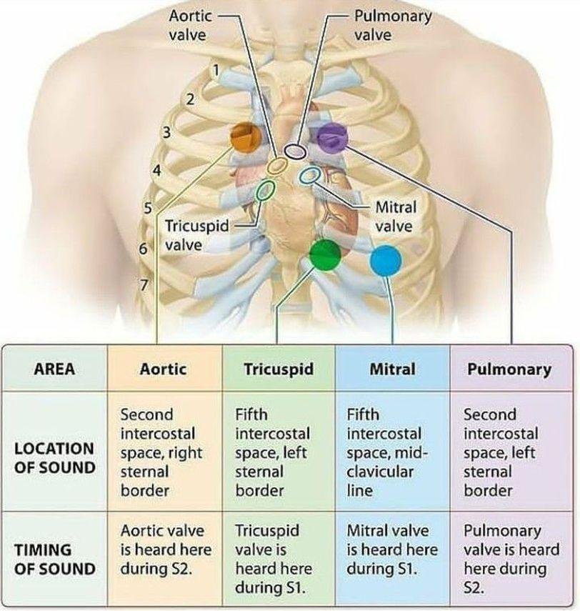
- Mitral valve = bicuspid valve
- Right AV valve = tricuspid valve
- Semilunar valves = aortic + pulmonary valves
- Lub = First heart sound (S1)
- Dub = Second heart sound (S2)
Basic Overview
- Atria receive blood and assist ventricular filling.
- Ventricles act as main pumping chambers.
- Valves open and close according to pressure differences.
- Electrical activity initiates mechanical contraction.
- Cardiac cycle has filling phase + ejection phase.
- Heart sounds correlate with valve closure.
- Proper coordination maintains effective circulation.
🔹 3️⃣ Core Learning — Curriculum Coverage
Cardiac Cycle: Sequential Events of One Heartbeat
🧠 CORE
- One cardiac cycle includes atrial systole, ventricular systole, and diastole.
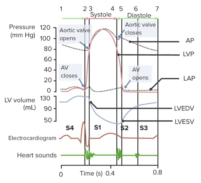
- Average duration at 75 bpm ≈ 0.8 seconds.
- Right and left sides function simultaneously.
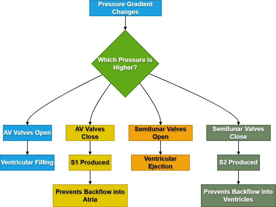
- Pressure changes determine valve opening and closure.
- Blood flows from high pressure → low pressure.
- ECG electrical events precede mechanical contraction.
- Ventricular systole ejects blood into arteries.
- Ventricular diastole allows chamber filling.
🔬 CONCEPT EXPLAINED
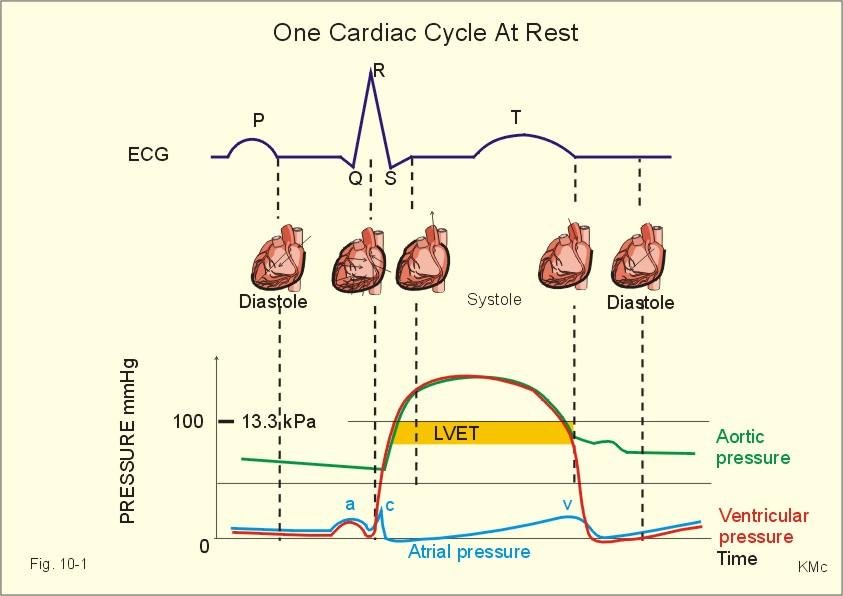
The cardiac cycle represents coordinated electrical and mechanical activity of the heart. Electrical depolarization initiates myocardial contraction, while repolarization leads to relaxation. The atria contract first to complete ventricular filling, followed by powerful ventricular contraction which ejects blood into pulmonary and systemic circulation.
The valves function passively according to pressure gradients. When atrial pressure exceeds ventricular pressure, AV valves open allowing ventricular filling. When ventricular pressure rises during contraction, AV valves close preventing backflow into atria. Similarly, semilunar valves open only when ventricular pressure exceeds arterial pressure.
The cycle ensures efficient unidirectional blood flow and continuous oxygen delivery to tissues.
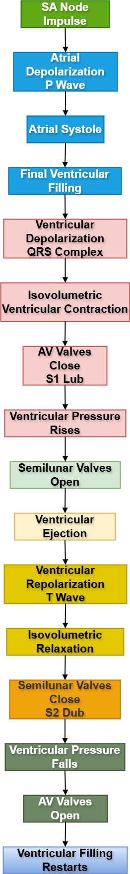
Sequential Events of Cardiac Cycle
1. Ventricular Filling Phase
- Ventricles relaxed
- AV valves open
- Semilunar valves closed
- Blood flows passively from atria to ventricles
- Rapid filling followed by reduced filling (diastasis)
2. Atrial Systole
Initiation
- SA node fires
- P wave appears on ECG
Muscle Activation
- Atrial myocardium contracts
Joint/Mechanical Movement
- Atrial contraction increases ventricular filling
Functional Outcome
- Final 20–30% ventricular filling occurs
3. Isovolumetric Ventricular Contraction
Initiation
- Ventricular depolarization begins
- QRS complex appears
Muscle Activation
- Ventricular myocardium contracts
Movement
- Ventricular pressure rises rapidly
Functional Outcome
- AV valves close producing S1
- All valves closed temporarily
- No change in ventricular volume
4. Ventricular Ejection Phase
- Ventricular pressure exceeds arterial pressure
- Semilunar valves open
- Blood ejected into aorta and pulmonary trunk
- Stroke volume expelled
5. Isovolumetric Relaxation
Initiation
- Ventricular repolarization
- T wave appears
Muscle Relaxation
- Ventricles relax
Movement
- Ventricular pressure falls
Functional Outcome
- Semilunar valves close producing S2
- All valves closed briefly
6. Ventricular Filling Begins Again
- Ventricular pressure becomes lower than atrial pressure
- AV valves reopen
- New cycle starts
⚠️ CLINICAL IMPORTANCE
- Tachycardia shortens diastole reducing ventricular filling.
- Heart failure decreases pumping efficiency.
- Loss of atrial contraction reduces ventricular preload.
- Impaired ventricular relaxation causes diastolic dysfunction.
- Hypertension increases ventricular workload.
Heart Valves and Their Functional Significance
🧠 CORE
- Four valves maintain one-way blood flow.
- AV valves: mitral and tricuspid.
- Semilunar valves: aortic and pulmonary.
- Valves open due to pressure differences.
- Papillary muscles stabilize AV valves.
- Chordae tendineae prevent valve prolapse.
- Valve closure produces heart sounds.
- Proper valve function maintains cardiac efficiency.
🔬 CONCEPT EXPLAINED
Heart valves ensure unidirectional blood movement through the heart chambers. AV valves lie between atria and ventricles, while semilunar valves lie between ventricles and great arteries.
Mitral and Tricuspid Valves
What They Are
Fibrous cusps attached to papillary muscles through chordae tendineae.
How They Work
They open during ventricular diastole when atrial pressure exceeds ventricular pressure. During ventricular systole, rising ventricular pressure closes the valves.
Why They Exist
They prevent backflow of blood into atria during ventricular contraction.
What Happens if They Fail
Regurgitation causes atrial dilation, pulmonary congestion, and reduced cardiac output.
Aortic and Pulmonary Valves
What They Are
Semilunar-shaped cusps located at ventricular outflow tracts.
How They Work
They open during ventricular systole when ventricular pressure exceeds arterial pressure.
Why They Exist
They prevent arterial blood from returning into ventricles during diastole.
What Happens if They Fail
Backflow increases ventricular workload and may lead to hypertrophy and heart failure.
Coordination of Valve Function During Cardiac Cycle
- AV valves open during ventricular filling.
- AV valves close at start of ventricular systole.
- Semilunar valves open during ventricular ejection.
- Semilunar valves close during ventricular relaxation.
This sequential valve movement maintains efficient forward circulation.
⚠️ CLINICAL IMPORTANCE
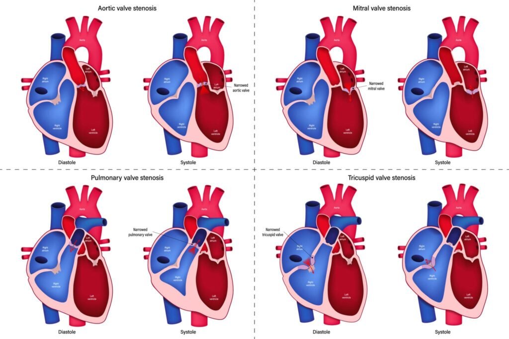
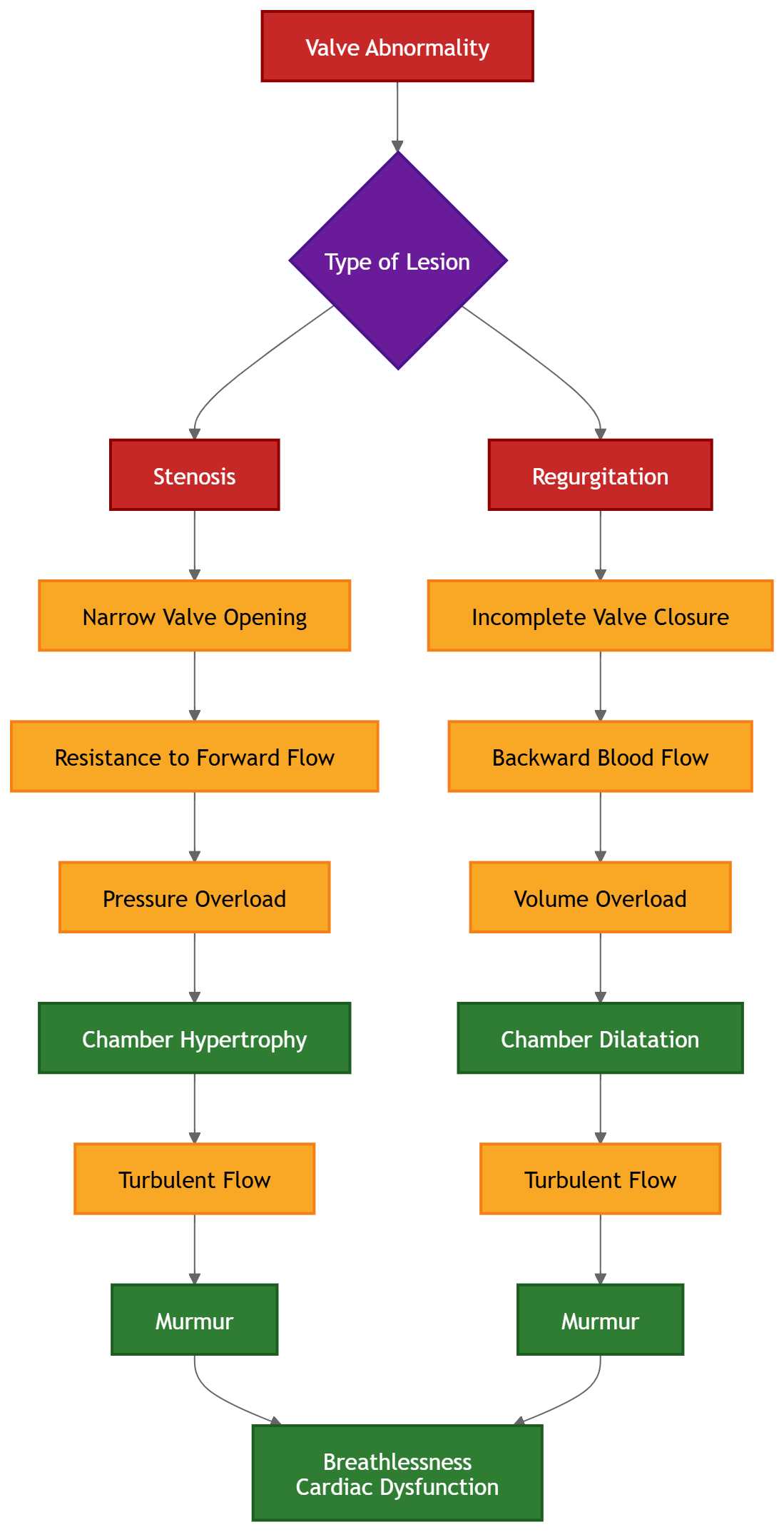
Valve Stenosis
- Valve opening becomes narrow
- Increases resistance to blood flow
- Causes pressure overload
- Leads to chamber hypertrophy
Valve Regurgitation
- Incomplete valve closure
- Causes backward blood flow
- Produces volume overload
- Leads to chamber dilation
Mitral Valve Disease
- Pulmonary congestion
- Breathlessness
- Atrial enlargement
Aortic Valve Disease
- Reduced systemic perfusion
- Syncope
- Left ventricular hypertrophy
Heart Sounds and Murmurs
🧠 CORE
- S1 produced by AV valve closure.
- S2 produced by semilunar valve closure.
- S3 may occur during rapid ventricular filling.
- S4 associated with atrial contraction against stiff ventricle.
- Murmurs result from turbulent blood flow.
- Heart sounds correlate with cardiac cycle phases.
- Auscultation helps diagnose valve disease.
🔬 CONCEPT EXPLAINED
Heart sounds arise mainly due to vibrations produced by valve closure and turbulent blood movement.
First Heart Sound (S1)
Initiation
Start of ventricular systole.
Mechanism
Closure of mitral and tricuspid valves.
Functional Outcome
Prevents backflow into atria.
Second Heart Sound (S2)
Initiation
Beginning of ventricular diastole.
Mechanism
Closure of aortic and pulmonary valves.
Functional Outcome
Prevents arterial backflow into ventricles.
Third Heart Sound (S3)
- Occurs during rapid ventricular filling.
- May be normal in children.
- In adults suggests volume overload.
Fourth Heart Sound (S4)
- Produced by atrial contraction against stiff ventricle.
- Associated with ventricular hypertrophy.
Murmurs
What They Are
Abnormal sounds produced by turbulent blood flow.
Why They Occur
- Stenosis
- Regurgitation
- Septal defects
- Increased flow states
Functional Outcome
Indicate abnormal hemodynamics.
⚠️ CLINICAL IMPORTANCE
- Mitral stenosis produces diastolic murmur.
- Mitral regurgitation causes pansystolic murmur.
- Aortic stenosis causes ejection systolic murmur.
- Aortic regurgitation produces early diastolic murmur.
- Heart sounds help localize valve pathology clinically.
Correlation of Cardiac Cycle with ECG
🧠 CORE
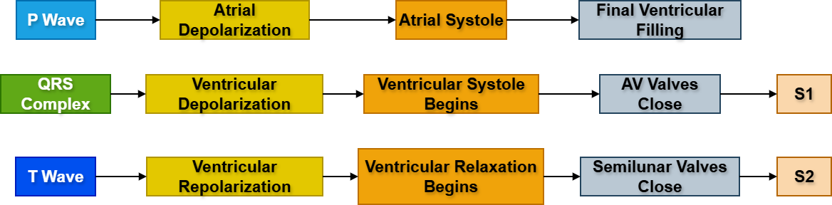
- Electrical events precede contraction.
- P wave represents atrial depolarization.
- QRS complex represents ventricular depolarization.
- T wave represents ventricular repolarization.
- Mechanical contraction follows depolarization.
- ECG helps assess rhythm and cardiac timing.
🔬 CONCEPT EXPLAINED
The ECG records electrical activity while the cardiac cycle represents mechanical activity. Electrical excitation stimulates myocardial contraction.
ECG and Mechanical Correlation
| ECG Event | Mechanical Event |
|---|---|
| P wave | Atrial systole |
| QRS complex | Ventricular systole |
| T wave | Ventricular relaxation |
Sequential Logic
- SA node initiates impulse.
- Atria depolarize and contract.
- AV node delays impulse.
- Ventricles depolarize and contract.
- Ventricles repolarize and relax.
This coordination ensures proper chamber filling before ventricular ejection.
⚠️ CLINICAL IMPORTANCE
- Arrhythmias disturb cardiac cycle coordination.
- AV block alters atrioventricular timing.
- Ventricular fibrillation abolishes effective pumping.
- ECG abnormalities may indicate ischemia or hypertrophy.
Metabolism and Oxygen Utilization of Cardiac Muscle
🧠 CORE
- Cardiac muscle has high metabolic demand.
- Aerobic metabolism predominates.
- Myocardium requires continuous oxygen supply.
- Coronary circulation supplies nutrients.
- ATP required for continuous contraction.
- Cardiac muscle contains many mitochondria.
🔬 CONCEPT EXPLAINED
Cardiac muscle works continuously throughout life and therefore requires enormous energy production. The myocardium primarily uses aerobic metabolism because oxygen efficiently generates ATP necessary for repeated contraction-relaxation cycles.
The heart extracts a very high percentage of oxygen from coronary blood. During exercise, increased oxygen demand is mainly met by increasing coronary blood flow rather than increasing oxygen extraction.
Functional Importance
- Continuous ATP production maintains pumping ability.
- Adequate oxygen prevents ischemia.
- Mitochondria support sustained contraction.
Failure of Oxygen Supply
Reduced coronary blood flow causes ischemia, impaired contraction, arrhythmias, and myocardial infarction.
⚠️ CLINICAL IMPORTANCE
- Coronary artery disease reduces oxygen delivery.
- Myocardial ischemia impairs ventricular contraction.
- Infarction causes irreversible muscle damage.
- Hypoxia may precipitate arrhythmias.
Regulation of Cardiac Cycle
🧠 CORE
- SA node acts as pacemaker.
- Autonomic nervous system regulates heart rate.
- Sympathetic stimulation increases cardiac activity.
- Parasympathetic stimulation slows heart rate.
- Hormones influence cardiac performance.
- Venous return affects stroke volume.
🔬 CONCEPT EXPLAINED
Cardiac activity is regulated intrinsically and extrinsically to meet body demands.
Intrinsic Regulation
Frank-Starling mechanism states that increased ventricular filling stretches myocardial fibers leading to stronger contraction.
Extrinsic Regulation
Sympathetic Stimulation
- Norepinephrine released
- SA node firing increases
- Contractility increases
- Cardiac output rises
Parasympathetic Stimulation
- Vagus nerve releases acetylcholine
- SA node activity decreases
- Heart rate slows
- Cardiac output decreases
Functional Outcome
The body adjusts cardiac output according to exercise, stress, and metabolic demand.
⚠️ CLINICAL IMPORTANCE
- Excess sympathetic activity may cause tachycardia.
- Vagal stimulation may cause bradycardia.
- Heart failure impairs regulatory compensation.
- Autonomic imbalance contributes to arrhythmias.
⚙️ 4️⃣ Functional Flow
Structure → Function → Outcome
AV Valves
Structure:
Thin cusps with chordae tendineae.
Function:
Prevent backflow during systole.
Outcome:
Efficient forward ventricular ejection.
Semilunar Valves
Structure:
Pocket-shaped cusps.
Function:
Prevent arterial backflow during diastole.
Outcome:
Maintains arterial circulation.
Ventricular Myocardium
Structure:
Thick muscular walls with abundant mitochondria.
Function:
Generate strong contraction.
Outcome:
Effective systemic and pulmonary circulation.
SA Node
Structure:
Specialized pacemaker cells.
Function:
Initiate rhythmic impulses.
Outcome:
Coordinated heartbeat.
🩺 5️⃣ Clinical Correlation
Mitral Stenosis
- Narrowed mitral valve obstructs ventricular filling.
- Causes left atrial enlargement and pulmonary congestion.
- Produces diastolic murmur.
Mitral Regurgitation
- Incomplete valve closure causes backflow.
- Leads to volume overload.
- Causes pansystolic murmur.
Aortic Stenosis
- Increased resistance to ventricular ejection.
- Causes left ventricular hypertrophy.
- May cause syncope and angina.
Aortic Regurgitation
- Blood flows back into ventricle during diastole.
- Causes ventricular dilation.
- Produces early diastolic murmur.
Heart Failure
- Reduced pumping efficiency.
- Leads to breathlessness and edema.
- Often associated with valvular disease.
📌 6️⃣ Summary Points
- Cardiac cycle consists of systole and diastole.
- Pressure gradients determine valve movement.
- S1 = closure of AV valves.
- S2 = closure of semilunar valves.
- QRS complex precedes ventricular contraction.
- Semilunar valves open during ventricular systole.
- Tachycardia shortens diastole most significantly.
- Murmurs are caused by turbulent blood flow.
- Mitral valve disease commonly causes pulmonary congestion.
- Coronary blood flow mainly occurs during diastole.
- Cardiac muscle depends heavily on aerobic metabolism.
- SA node is the normal pacemaker of the heart.