
“`html
🩺 Station 3 — Chambers of the Heart
AIM OSPE/OSCE Lab — Practical Station | KMU Style | MBBS Practical + Viva
📌 Station Overview
Module: Cardiovascular System
Year: 1st Year MBBS
Focus: Identification • Procedure • Interpretation • Viva
Total Marks: 5
📋 Complete OSPE Station Content
Learning Target
By the end of this station, the student should be able to:
- Identify the four chambers of the heart on an anatomical model or diagram.
- Relate each chamber with its receiving and outgoing blood pathway.
Required Material
- Anatomical heart model showing internal chambers
- Opened heart model or labelled/unlabelled diagram
- Pointer
- Answer sheet
- Station instruction card
Student Task / Procedure
- Observe the given heart model carefully.
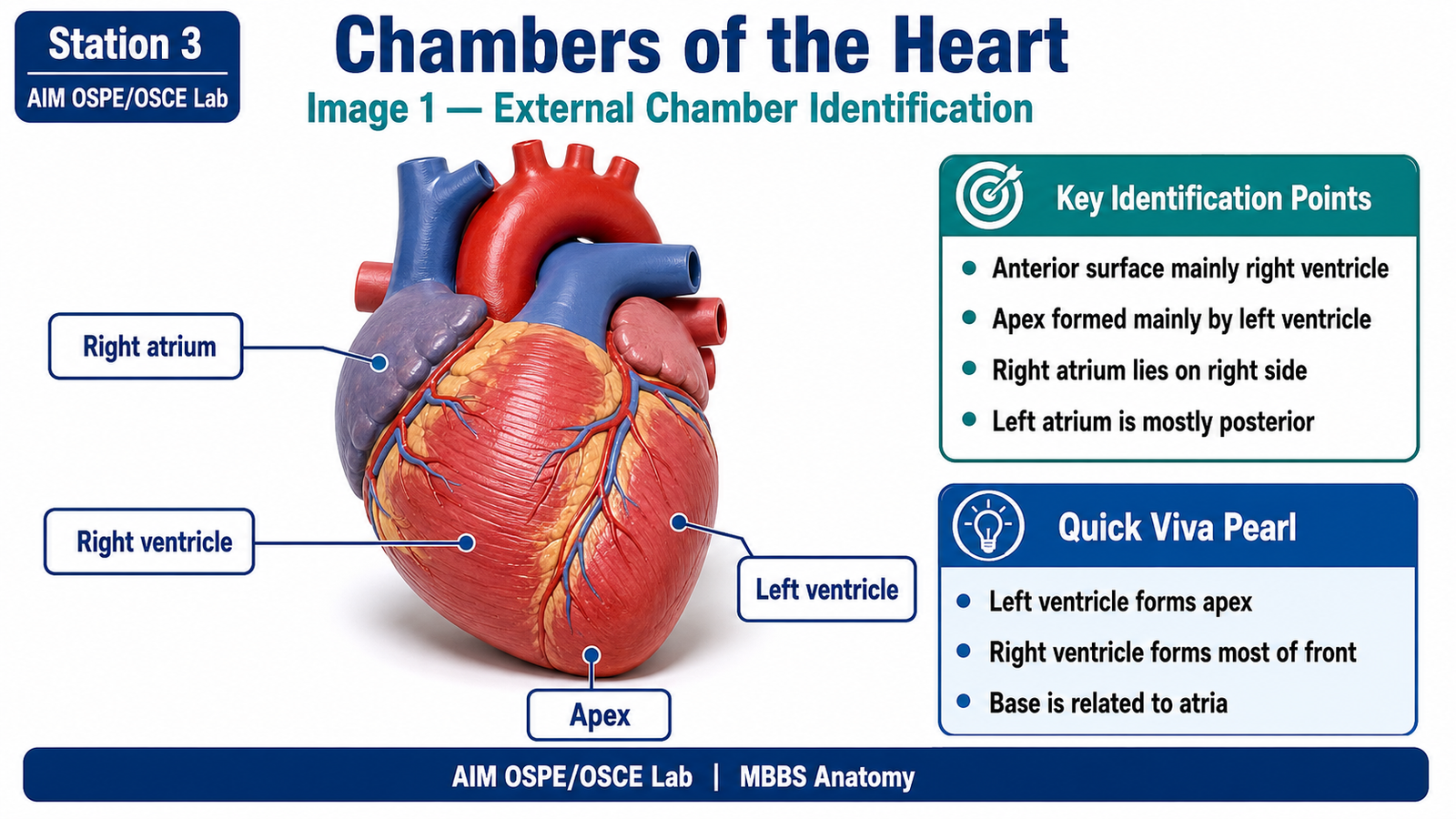
- Identify the following chambers:
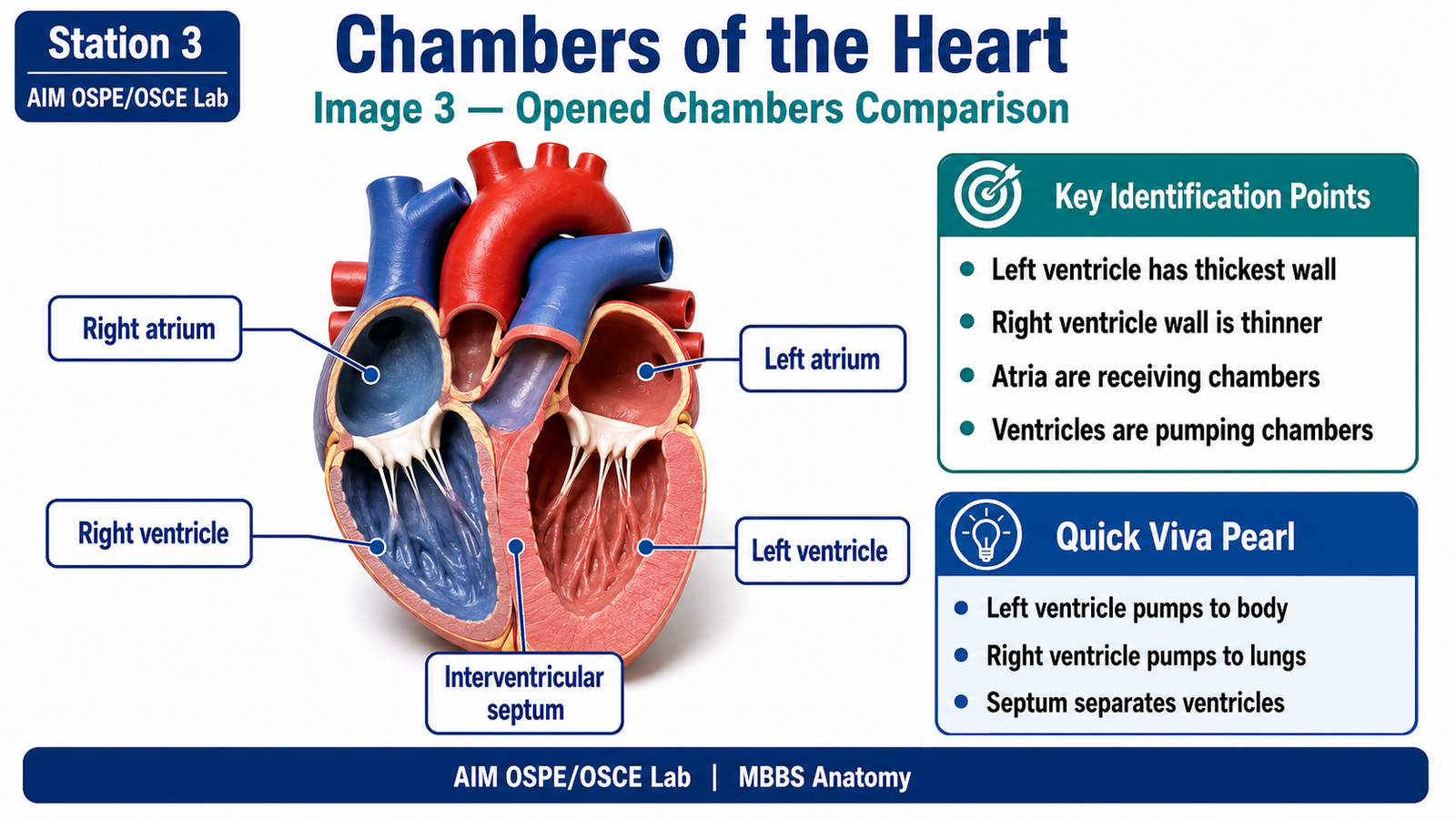
- Right atrium
- Right ventricle
- Left atrium
- Left ventricle
- Point to each chamber using the pointer.
- State which chamber receives venous blood from the body.
- State which chamber pumps oxygenated blood to the body.
Observation / Identification Points
Students should identify or demonstrate:
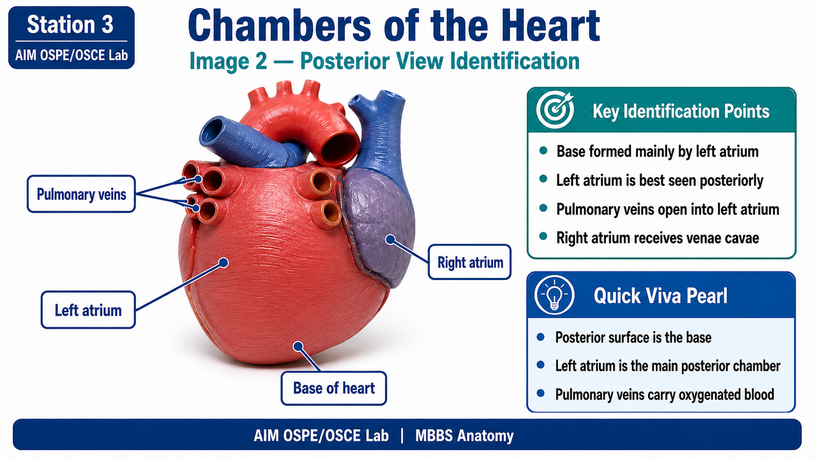
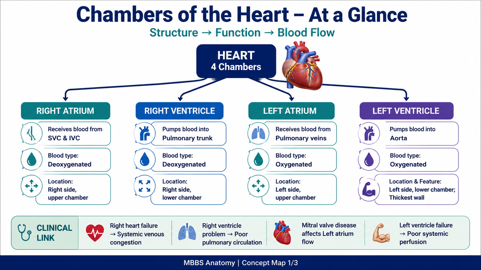
- Right atrium receives blood from superior and inferior vena cava
- Right ventricle pumps blood into the pulmonary trunk
- Left atrium receives oxygenated blood from pulmonary veins
- Left ventricle pumps blood into the aorta
- Right ventricle forms most of the anterior surface of the heart
- Left atrium forms most of the base of the heart
- Left ventricle has the thickest wall
- Interventricular septum separates right and left ventricles
Result / Interpretation
The heart has four chambers arranged into right and left functional pumps.
The right side receives deoxygenated blood and sends it to the lungs.
The left side receives oxygenated blood and pumps it to the systemic circulation.
The left ventricle has the thickest wall because it pumps blood to the whole body.
Viva Questions
1. Which chamber receives blood from the superior and inferior vena cava?
Answer: Right atrium.
2. Which chamber pumps blood into the pulmonary trunk?
Answer: Right ventricle.
3. Which chamber receives blood from pulmonary veins?
Answer: Left atrium.
4. Which chamber has the thickest wall?
Answer: Left ventricle.
5. Why is the wall of the left ventricle thicker than the right ventricle?
Answer: Because it pumps blood to the systemic circulation under higher pressure.
Marking Scheme
Total Marks: 5
| Component | Marks |
|---|---|
| Correct identification / performance | 2 |
| Key observation / procedure steps | 1 |
| Interpretation / principle | 1 |
| Viva answer | 1 |
Common Student Mistakes
- Confusing right and left sides of the heart on the model.
- Forgetting that the left ventricle has the thickest wall.
- Confusing atria with ventricles during internal chamber identification.
AIM Feedback
Revise the heart chambers by linking each chamber with its blood pathway. Remember: right atrium receives venae cavae, right ventricle sends blood to lungs, left atrium receives pulmonary veins, and left ventricle sends blood to body through the aorta. Wall thickness is an important identification clue, especially for recognizing the left ventricle.
Short Caption
Identify the four chambers and remember their blood flow: right atrium → right ventricle → lungs → left atrium → left ventricle → body.
🖼️ Visual / Image Support
🧩 Concept Map / Interpretation Support
🎥 Video Demonstration / Procedure Support
Watch this heart model video before attempting Station 3. Focus on identifying the four chambers: right atrium, right ventricle, left atrium, and left ventricle.