
🩺 Station 2 — Heart and Major Blood Vessels
AIM OSPE/OSCE Lab — Practical Station | KMU Style | MBBS Practical + Viva
📌 Station Overview
Module: Cardiovascular System
Year: 1st Year MBBS
Focus: Identification • Procedure • Interpretation • Viva
Total Marks: 5
📋 Complete OSPE Station Content
Learning Target
By the end of this station, the student should be able to:
- Identify the major blood vessels attached to the heart on a model or diagram.
- State the direction of blood flow and basic functional significance of each vessel.
Required Material
- Anatomical heart model showing major vessels
- Unlabelled heart diagram/image
- Pointer
- Answer sheet
- Station instruction card
Student Task / Procedure
- Observe the given heart model carefully.
- Identify the following major blood vessels:
- Aorta
- Pulmonary trunk
- Superior vena cava
- Inferior vena cava
- Pulmonary veins
- Point to each vessel using the pointer.
- State whether each vessel carries blood to or away from the heart.
- Mention whether the vessel carries mainly oxygenated or deoxygenated blood.
Observation / Identification Points
Students should identify or demonstrate:
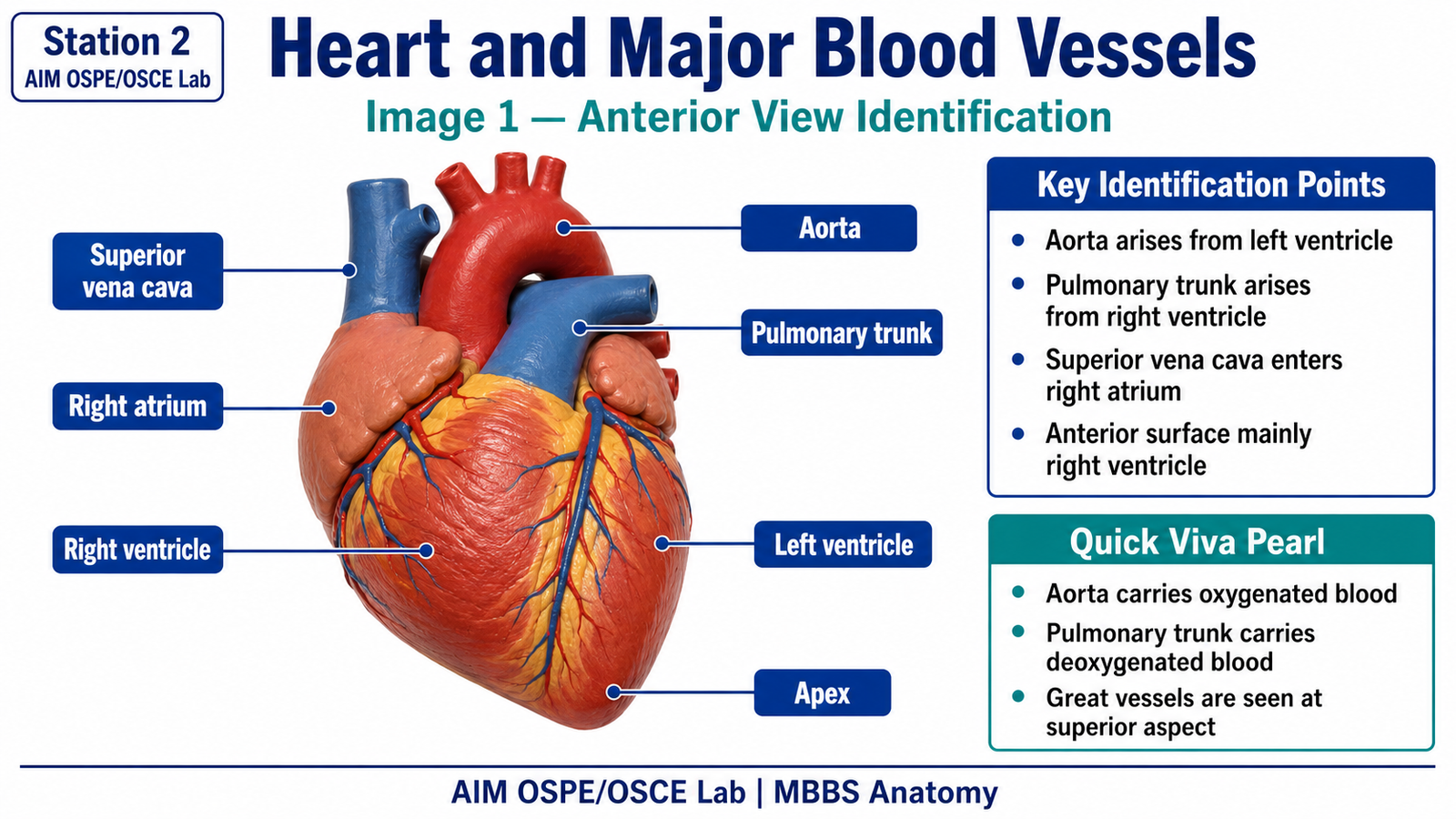
- Aorta arising from the left ventricle
- Pulmonary trunk arising from the right ventricle
- Superior vena cava entering the right atrium from above
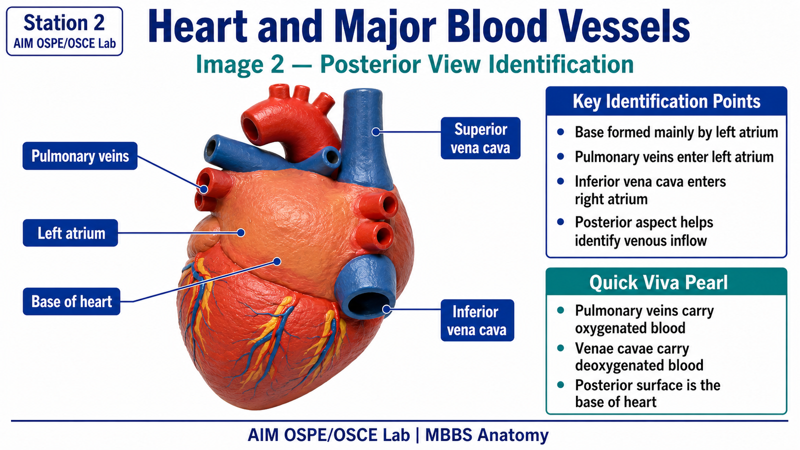
- Inferior vena cava entering the right atrium from below
- Pulmonary veins entering the left atrium
- Aorta carries oxygenated blood to the systemic circulation
- Pulmonary trunk carries deoxygenated blood to the lungs
- Pulmonary veins carry oxygenated blood from lungs to left atrium
- Venae cavae carry deoxygenated blood to right atrium
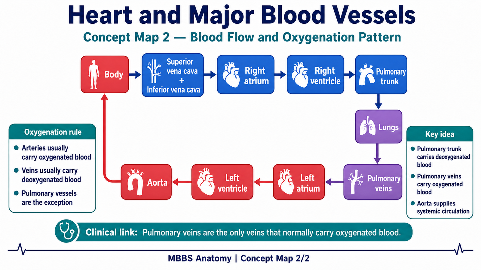
Result / Interpretation
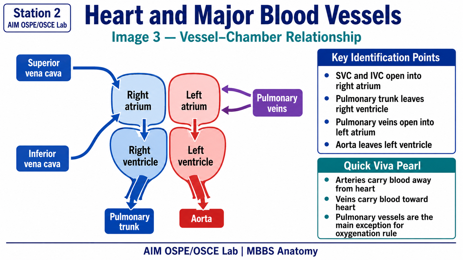
The major blood vessels of the heart maintain the direction of circulation.
The right side of the heart receives deoxygenated blood and sends it to the lungs through the pulmonary trunk.
The left side of the heart receives oxygenated blood through the pulmonary veins and pumps it to the body through the aorta.
Viva Questions
1. Which vessel arises from the left ventricle?
Answer: Aorta.
2. Which vessel carries deoxygenated blood from the right ventricle to the lungs?
Answer: Pulmonary trunk.
3. Which chamber receives the superior and inferior vena cava?
Answer: Right atrium.
4. Which vessels bring oxygenated blood from the lungs to the heart?
Answer: Pulmonary veins.
5. Why are pulmonary veins unusual?
Answer: They are veins but carry oxygenated blood.
Marking Scheme
Total Marks: 5
| Component | Marks |
|---|---|
| Correct identification / performance | 2 |
| Key observation / procedure steps | 1 |
| Interpretation / principle | 1 |
| Viva answer | 1 |
Common Student Mistakes
- Confusing pulmonary trunk with aorta.
- Thinking all arteries carry oxygenated blood and all veins carry deoxygenated blood.
- Forgetting that pulmonary veins enter the left atrium.
AIM Feedback
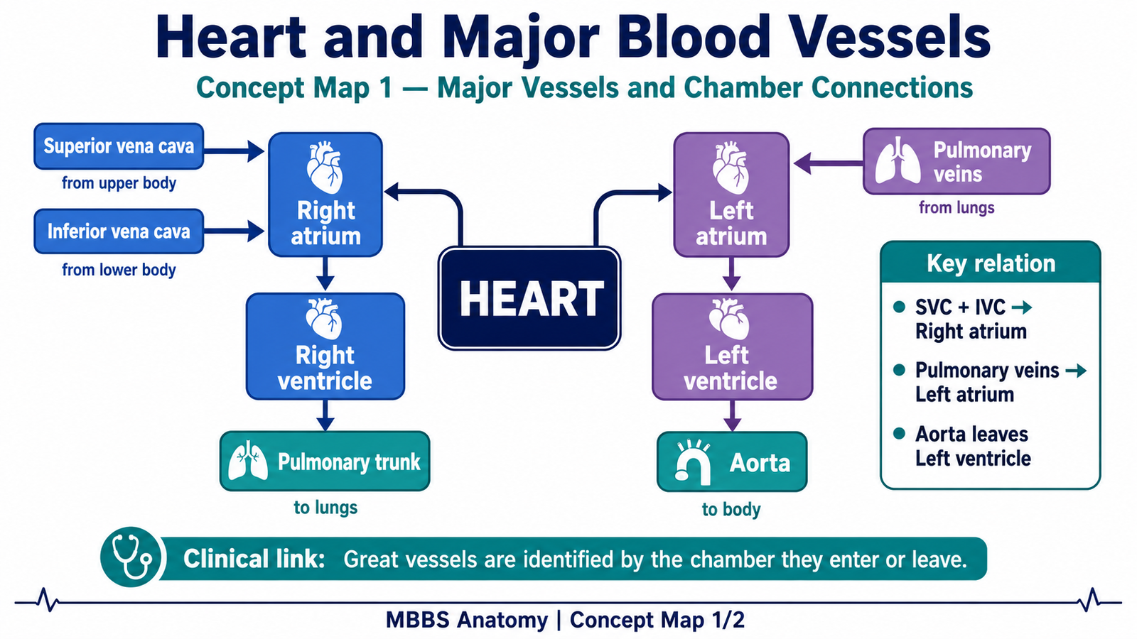
Revise the major vessels by linking each vessel with its chamber and blood direction. Remember: aorta leaves left ventricle, pulmonary trunk leaves right ventricle, vena cavae enter right atrium, and pulmonary veins enter left atrium. This chamber-vessel relationship is the key to identifying them confidently in OSPE.
Short Caption
Identify each major vessel by its chamber connection: aorta from left ventricle, pulmonary trunk from right ventricle, venae cavae to right atrium, and pulmonary veins to left atrium.
🖼️ Visual / Image Support
🧩 Concept Map / Interpretation Support
🎥 Video Demonstration / Procedure Support