
🩺 Station 4 — Internal Structures of Heart Chambers
AIM OSPE/OSCE Lab — Practical Station | KMU Style | MBBS Practical + Viva
📌 Station Overview
Module: Cardiovascular System
Year: 1st Year MBBS
Focus: Identification • Procedure • Interpretation • Viva
Total Marks: 5
📋 Complete OSPE Station Content
Learning Target
By the end of this station, the student should be able to:
- Identify the major internal structures of heart chambers on a heart model or specimen.
- Explain the functional and clinical importance of papillary muscles, chordae tendineae, trabeculae carneae, pectinate muscles, interventricular septum, and valves.
Required Material
- Anatomical heart model or dissected heart specimen
- Pointer
- Labeled/unlabeled diagram of internal heart chambers
- Station instruction sheet
- Marking checklist
Student Task / Procedure
- Observe the internal aspect of the heart model/specimen.
- Identify the following structures:
- Papillary muscles
- Chordae tendineae
- Trabeculae carneae
- Pectinate muscles
- Interventricular septum
- Heart valves
- Point out the chamber where each structure is mainly found.
- State one function or clinical importance of any two identified structures.
Observation / Identification Points
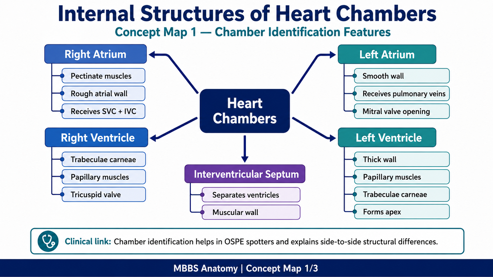
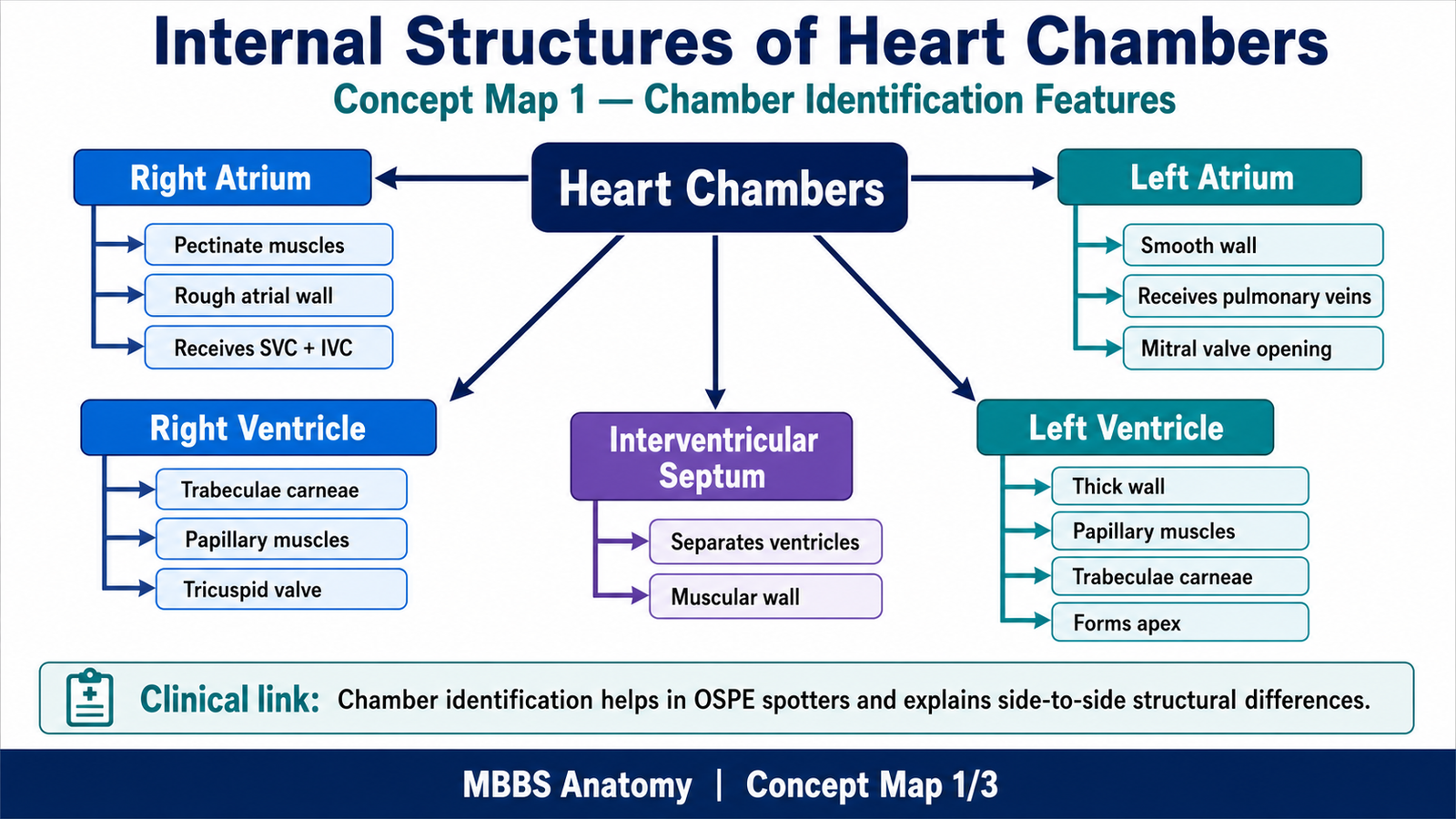
The student should identify and observe:
- Papillary muscles: Muscular projections in ventricles attached to chordae tendineae.
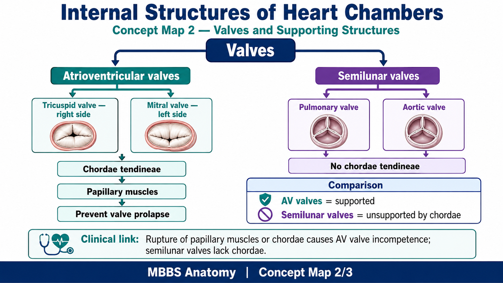
- Chordae tendineae: Fibrous cords connecting AV valve cusps to papillary muscles.
- Trabeculae carneae: Irregular muscular ridges inside ventricles.
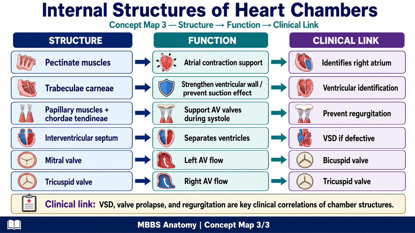
- Pectinate muscles: Comb-like muscular ridges mainly in atria, especially right atrium.
- Interventricular septum: Wall separating right and left ventricles.
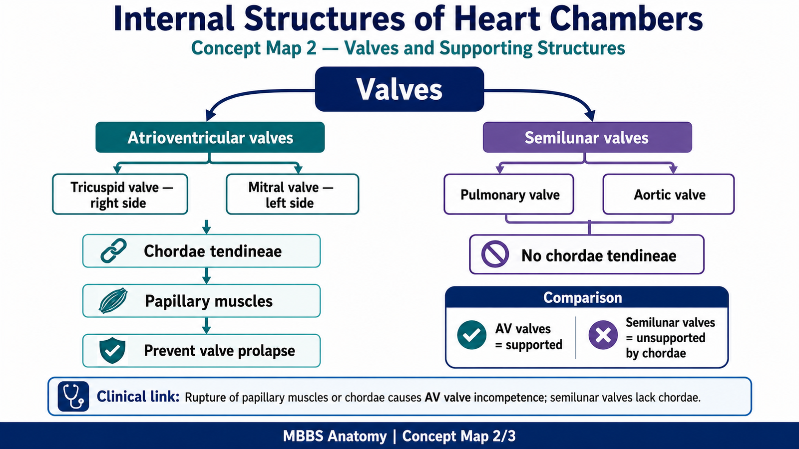
- Valves: AV valves and semilunar valves controlling one-way blood flow.
Result / Interpretation
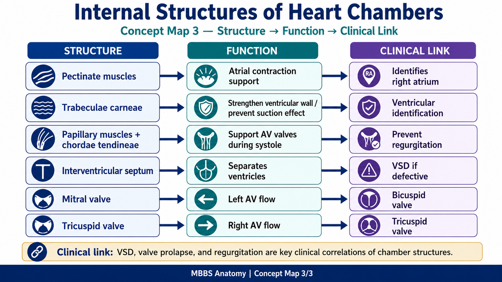
Internal structures of heart chambers help maintain efficient one-way blood flow.
Papillary muscles and chordae tendineae prevent prolapse of AV valves during ventricular systole.
Trabeculae carneae increase internal muscular surface of ventricles.
Pectinate muscles are important identifying features of atria.
The interventricular septum separates the two ventricles and contains part of the conducting system.
Clinically, damage to papillary muscles or chordae tendineae may cause valve incompetence and regurgitation.
Viva Questions
1. What is the function of chordae tendineae?
They prevent prolapse of AV valve cusps during ventricular contraction.
2. Where are papillary muscles found?
They are found in the ventricles.
3. What are trabeculae carneae?
They are irregular muscular ridges present inside the ventricles.
4. Which chamber commonly shows pectinate muscles clearly?
Right atrium, especially in the auricle and anterior wall.
5. What is the clinical importance of the interventricular septum?
It separates the ventricles and may be involved in ventricular septal defect.
Marking Scheme
Total Marks: 5
| Component | Marks |
|---|---|
| Correct identification / performance | 2 |
| Key observation / procedure steps | 1 |
| Interpretation / principle | 1 |
| Viva answer | 1 |
Common Student Mistakes
- Confusing pectinate muscles with trabeculae carneae.
- Identifying chordae tendineae as valve cusps.
- Forgetting that papillary muscles are ventricular structures.
- Not relating papillary muscles and chordae tendineae to prevention of valve prolapse.
AIM Feedback
Revise the internal structures chamber-wise. Remember that atria mainly show pectinate muscles, while ventricles contain papillary muscles, chordae tendineae, and trabeculae carneae. Always connect structure with function: AV valves need chordae tendineae and papillary muscles to prevent backward flow during ventricular systole.
Short Caption
Use this labeled image to revise the internal structures of heart chambers. Focus on differentiating atrial pectinate muscles from ventricular trabeculae carneae and understanding how papillary muscles and chordae tendineae support AV valves.
🖼️ Visual / Image Support
🧩 Concept Map / Interpretation Support
🎥 Video Demonstration / Procedure Support