🩺 Station 17 — Basic Life Support
AIM OSPE/OSCE Lab — Practical Station | KMU Style | MBBS Practical + Viva
📌 Station Overview
Module: Cardiovascular System
Year: 1st Year MBBS
Focus: Identification • Procedure • Interpretation • Viva
Total Marks: 5
📋 Complete OSPE Station Content
Learning Target
By the end of this station, the student should be able to:
- Demonstrate the initial adult BLS sequence: check response, call for help, assess airway and breathing, and start chest compressions.
- Explain the basic principle of chest compressions in maintaining temporary circulation during cardiac arrest.
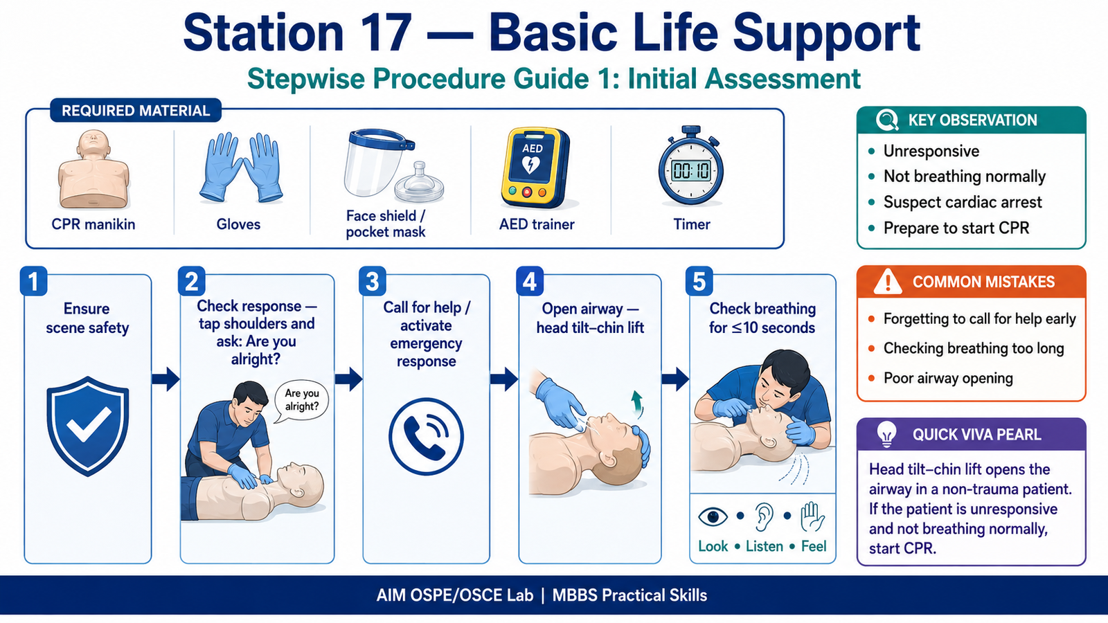
Required Material
- CPR manikin / simulator
- Clean floor or firm examination couch
- Gloves
- Face shield / pocket mask, if available
- AED trainer, if available
- Stopwatch or timer
- Checklist for examiner
Student Task / Procedure
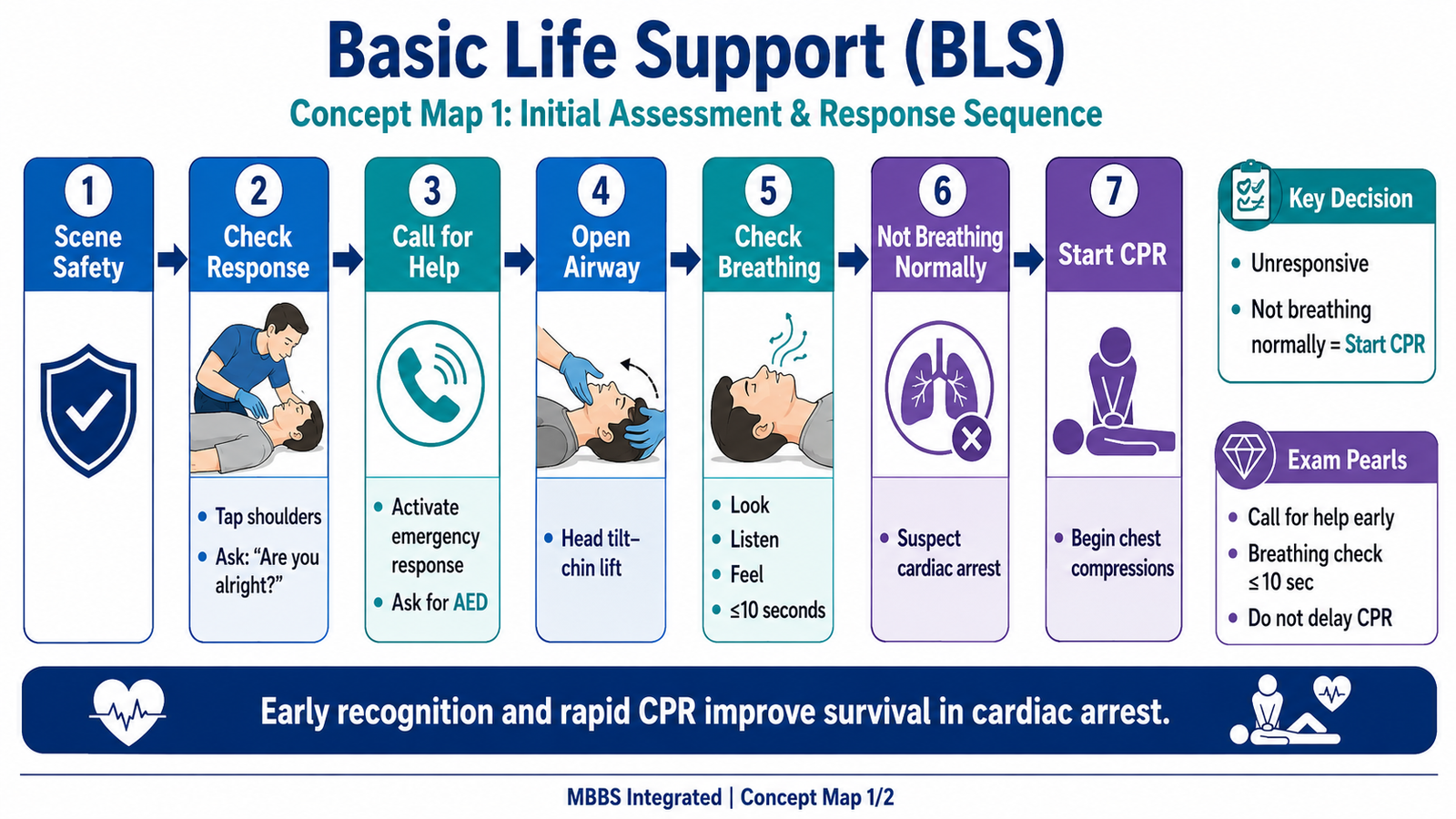
- Ensure the scene is safe.
- Approach the patient/manikin and check response by tapping shoulders and speaking loudly: “Are you alright?”
- If no response, call for help loudly and ask someone to activate emergency response.
- Position the patient on a firm surface.
- Open the airway using head tilt–chin lift if no trauma is suspected.
- Check breathing for no more than 10 seconds by looking for chest movement and listening/feeling for breathing.
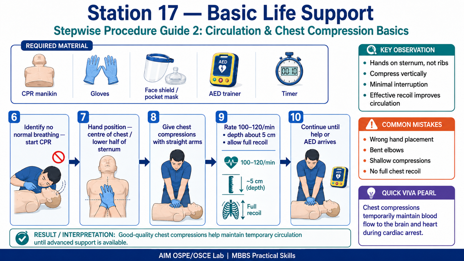
- If not breathing normally, identify cardiac arrest and prepare for CPR.
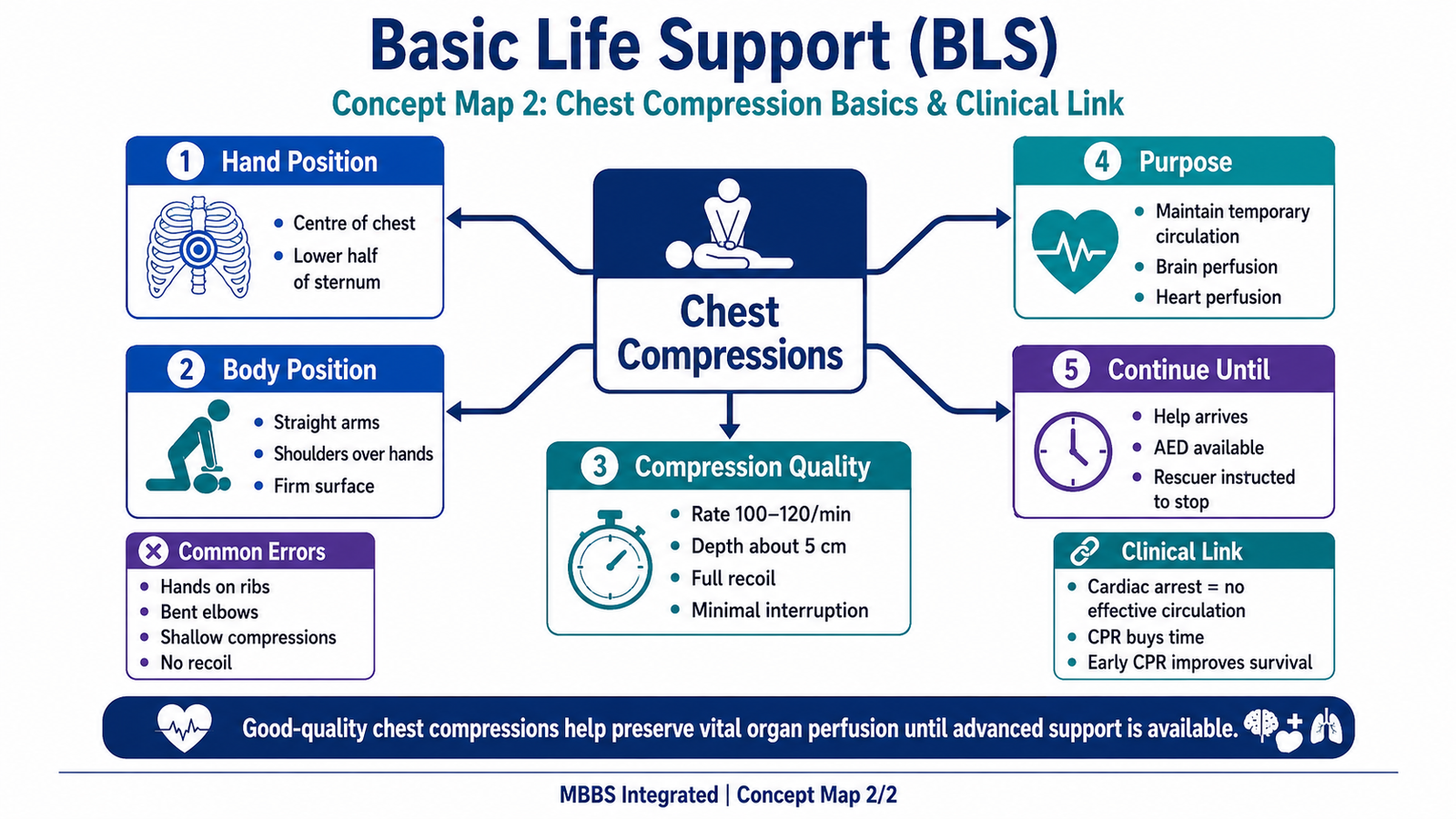
- Place heel of one hand on the centre of the chest/lower half of sternum.
- Place the other hand on top, interlock fingers, keep elbows straight.
- Give chest compressions vertically with proper depth, rate, recoil, and minimal interruption.
- Continue CPR until help/AED arrives or examiner stops the station.
Adult BLS guidance emphasizes early recognition, calling for help, checking breathing, and starting CPR when normal breathing is absent; adult chest compressions are commonly taught at 100–120/min with about 5 cm depth in an average adult.
Observation / Identification Points
The student should demonstrate:
- Safe approach to the patient
- Correct response check
- Clear call for help
- Correct airway opening technique
- Breathing check within 10 seconds
- Correct recognition of absent/abnormal breathing
- Correct hand position on sternum
- Straight arms and vertical compression technique
- Adequate compression rate and depth
- Full chest recoil after each compression
- Minimal interruption during compressions
Result / Interpretation
If an adult patient is unresponsive and not breathing normally, the student should interpret this as possible cardiac arrest and start Basic Life Support immediately.
Clinical significance:
Chest compressions provide temporary blood flow to the brain and heart until definitive help, AED, or advanced resuscitation becomes available.
Viva Questions
Q1. What is the first step before approaching a collapsed patient?
A: Ensure scene safety.
Q2. How do you check response in BLS?
A: Tap the shoulders and speak loudly, such as “Are you alright?”
Q3. Why is the airway opened before checking breathing?
A: To remove tongue-related airway obstruction and allow proper assessment of breathing.
Q4. Where should hands be placed for adult chest compressions?
A: On the centre of the chest, over the lower half of the sternum.
Q5. What is the purpose of chest compressions?
A: To maintain temporary circulation to vital organs, especially the brain and heart.
Marking Scheme
Total Marks: 5
| Component | Marks |
|---|---|
| Correct identification / performance | 2 |
| Key observation / procedure steps | 1 |
| Interpretation / principle | 1 |
| Viva answer | 1 |
Checklist breakdown for examiner:
- Checks response and calls for help correctly
- Opens airway and checks breathing properly
- Recognizes absent/abnormal breathing
- Places hands correctly on sternum
- Performs basic chest compression technique correctly
Common Student Mistakes
- Forgetting to call for help early
- Checking breathing for too long
- Wrong hand placement on ribs or xiphisternum
- Bending elbows during compressions
- Not allowing full chest recoil
- Performing shallow or very slow compressions
AIM Feedback
In BLS, speed and sequence matter. First confirm unresponsiveness, call for help, open the airway, check breathing quickly, and start effective chest compressions if breathing is absent or abnormal. Good compressions need correct hand position, straight arms, adequate depth, proper rate, and full recoil.
🖼️ Visual / Image Support
🧩 Concept Map / Interpretation Support