🩺 Station 9 — Surface Marking of Heart Borders and Valves
AIM OSPE/OSCE Lab — Practical Station | KMU Style | MBBS Practical + Viva
📌 Station Overview
Module: Cardiovascular System
Year: 1st Year MBBS
Focus: Identification • Procedure • Interpretation • Viva
Total Marks: 5
📋 Complete OSPE Station Content
Learning Target
By the end of this station, the student should be able to:
- Mark the surface borders of the heart correctly on a chest model/simulator.
- Identify the clinical valve auscultation areas and explain their anatomical importance.
Required Material
- Chest wall model / torso simulator
- Washable marker or skin pencil
- Pointer
- Measuring tape or ruler
- Anterior chest wall landmark diagram
- Examiner checklist
Student Task / Procedure
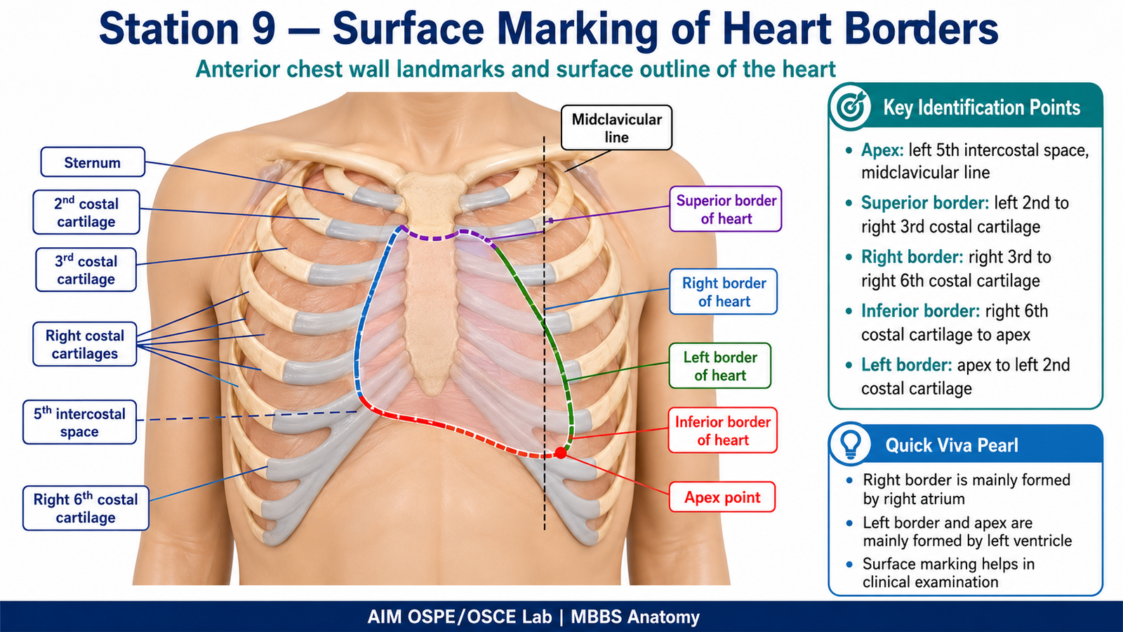
- Identify the sternum, costal cartilages, intercostal spaces, and midclavicular line on the model.
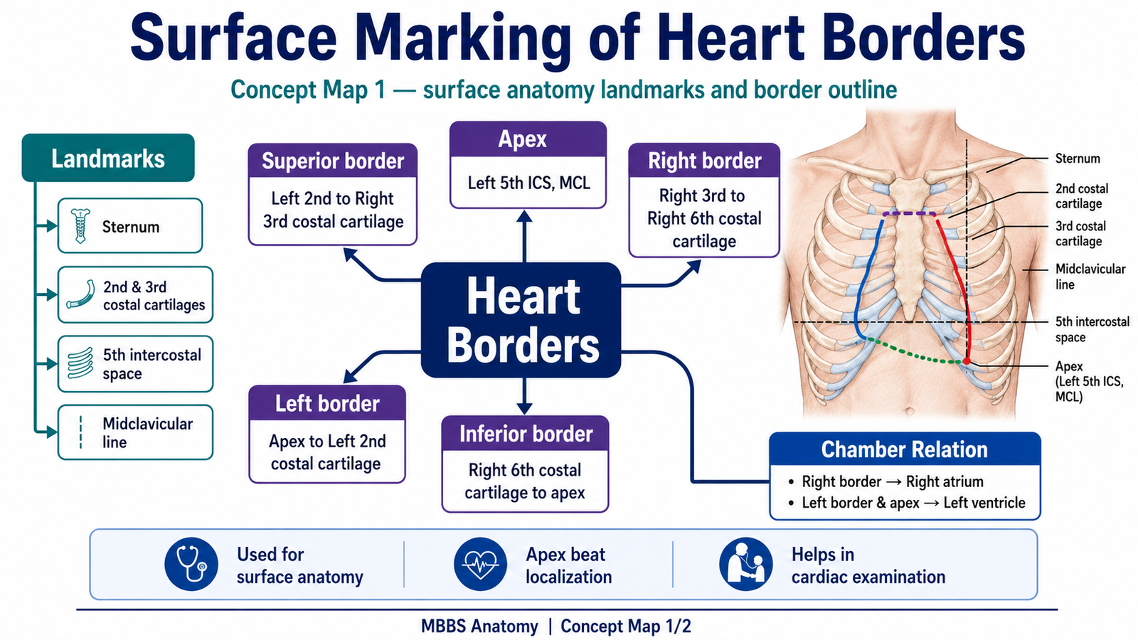
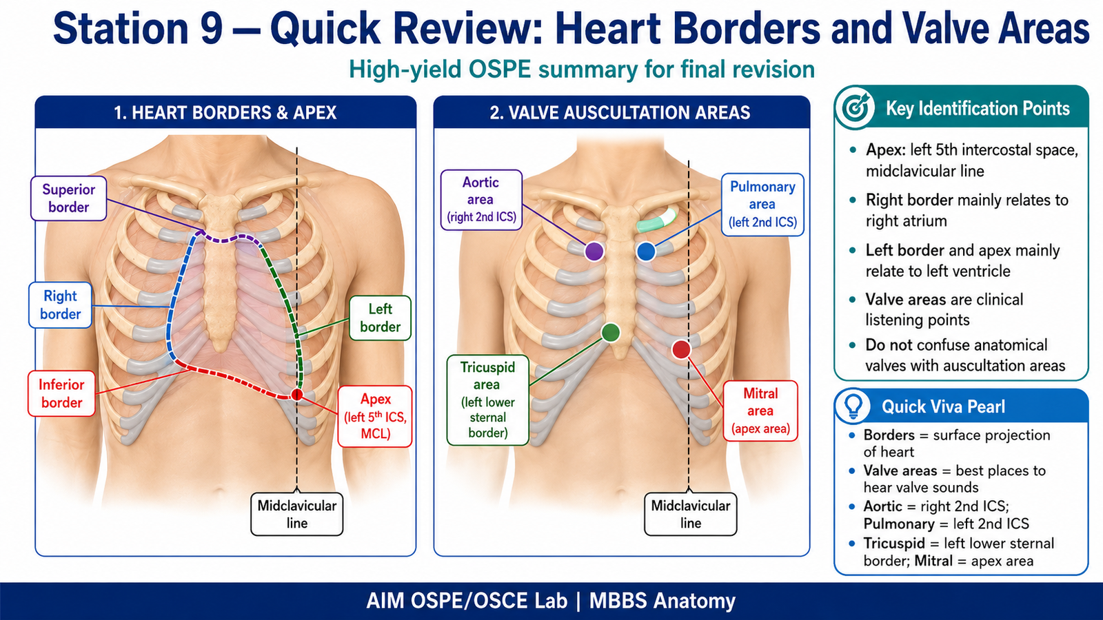
- Mark the apex of the heart at the left 5th intercostal space in the midclavicular line.
- Mark the superior border from the left 2nd costal cartilage to the right 3rd costal cartilage near the sternum.
- Mark the right border from the right 3rd costal cartilage to the right 6th costal cartilage near the sternum.
- Mark the inferior border from the right 6th costal cartilage to the apex point.
- Mark the left border from the apex point to the left 2nd costal cartilage.
- Mark the four valve auscultation areas:
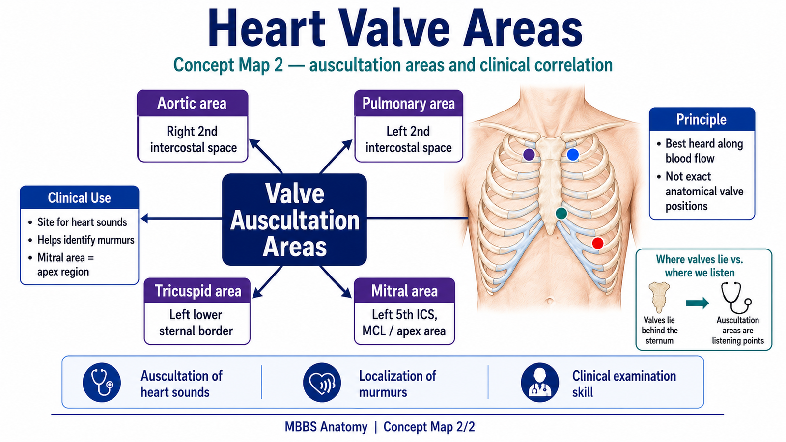
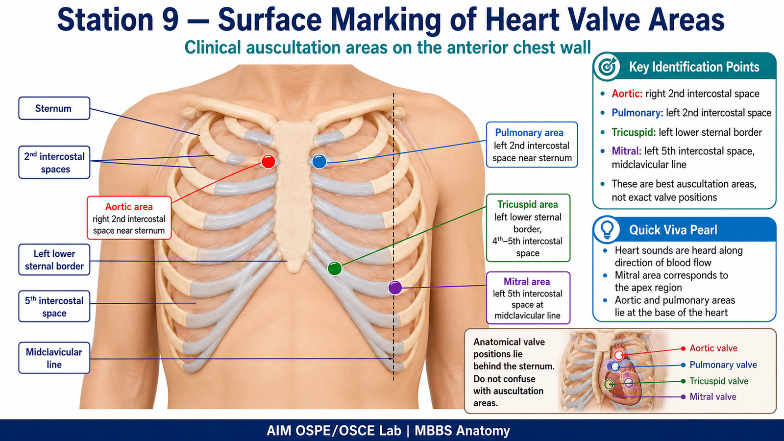
- Aortic area: Right 2nd intercostal space near sternum
- Pulmonary area: Left 2nd intercostal space near sternum
- Tricuspid area: Left lower sternal border, 4th–5th intercostal space
- Mitral area: Left 5th intercostal space at midclavicular line
Observation / Identification Points
The student should correctly demonstrate:
- Identification of key anterior chest wall landmarks
- Correct marking of the apex point
- Correct outline of the superior, right, inferior, and left borders of the heart
- Correct identification of aortic, pulmonary, tricuspid, and mitral auscultation areas
- Understanding that valve auscultation areas are best listening points, not exact anatomical valve positions
Result / Interpretation
The surface marking represents the approximate projection of the heart on the anterior chest wall.
The valve areas are clinically important because heart sounds and murmurs are best heard where sound is transmitted along the direction of blood flow.
This station integrates surface anatomy with cardiovascular clinical examination.
Viva Questions
Q1. Where is the apex of the heart marked?
A: Left 5th intercostal space in the midclavicular line.
Q2. Which chamber mainly forms the right border of the heart?
A: Right atrium.
Q3. Which chamber mainly forms the left border and apex?
A: Left ventricle.
Q4. Where is the aortic area auscultated?
A: Right 2nd intercostal space near the sternum.
Q5. Why are valve sounds not heard exactly over anatomical valve positions?
A: Because sounds are transmitted in the direction of blood flow.
Marking Scheme
Total Marks: 5
| Component | Marks |
|---|---|
| Correct identification / performance | 2 |
| Key observation / procedure steps | 1 |
| Interpretation / principle | 1 |
| Viva answer | 1 |
Common Student Mistakes
- Marking the apex too medial, too lateral, or too low.
- Confusing aortic and pulmonary auscultation areas.
- Confusing anatomical valve positions with clinical auscultation areas.
AIM Feedback
First identify chest wall landmarks before marking the heart.
Remember the key OSPE anchor point: apex = left 5th intercostal space at midclavicular line.
For valve areas, revise clinically: Aortic right 2nd ICS, Pulmonary left 2nd ICS, Tricuspid left lower sternal border, Mitral apex area.
Short Caption
This labeled image summarizes the OSPE landmarks for surface marking of heart borders and cardiac valve auscultation areas.
🖼️ Visual / Image Support
🧩 Concept Map / Interpretation Support