🩺 Station 12 — Blood Pressure Measurement
AIM OSPE/OSCE Lab — Practical Station | KMU Style | MBBS Practical + Viva
📌 Station Overview
Module: Cardiovascular System
Year: 1st Year MBBS
Focus: Identification • Procedure • Interpretation • Viva
Total Marks: 5
📋 Complete OSPE Station Content
Learning Target
By the end of this station, the student should be able to:
- Measure blood pressure correctly using palpatory and auscultatory methods.
- Identify systolic and diastolic blood pressure and explain their physiological significance.
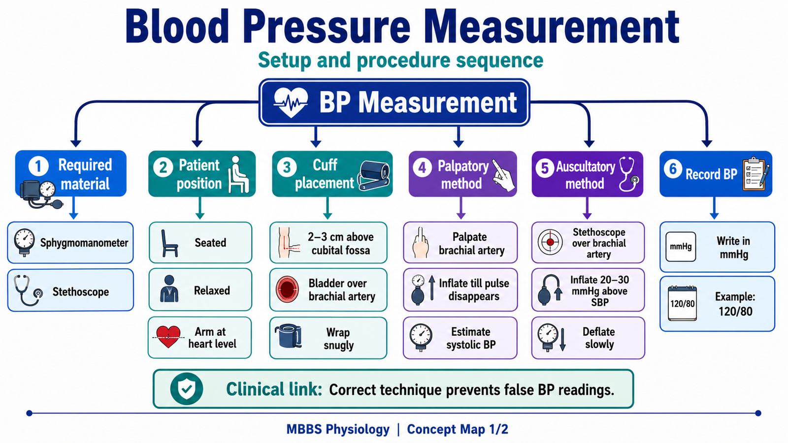
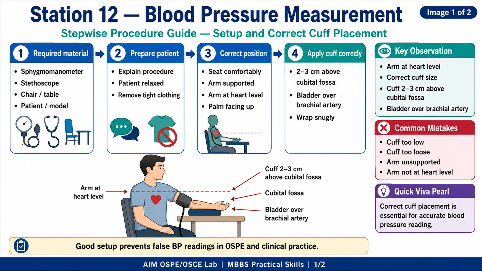
Required Material
- Sphygmomanometer
- Stethoscope
- Chair / examination couch
- Table or arm support
- Simulated patient / peer model
- Station instruction card
Student Task / Procedure
- Explain the procedure to the patient and ensure the patient is relaxed.
- Seat the patient comfortably with the arm supported at heart level.
- Wrap the cuff around the upper arm, about 2–3 cm above the cubital fossa.
- Ensure the cuff bladder is placed over the brachial artery.
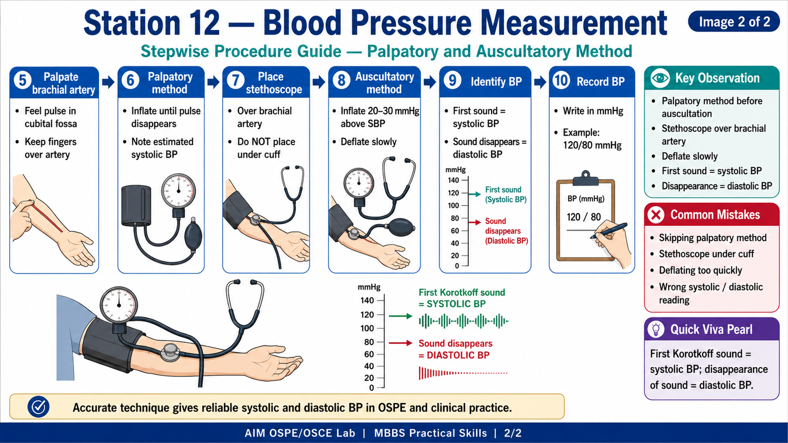
- Palpate the brachial artery in the cubital fossa.
- Use the palpatory method to estimate systolic blood pressure.
- Place the stethoscope over the brachial artery.
- Inflate the cuff above the estimated systolic pressure.
- Deflate slowly and listen for Korotkoff sounds.
- Record:
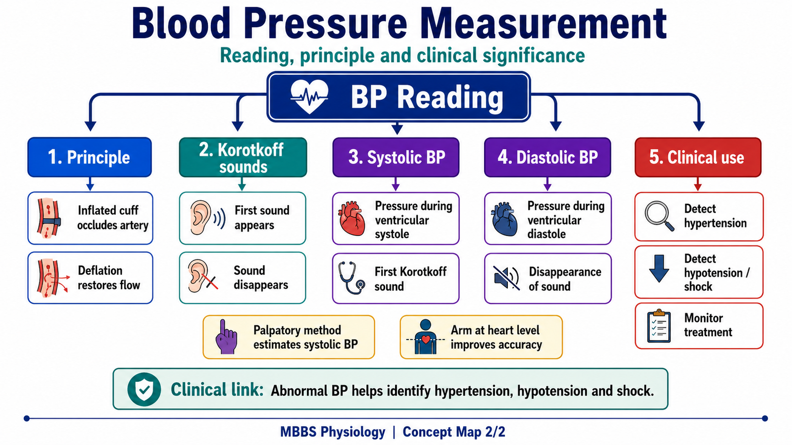
- Systolic BP: First clear Korotkoff sound
- Diastolic BP: Disappearance of Korotkoff sounds
- State the BP reading in mmHg.
Observation / Identification Points
The student should correctly demonstrate:
- Correct patient position: seated, relaxed, arm supported
- Correct cuff placement on upper arm
- Cuff lower edge 2–3 cm above cubital fossa
- Cuff bladder aligned over brachial artery
- Palpatory estimation of systolic BP before auscultation
- Stethoscope placed over brachial artery, not under cuff
- Slow cuff deflation during auscultatory method
- Correct identification of systolic and diastolic BP
Result / Interpretation
- Systolic blood pressure is the pressure in arteries during ventricular systole.
- Diastolic blood pressure is the pressure in arteries during ventricular diastole.
- In auscultatory BP measurement:
- First Korotkoff sound indicates systolic BP.
- Disappearance of Korotkoff sounds indicates diastolic BP.
- Correct BP measurement is important for screening and monitoring hypertension, hypotension, shock, and cardiovascular risk.
Viva Questions
1. Which artery is used for routine blood pressure measurement?
Answer: Brachial artery.
2. Why is the palpatory method performed before auscultatory measurement?
Answer: To estimate systolic BP and avoid missing the auscultatory gap.
3. What indicates systolic blood pressure during auscultation?
Answer: The first clear Korotkoff sound.
4. What indicates diastolic blood pressure during auscultation?
Answer: Disappearance of Korotkoff sounds.
5. At what level should the arm be supported during BP measurement?
Answer: At the level of the heart.
Marking Scheme
Total Marks: 5
| Component | Marks |
|---|---|
| Correct identification / performance | 2 |
| Key observation / procedure steps | 1 |
| Interpretation / principle | 1 |
| Viva answer | 1 |
Common Student Mistakes
- Placing the cuff too low or too loose.
- Putting the stethoscope under the cuff.
- Deflating the cuff too quickly.
- Skipping the palpatory method.
- Not supporting the arm at heart level.
AIM Feedback
To improve, revise the correct sequence of BP measurement: position patient → apply cuff → palpate brachial artery → estimate systolic BP by palpatory method → auscultate Korotkoff sounds. Always remember that the first Korotkoff sound is systolic BP, and the disappearance of sound is diastolic BP. Accurate technique prevents false BP readings.
Short Caption:
Watch this short procedure video to revise correct cuff placement, palpatory estimation, auscultatory method, and identification of systolic and diastolic blood pressure.
🖼️ Visual / Image Support
🧩 Concept Map / Interpretation Support