
🩺 Station 14 — Examination of Arterial Pulse
AIM OSPE/OSCE Lab — Practical Station | KMU Style | MBBS Practical + Viva
📌 Station Overview
Module: Cardiovascular System
Year: 1st Year MBBS
Focus: Identification • Procedure • Interpretation • Viva
Total Marks: 5
📋 Complete OSPE Station Content
Learning Target
By the end of this station, the student should be able to:
- Examine the arterial pulse correctly for rate, rhythm, volume, and character.
- Interpret common pulse findings and relate them to basic cardiovascular physiology.
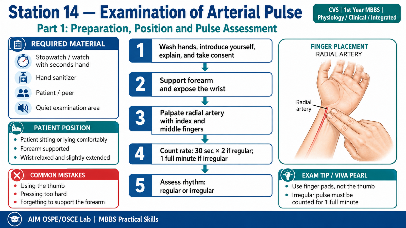
Required Material
- Examination couch or chair
- Stopwatch / watch with seconds hand
- Hand sanitizer
- Simulated patient / peer
- Clean, quiet examination area
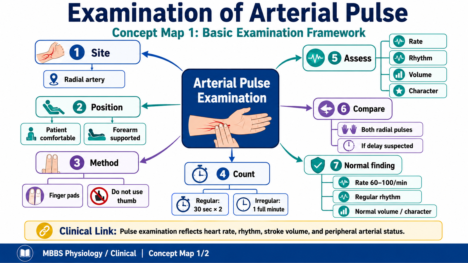
Student Task / Procedure
- Wash hands and introduce yourself to the patient.
- Explain the procedure and take permission.
- Ask the patient to sit or lie comfortably with forearm supported.
- Locate the radial artery at the wrist using the pads of index and middle fingers.
- Do not use the thumb.
- Count pulse rate for 30 seconds and multiply by 2 if rhythm is regular.
- Count for 1 full minute if rhythm is irregular.
- Assess rhythm as regular or irregular.
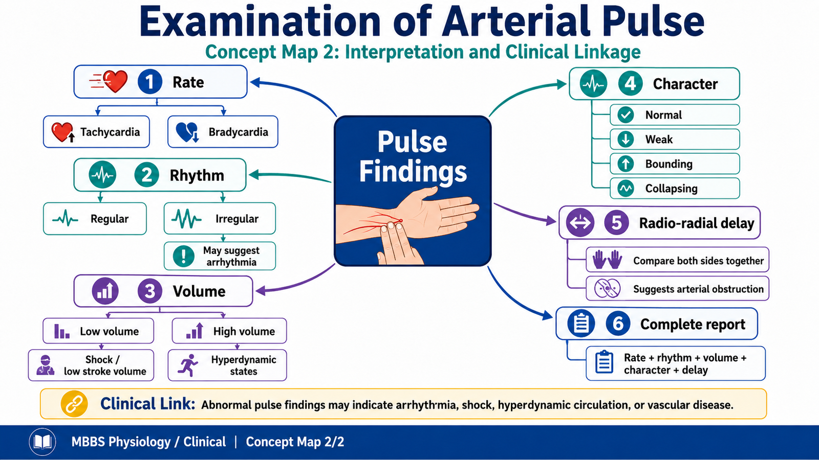
- Assess volume as normal, low volume, or high volume.
- Assess character of pulse, such as normal, weak, bounding, or collapsing if specifically asked.
- Compare both radial pulses together if radio-radial delay is required.
- Report findings clearly.
Observation / Identification Points
The student should demonstrate:
- Correct identification of the radial pulse
- Use of correct fingers and gentle pressure
- Proper counting method
- Assessment of:
- Rate
- Rhythm
- Volume
- Character
- Comparison of both radial pulses for radio-radial delay if asked
- Clear verbal reporting of findings
Result / Interpretation
A normal adult radial pulse is usually:
- Rate: 60–100 beats/minute
- Rhythm: Regular
- Volume: Normal
- Character: Normal pulse wave
- Radio-radial delay: Absent
Clinical significance:
Arterial pulse examination gives quick information about cardiac rate, rhythm, stroke volume, arterial wall condition, and peripheral circulation. An abnormal pulse may suggest arrhythmia, shock, fever, anemia, hyperdynamic circulation, or vascular obstruction.
Viva Questions
Q1. Which artery is commonly used for pulse examination at the wrist?
A: Radial artery.
Q2. Why should the thumb not be used to feel the pulse?
A: The thumb has its own pulsation, which may confuse the examiner.
Q3. What is the normal adult pulse rate?
A: About 60–100 beats per minute.
Q4. When should pulse be counted for one full minute?
A: When the rhythm is irregular.
Q5. What is radio-radial delay?
A: Delay between right and left radial pulses when both are palpated simultaneously.
Marking Scheme
Total Marks: 5
| Component | Marks |
|---|---|
| Correct identification / performance | 2 |
| Key observation / procedure steps | 1 |
| Interpretation / principle | 1 |
| Viva answer | 1 |
Common Student Mistakes
- Using the thumb to feel the pulse
- Counting for too short a time, especially in irregular rhythm
- Reporting only pulse rate and forgetting rhythm, volume, and character
AIM Feedback
When examining the pulse, do not rush directly to rate. A complete pulse examination includes rate, rhythm, volume, and character. Always support the patient’s forearm, use finger pads gently, and count for one full minute if rhythm is irregular. In OSPE, marks are gained by demonstrating the method clearly and reporting findings in an organized way.
🖼️ Visual / Image Support

🧩 Concept Map / Interpretation Support
🎥 Video Demonstration / Procedure Support