🩺 Station 7 — Large Artery and Large Vein
AIM OSPE/OSCE Lab — Practical Station | KMU Style | MBBS Practical + Viva
📌 Station Overview
Module: Cardiovascular System
Year: 1st Year MBBS
Focus: Identification • Procedure • Interpretation • Viva
Total Marks: 5
📋 Complete OSPE Station Content
Learning Target
By the end of this station, the student should be able to:
- Identify histological differences between a large artery and a large vein.
- Link the structure of large vessels with their function in circulation.
Required Material
- Histology slide/image of large artery and large vein
- Light microscope or digital histology image
- Pointer or cursor
- Station instruction sheet
- Marking checklist
Student Task / Procedure
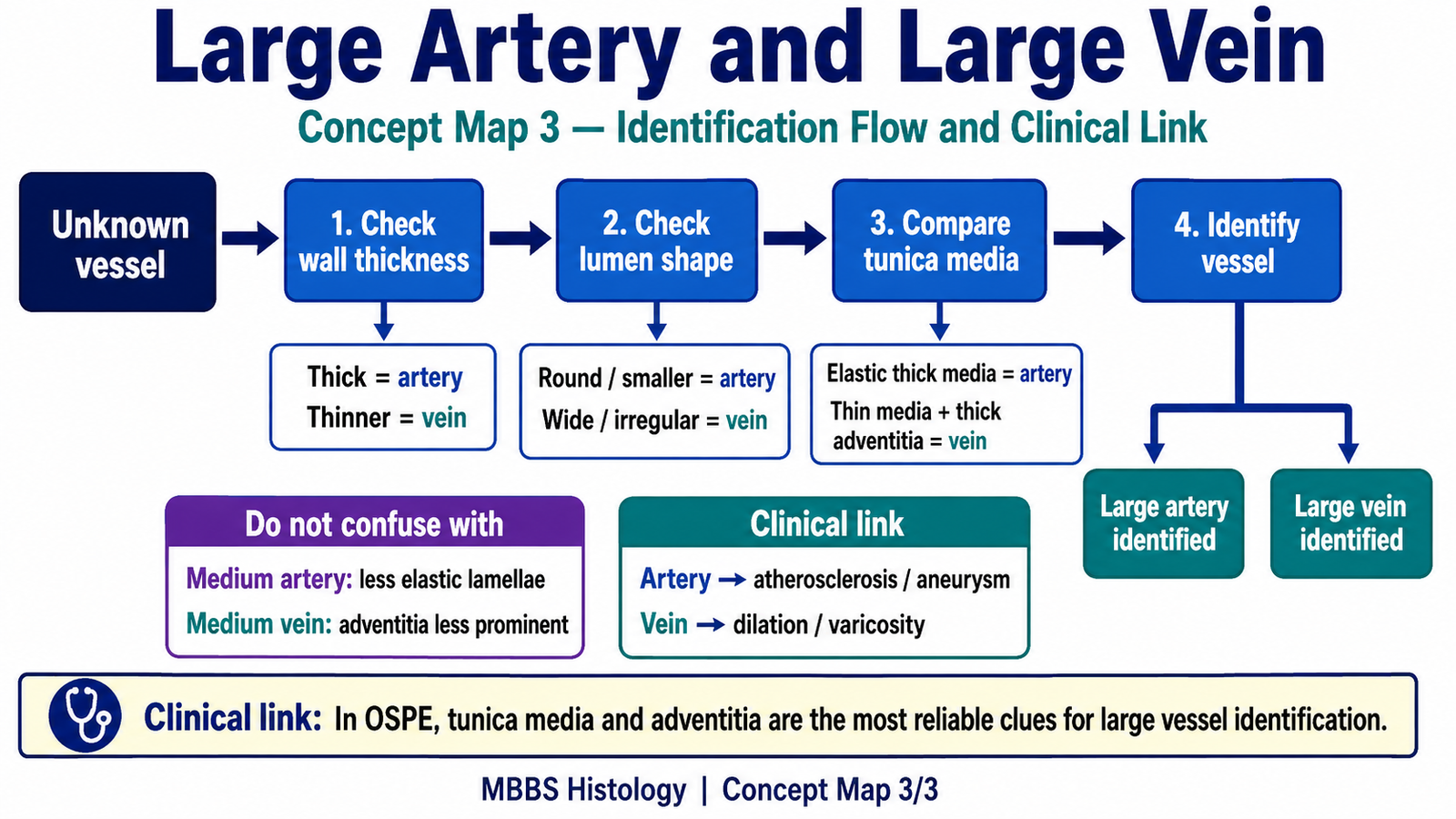
- Observe the given histology slide/image.
- Identify the large artery and large vein.
- Compare their:
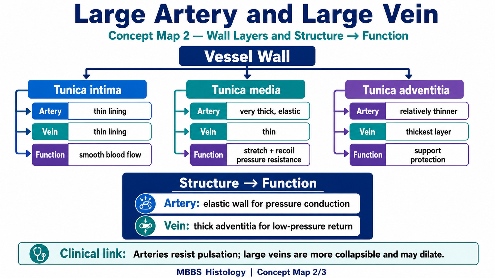
- Wall thickness
- Lumen shape
- Tunica media
- Tunica adventitia
- State one structure–function relationship for each vessel.
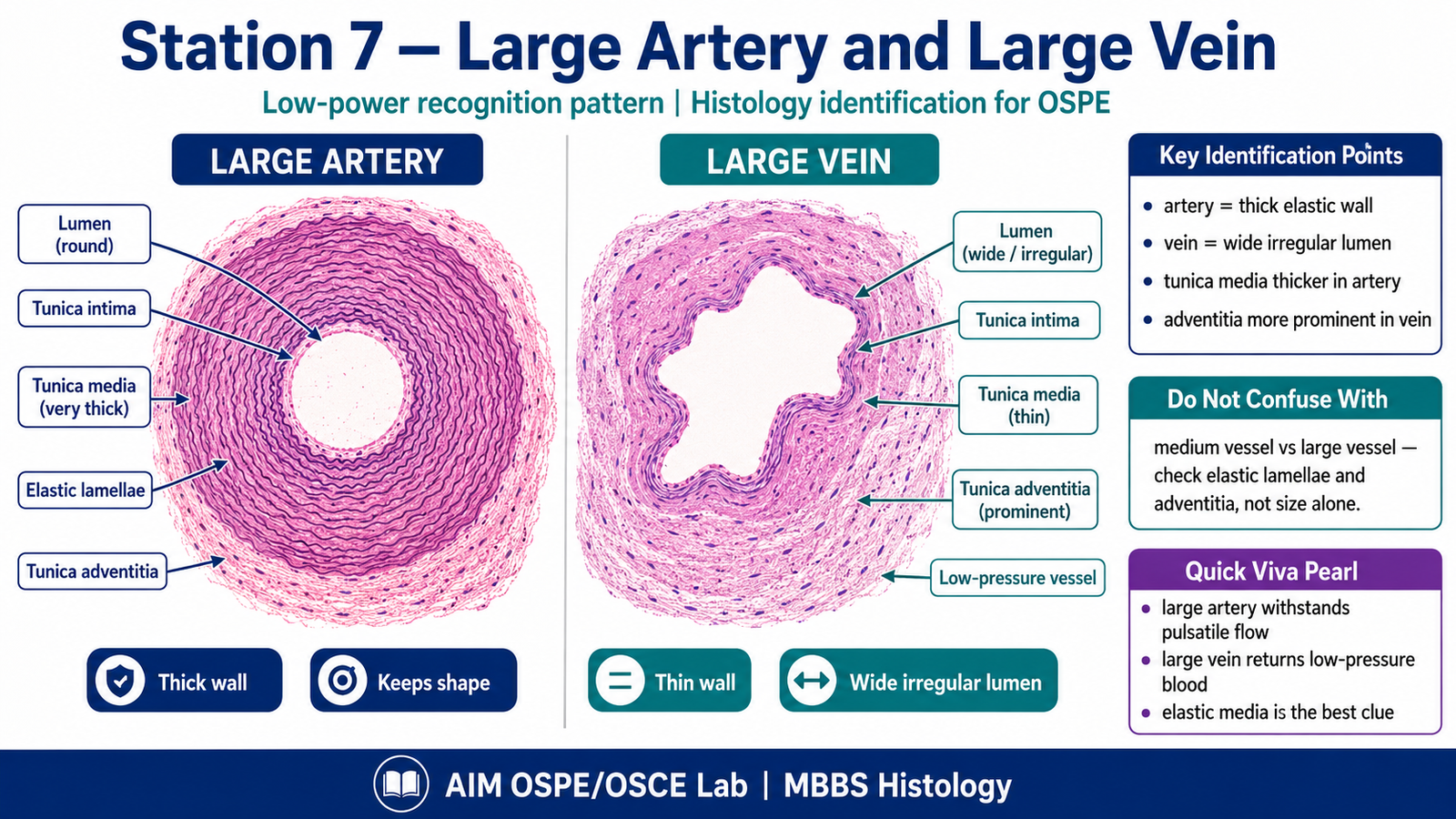
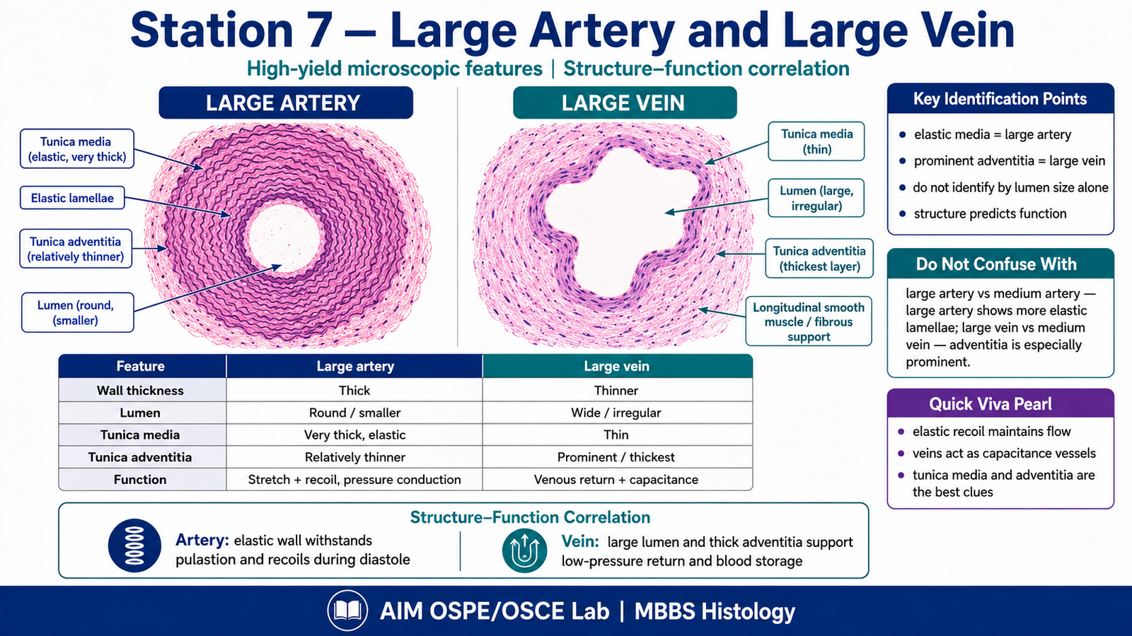
Observation / Identification Points
The student should observe and identify:
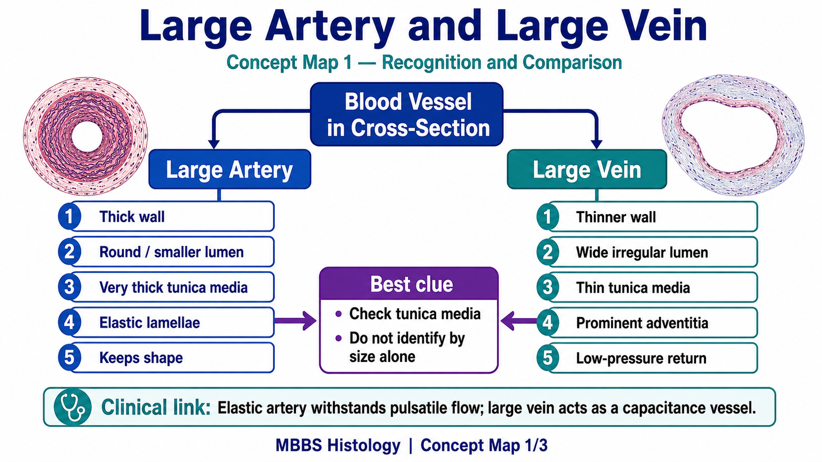
Large Artery
- Thick wall
- Relatively round lumen
- Very thick tunica media
- Many elastic lamellae in tunica media
- Designed to withstand high pulsatile pressure
Large Vein
- Thinner wall than artery
- Large, irregular or collapsed lumen
- Thin tunica media
- Thick tunica adventitia
- Designed for low-pressure venous return and blood storage
Key Comparison
| Feature | Large Artery | Large Vein |
|---|---|---|
| Wall | Thick | Thin |
| Lumen | Smaller, round | Large, irregular |
| Tunica media | Very thick, elastic lamellae | Thin |
| Tunica adventitia | Relatively thinner | Thickest layer |
| Function | Withstands pulsatile pressure | Low-pressure return / capacitance |
Result / Interpretation
A large artery is identified by its very thick tunica media containing many elastic lamellae. This allows it to stretch during systole and recoil during diastole, helping maintain continuous blood flow.
A large vein is identified by its large irregular lumen, thinner tunica media, and prominent tunica adventitia. This structure supports low-pressure venous return and allows veins to act as capacitance vessels.
Viva Questions
1. What is the most important histological feature of a large artery?
A very thick tunica media with many elastic lamellae.
2. Which layer is thickest in a large vein?
Tunica adventitia.
3. Why does a large artery contain elastic lamellae?
To allow stretch and recoil during pulsatile blood flow.
4. Why is the lumen of a large vein usually large and irregular?
Because veins carry blood under low pressure and collapse easily.
5. Give one functional difference between large artery and large vein.
Large arteries conduct high-pressure blood away from the heart, while large veins return low-pressure blood to the heart and store blood.
Marking Scheme
Total Marks: 5
| Component | Marks |
| Correct identification / performance | 2 |
| Key observation / procedure steps | 1 |
| Interpretation / principle | 1 |
| Viva answer | 1 |
Common Student Mistakes
- Confusing large vein with artery because of its larger lumen.
- Forgetting that large arteries have abundant elastic lamellae.
- Identifying only by vessel size instead of comparing wall layers.
- Missing that tunica adventitia is prominent in large veins.
AIM Feedback
For large vessels, first compare the tunica media and tunica adventitia. A large artery has a very thick elastic tunica media for pressure handling. A large vein has a thinner media, wider irregular lumen, and prominent adventitia for low-pressure venous return. In OSPE, use the sequence: lumen → wall thickness → tunica media → tunica adventitia → function.
Short Caption
Use this labeled image to revise large artery and large vein identification. Focus on elastic lamellae in the arterial tunica media and prominent tunica adventitia in the large vein.
🖼️ Visual / Image Support
🧩 Concept Map / Interpretation Support