
🩺 Station 1 — Heart and Its Coverings
AIM OSPE/OSCE Lab — Practical Station
OSPE Station Name
Station 1 — Heart and Its Coverings
Learning Target
By the end of this station, the student should be able to:
- Identify the heart and its coverings on a model/specimen/image.
- Explain the basic arrangement and clinical importance of the pericardium.
Required Material
- Anatomical model of heart
- Model/image showing pericardium
- Pointer
- Labelled/unlabelled diagram of heart and pericardium
- Answer sheet
Student Task / Procedure
- Observe the given heart model/specimen carefully.
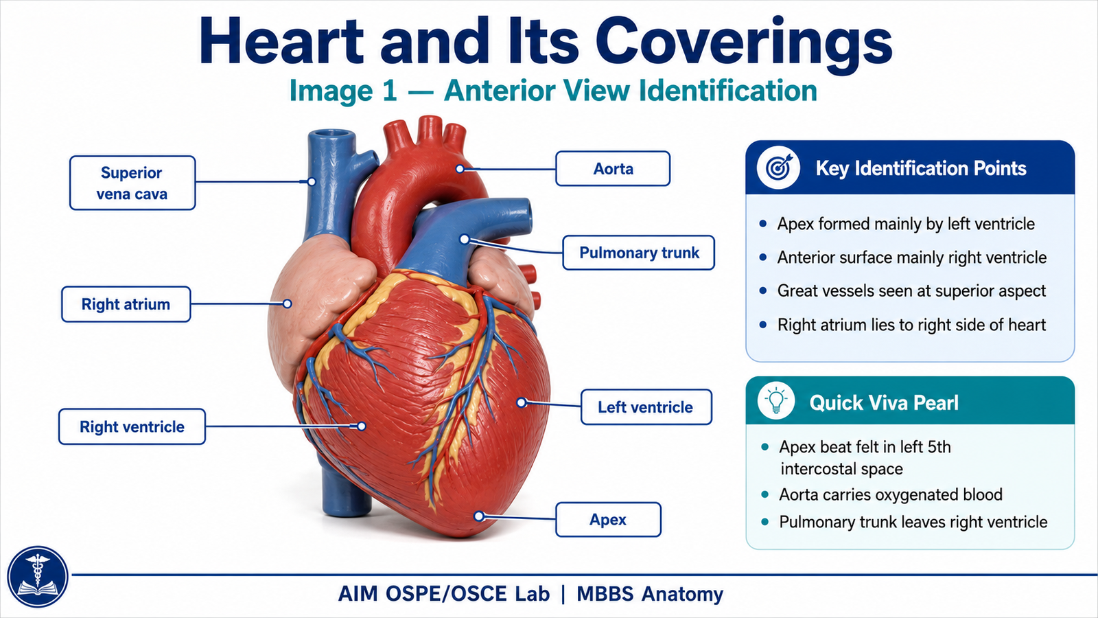
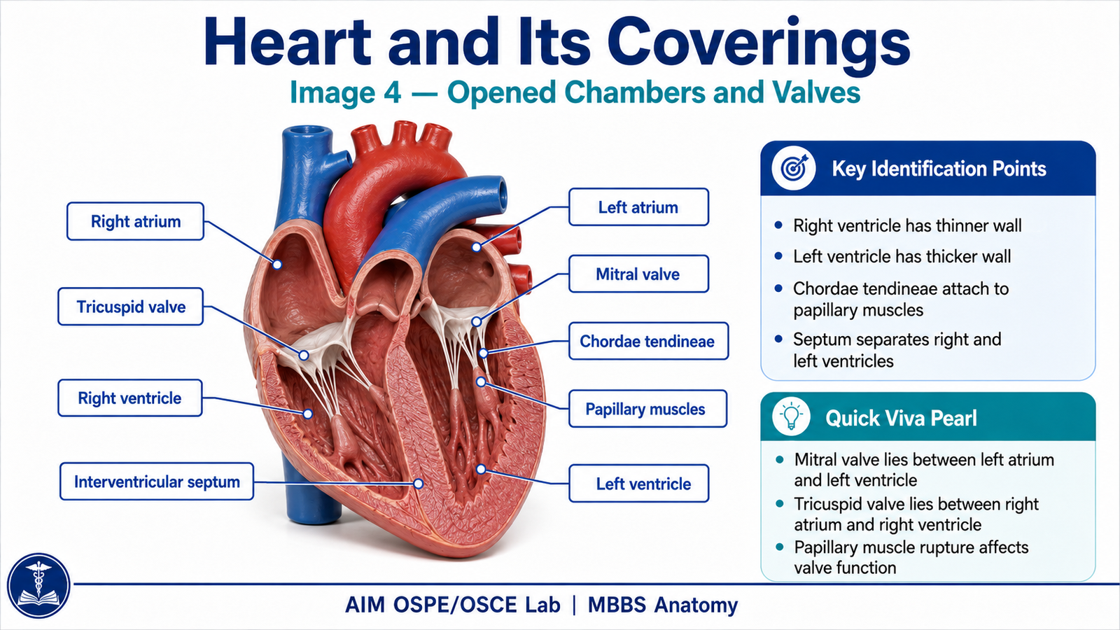
- Identify the heart and its main external features.
- Identify the coverings of the heart.
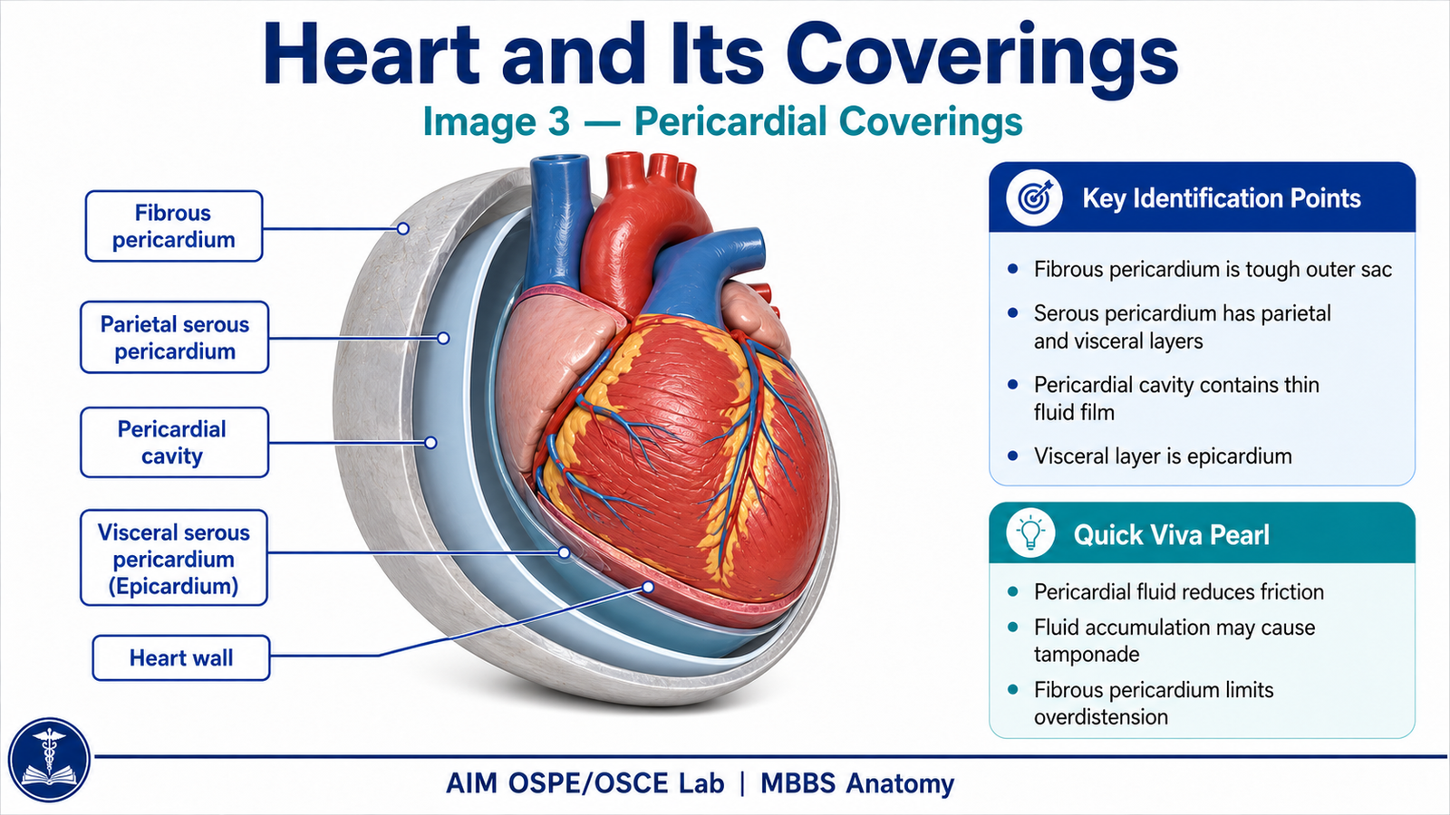
- Point out the fibrous pericardium, parietal layer of serous pericardium, and visceral layer of serous pericardium / epicardium.
- State one function and one clinical significance of the pericardium.
Observation / Identification Points
Students should identify or demonstrate:
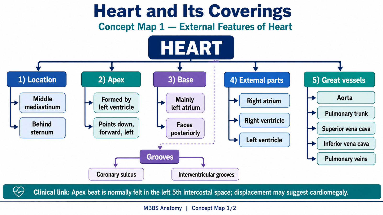
- Heart located in the middle mediastinum
- Apex of heart directed downwards, forwards, and to the left
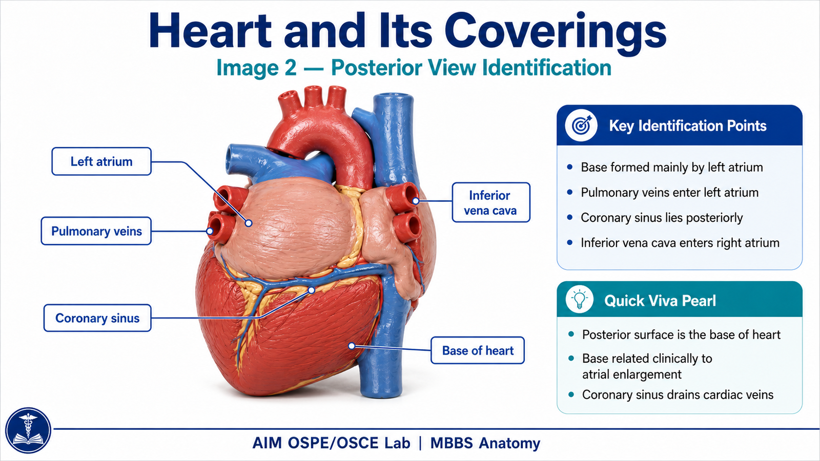
- Base mainly formed by left atrium
- External grooves indicating chambers and vessels
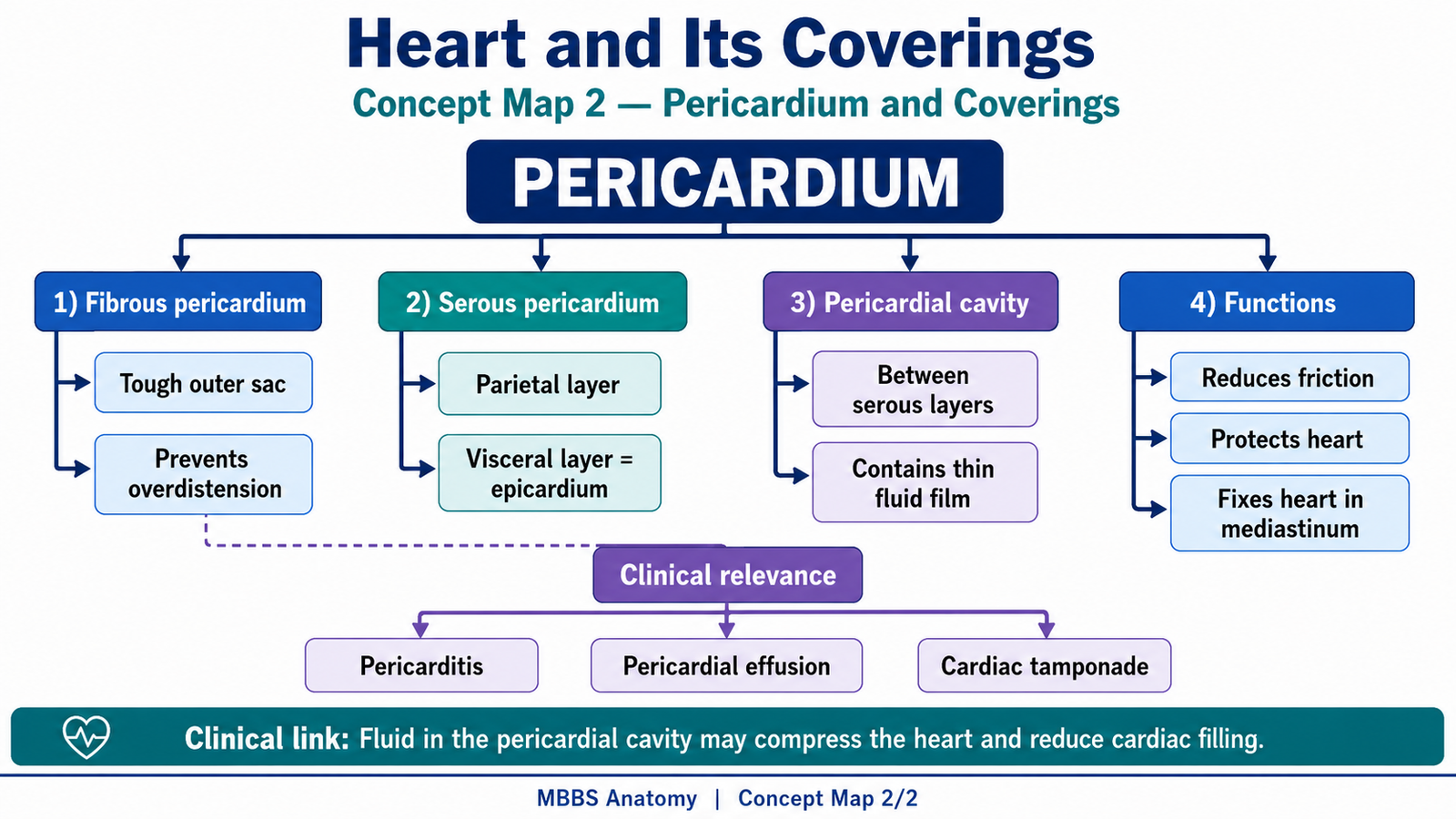
- Fibrous pericardium as the tough outer covering
- Serous pericardium with:
- Parietal layer
- Visceral layer / epicardium
- Pericardial cavity between parietal and visceral layers
- Small amount of pericardial fluid reducing friction during heart movement
Result / Interpretation
The heart is enclosed in a protective pericardial sac.
The fibrous pericardium prevents overdistension of the heart, while the serous pericardium allows smooth movement of the beating heart.
Clinically, inflammation or fluid accumulation in the pericardial cavity may cause pericarditis, pericardial effusion, or cardiac tamponade.
Viva Questions
1. What is the outermost covering of the heart?
Answer: Fibrous pericardium.
2. What is the visceral layer of serous pericardium also called?
Answer: Epicardium.
3. What is present between the parietal and visceral layers of serous pericardium?
Answer: Pericardial cavity containing a small amount of pericardial fluid.
4. What is the main function of pericardial fluid?
Answer: It reduces friction during heart movements.
5. What is cardiac tamponade?
Answer: Compression of the heart due to excessive fluid or blood in the pericardial cavity.
Marking Scheme
Total Marks: 5
| Component | Marks |
|---|---|
| Correct identification / performance | 2 |
| Key observation / procedure steps | 1 |
| Interpretation / principle | 1 |
| Viva answer | 1 |
Common Student Mistakes
- Confusing fibrous pericardium with serous pericardium.
- Forgetting that visceral serous pericardium is epicardium.
- Not identifying the pericardial cavity between parietal and visceral layers.
AIM Feedback
Revise the pericardium as a three-layer covering system: fibrous pericardium outside, parietal serous pericardium lining it, and visceral serous pericardium covering the heart surface. Remember that the small space between the serous layers is clinically important because fluid accumulation here can compress the heart.
Additional AIM Learning Support
1. Concept Maps
2. LMS-Friendly Procedure Video
3. LMS-Friendly Stepwise Image Guide
Short Caption for LMS
Identify the coverings of the heart from outside to inside: fibrous pericardium, parietal serous pericardium, pericardial cavity, and visceral serous pericardium / epicardium.