
🩺 Station 10 — Apex Beat and Auscultatory Areas
AIM OSPE/OSCE Lab — Practical Station | KMU Style | MBBS Practical + Viva
📌 Station Overview
Module: Cardiovascular System
Year: 1st Year MBBS
Focus: Identification • Procedure • Interpretation • Viva
Total Marks: 5
📋 Complete OSPE Station Content
Learning Target
By the end of this station, the student should be able to:
- Locate the normal position of the apex beat on the anterior chest wall.
- Identify the main cardiac auscultatory areas and state their clinical significance.
Required Material
- Chest wall mannequin / simulator / peer model
- Stethoscope
- Skin-safe marker or pointer
- Surface anatomy chart of thorax
- Station instruction card
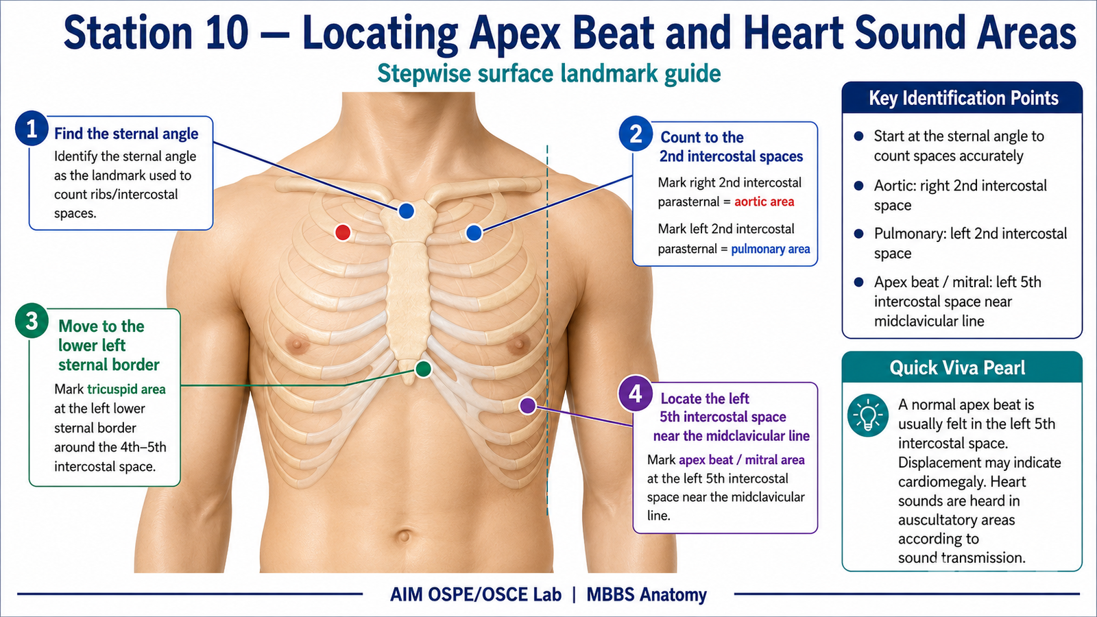
Student Task / Procedure
- Identify the left 5th intercostal space.
- Locate the midclavicular line.
- Mark the normal site of the apex beat at the left 5th intercostal space, near the midclavicular line.
- Identify the four main auscultatory areas:
- Mitral area
- Tricuspid area
- Pulmonary area
- Aortic area
- State the clinical importance of apex beat and auscultatory areas.
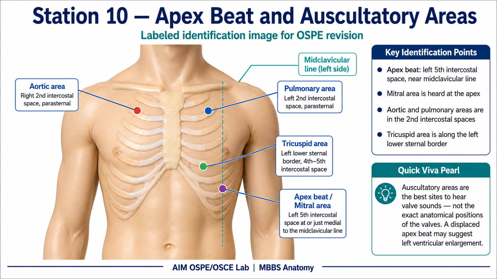
Observation / Identification Points
The student should correctly identify:
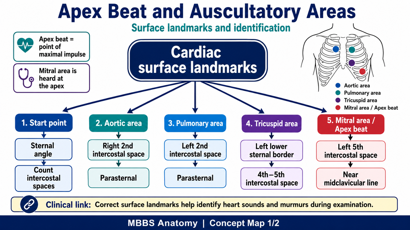
- Apex beat: Left 5th intercostal space at or just medial to the midclavicular line
- Mitral area: Left 5th intercostal space, midclavicular line
- Tricuspid area: Lower left sternal border, usually 4th–5th intercostal space
- Pulmonary area: Left 2nd intercostal space near the sternum
- Aortic area: Right 2nd intercostal space near the sternum
Important point:
Auscultatory areas are not the exact anatomical positions of valves. They are the best areas where valve sounds are heard.
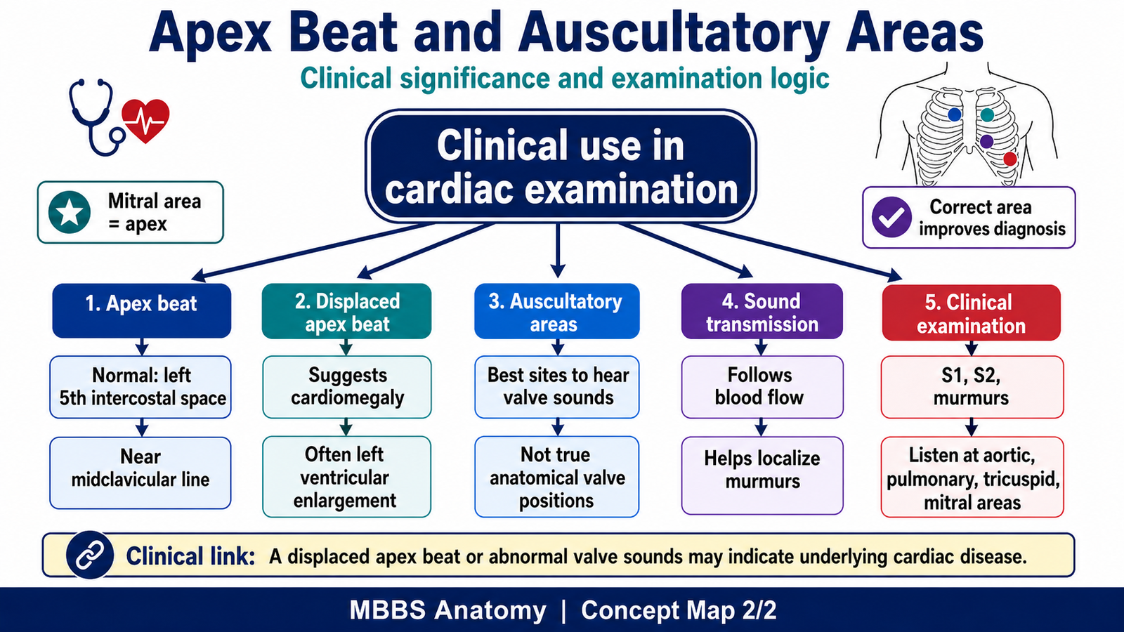
Result / Interpretation
- The apex beat represents the point of maximum cardiac impulse, mainly produced by the left ventricle.
- A displaced apex beat may suggest cardiac enlargement, especially left ventricular enlargement.
- Cardiac valve sounds and murmurs are best heard at specific auscultatory areas because sound is conducted along the direction of blood flow.
- Correct identification of auscultatory areas is essential for clinical examination of heart sounds and murmurs.
Viva Questions
1. Where is the normal apex beat located?
Answer: In the left 5th intercostal space at or just medial to the midclavicular line.
2. Which valve is best heard at the apex?
Answer: Mitral valve.
3. Where is the aortic area located?
Answer: Right 2nd intercostal space close to the sternum.
4. Where is the pulmonary area located?
Answer: Left 2nd intercostal space close to the sternum.
5. What is the clinical significance of a displaced apex beat?
Answer: It may indicate cardiac enlargement, commonly left ventricular enlargement.
Marking Scheme
Total Marks: 5
| Component | Marks |
|---|---|
| Correct identification / performance | 2 |
| Key observation / procedure steps | 1 |
| Interpretation / principle | 1 |
| Viva answer | 1 |
Checklist Detail
| Checklist Item | Marks |
| Correctly locates apex beat at left 5th intercostal space near midclavicular line | 1 |
| Correctly identifies mitral, tricuspid, pulmonary, and aortic auscultatory areas | 1 |
| Uses correct surface landmarks: intercostal spaces, sternum, midclavicular line | 1 |
| Explains that auscultatory areas are best hearing areas, not exact valve positions | 1 |
| Answers viva question correctly | 1 |
Common Student Mistakes
- Marking the apex beat in the wrong intercostal space.
- Confusing anatomical valve positions with auscultatory areas.
- Reversing aortic and pulmonary areas.
- Forgetting that mitral area corresponds to the apex beat region.
AIM Feedback
Review the chest wall landmarks before attempting this station again. First find the sternal angle, then count intercostal spaces accurately. Remember: Aortic and pulmonary areas are in the 2nd intercostal space, while mitral area is at the apex. Auscultatory areas are clinically important because they help detect abnormal heart sounds and murmurs.
🖼️ Visual / Image Support
🧩 Concept Map / Interpretation Support
🎥 Video Demonstration / Procedure Support