
🩺 Station 16 — Use of Stethoscope
AIM OSPE/OSCE Lab — Practical Station | KMU Style | MBBS Practical + Viva
📌 Station Overview
Module: Cardiovascular System
Year: 1st Year MBBS
Focus: Identification • Procedure • Interpretation • Viva
Total Marks: 5
📋 Complete OSPE Station Content
Learning Target
By the end of this station, the student should be able to:
- Identify and use the diaphragm and bell of the stethoscope correctly.
- Demonstrate proper stethoscope handling and placement during cardiovascular examination.
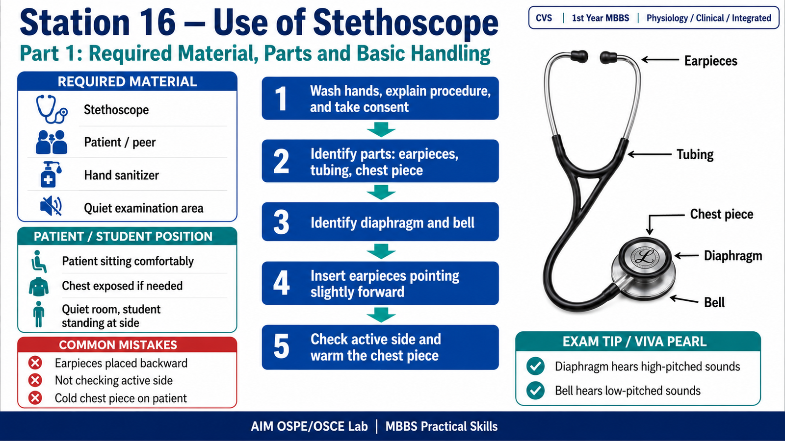
Required Material
- Stethoscope
- Simulated patient / peer
- Examination couch or chair
- Hand sanitizer
- Quiet examination area
Student Task / Procedure
- Wash hands and introduce yourself.
- Explain the procedure and take permission.
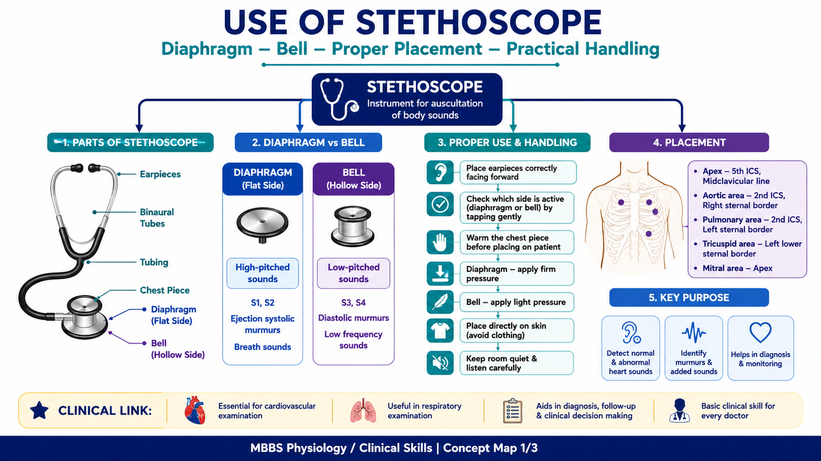
- Identify the main parts of the stethoscope:
- Earpieces
- Tubing
- Chest piece
- Diaphragm
- Bell
- Place earpieces correctly in the ears, pointing slightly forward.
- Check which side of the chest piece is active.
- Warm the chest piece before placing it on the patient.
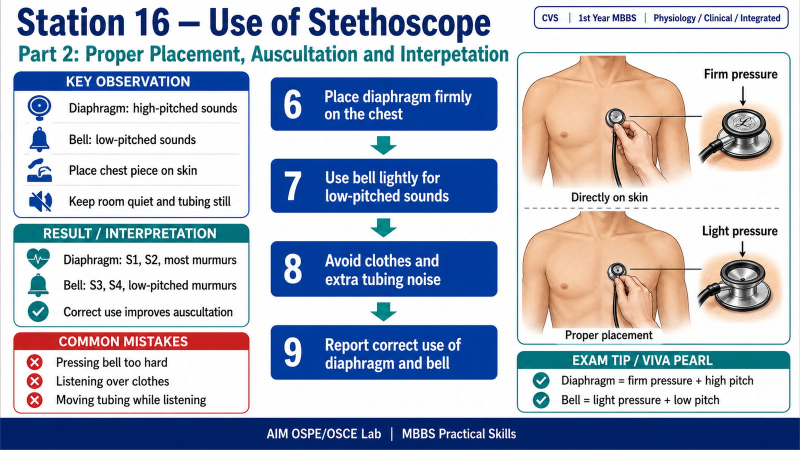
- Place the diaphragm firmly on the chest to hear high-pitched sounds.
- Place the bell lightly on the chest to hear low-pitched sounds.
- Avoid placing the stethoscope over clothes.
- Keep the room quiet and listen carefully.
- Remove the stethoscope respectfully and report the purpose of diaphragm and bell.
Observation / Identification Points
The student should demonstrate:
- Correct identification of diaphragm and bell
- Correct orientation of earpieces
- Proper handling of the chest piece
- Correct pressure:
- Diaphragm: firm pressure
- Bell: light pressure
- Proper placement directly on the skin
- Awareness that diaphragm and bell detect different sound frequencies
Result / Interpretation
The stethoscope is used to amplify body sounds during clinical examination.
- Diaphragm: Best for high-pitched sounds, such as normal heart sounds S1 and S2.
- Bell: Best for low-pitched sounds, such as S3, S4, and some low-frequency murmurs.
- Correct placement and correct pressure are essential for accurate auscultation.
Clinical significance:
Proper use of the stethoscope helps detect heart sounds, added sounds, murmurs, and other cardiovascular findings during clinical examination.
Viva Questions
Q1. What are the two main listening parts of a stethoscope chest piece?
A: Diaphragm and bell.
Q2. Which part is used for high-pitched sounds?
A: Diaphragm.
Q3. Which part is used for low-pitched sounds?
A: Bell.
Q4. How should the bell be placed on the chest?
A: Lightly, without pressing too hard.
Q5. Why should the stethoscope not be placed over clothes?
A: Clothes can reduce sound clarity and create extra noise.
Marking Scheme
Total Marks: 5
| Component | Marks |
|---|---|
| Correct identification / performance | 2 |
| Key observation / procedure steps | 1 |
| Interpretation / principle | 1 |
| Viva answer | 1 |
Common Student Mistakes
- Placing earpieces in the wrong direction
- Pressing the bell too firmly
- Using the stethoscope over clothes instead of directly on skin
AIM Feedback
A stethoscope is not just placed randomly on the chest. Good clinical examination depends on correct handling, correct earpiece direction, and correct use of the diaphragm and bell. Remember: diaphragm = high-pitched sounds with firm pressure, while bell = low-pitched sounds with light pressure.
🖼️ Visual / Image Support
🧩 Concept Map / Interpretation Support
🎥 Video Demonstration / Procedure Support