
🩺 Station 15 — Auscultation of Heart Sounds
AIM OSPE/OSCE Lab — Practical Station | KMU Style | MBBS Practical + Viva
📌 Station Overview
Module: Cardiovascular System
Year: 1st Year MBBS
Focus: Identification • Procedure • Interpretation • Viva
Total Marks: 5
📋 Complete OSPE Station Content
Learning Target
- Demonstrate correct use of stethoscope for auscultation of basic heart sounds.
- Identify S1 and S2 at the correct auscultatory areas and give basic physiological interpretation.
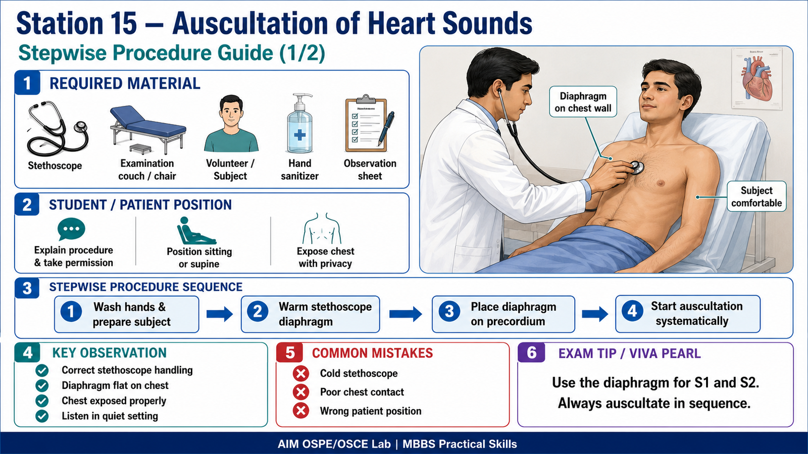
Required Material
- Stethoscope
- Examination couch / chair
- Clean teaching model / volunteer subject
- Hand sanitizer
- Chest auscultation area diagram
- Checklist / observation sheet
Student Task / Procedure
- Wash hands and introduce yourself.
- Explain the procedure to the subject and take permission.
- Position the subject comfortably in sitting or supine position.
- Expose the precordium appropriately while maintaining privacy.
- Warm the stethoscope diaphragm before use.
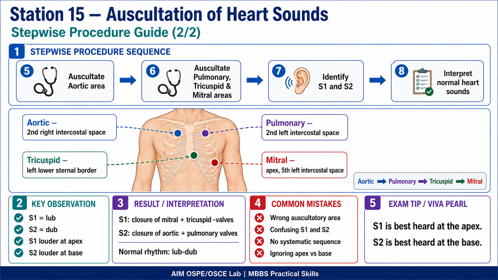
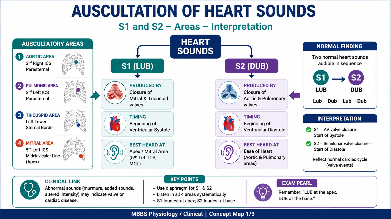
- Place the diaphragm on the main auscultatory areas in sequence:
- Aortic area
- Pulmonary area
- Tricuspid area
- Mitral area
- Listen carefully for S1 and S2.
- Identify where S1 and S2 are heard best.
- Give a basic interpretation of the heart sounds.
Observation / Identification Points
- Correct handling and placement of stethoscope
- Diaphragm used properly on chest wall
- Correct auscultatory areas identified
- S1 and S2 recognized as “lub–dub” sounds
- S1 heard louder at the mitral area / apex
- S2 heard louder at the aortic and pulmonary areas / base
- Student relates S1 and S2 to valve closure
Result / Interpretation
Normal finding:
Two normal heart sounds are heard: S1 and S2.
S1:
Produced mainly by closure of mitral and tricuspid valves at the beginning of ventricular systole. It is best heard at the apex / mitral area.
S2:
Produced by closure of aortic and pulmonary valves at the beginning of ventricular diastole. It is best heard at the base of the heart, especially aortic and pulmonary areas.
Clinical significance:
Auscultation helps assess normal heart sounds and may detect abnormal sounds such as murmurs, added sounds, or altered intensity in later clinical years.
Viva Questions
Q1. What are the two normal heart sounds?
A: S1 and S2.
Q2. What produces S1?
A: Closure of mitral and tricuspid valves.
Q3. What produces S2?
A: Closure of aortic and pulmonary valves.
Q4. Where is S1 heard best?
A: At the mitral area / apex of the heart.
Q5. Where is S2 heard best?
A: At the base of the heart, especially aortic and pulmonary areas.
Marking Scheme
Total Marks: 5
| Component | Marks |
|---|---|
| Correct identification / performance | 2 |
| Key observation / procedure steps | 1 |
| Interpretation / principle | 1 |
| Viva answer | 1 |
Common Student Mistakes
- Placing the stethoscope on incorrect auscultatory areas
- Not maintaining subject privacy during chest exposure
- Confusing S1 with S2
- Forgetting that S1 is best heard at the apex
- Forgetting that S2 is best heard at the base
AIM Feedback
In this station, focus on where to listen and what you are listening for.
Remember: S1 = AV valve closure = systole = best at apex.
S2 = semilunar valve closure = diastole = best at base.
Practice the auscultatory areas repeatedly because correct placement is the most important OSPE skill.
🖼️ Visual / Image Support
🧩 Concept Map / Interpretation Support
🎥 Video Demonstration / Procedure Support