🩺 Station 11 — Normal vs Displaced Apex Beat
AIM OSPE/OSCE Lab — Practical Station | KMU Style | MBBS Practical + Viva
📌 Station Overview
Module: Cardiovascular System
Year: 1st Year MBBS
Focus: Identification • Procedure • Interpretation • Viva
Total Marks: 5
📋 Complete OSPE Station Content
Learning Target
By the end of this station, the student should be able to:
- Identify the normal anatomical position of the apex beat on the anterior chest wall.
- Recognize displacement of the apex beat and explain its basic clinical significance.
Required Material
- Male/female chest model or standardized patient simulator
- Marker or skin pencil
- Measuring tape
- Anatomical landmark chart
- Gloves / hand sanitizer
- Station instruction card
Student Task / Procedure
- Introduce yourself and explain the procedure briefly.
- Ask the patient/model to lie comfortably at 45°.
- Expose the anterior chest appropriately.
- Identify the midclavicular line.
- Count down to the left 5th intercostal space.
- Palpate gently with the fingertips to locate the apex beat.
- State whether the apex beat is normal or displaced.
- Explain the clinical significance of a displaced apex beat.
Observation / Identification Points
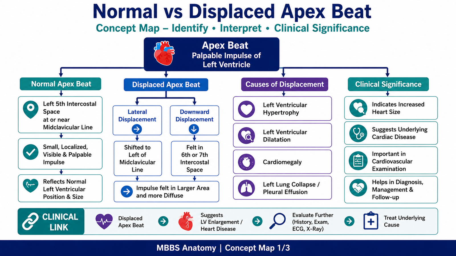
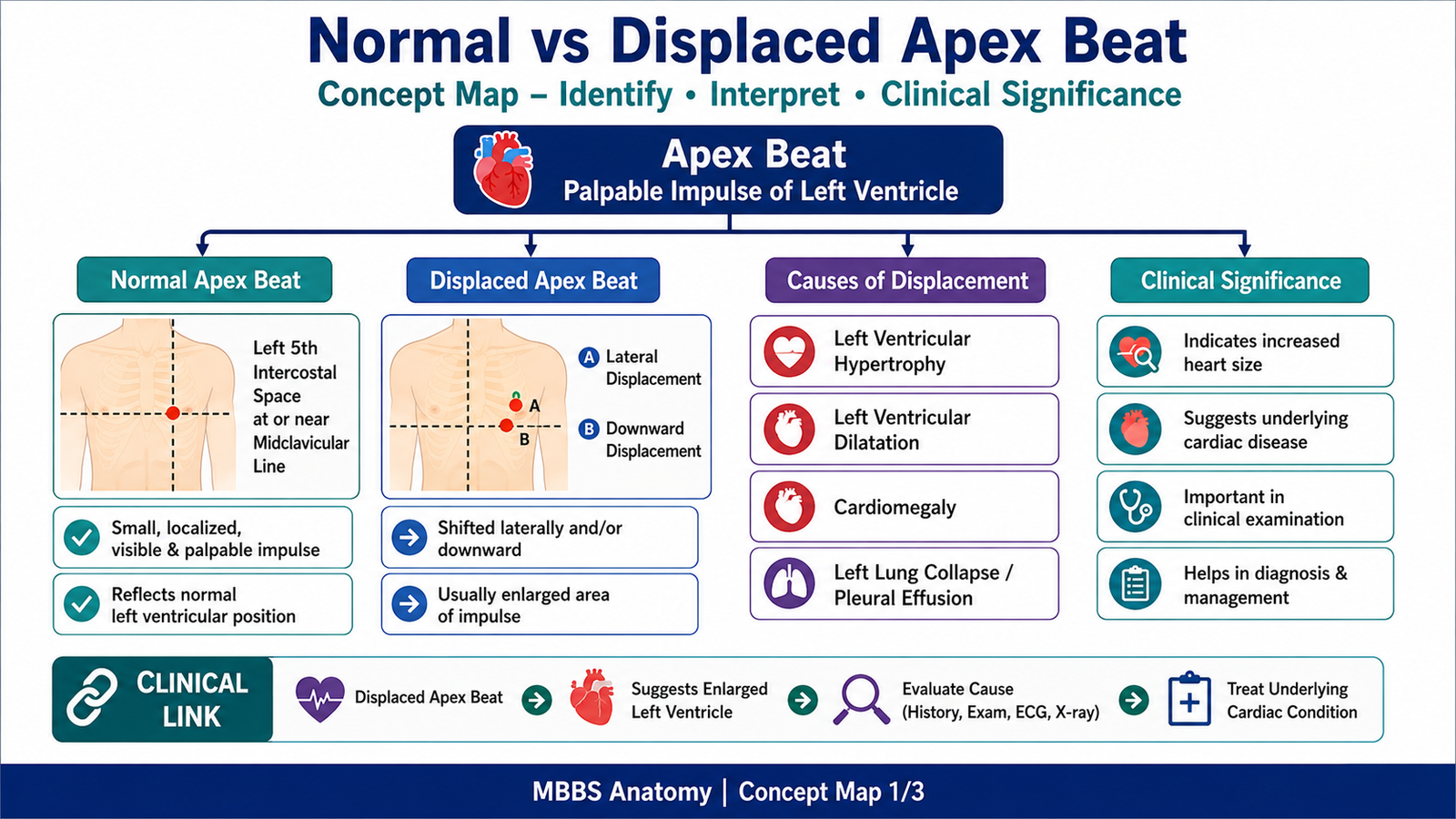
- Normal apex beat is usually felt in the left 5th intercostal space.
- It lies near the left midclavicular line.
- It is normally a small, localized impulse.
- Displacement means the apex beat is felt away from its normal position.
- Lateral and downward displacement may suggest cardiac enlargement, especially left ventricular enlargement.
Result / Interpretation
Normal finding:
Apex beat present in the left 5th intercostal space at or near the midclavicular line.
Displaced apex beat:
Apex beat shifted laterally and/or downward, suggesting possible cardiomegaly or ventricular enlargement.
Clinical significance:
A displaced apex beat is an important surface sign that may indicate enlarged heart size, commonly due to left ventricular hypertrophy or dilatation.
Viva Questions
Q1. What is the apex beat?
A: It is the palpable impulse produced mainly by the left ventricular apex against the chest wall.
Q2. What is the normal position of the apex beat?
A: Left 5th intercostal space near the midclavicular line.
Q3. Which chamber mainly forms the anatomical apex of the heart?
A: The left ventricle.
Q4. What does a laterally displaced apex beat suggest?
A: It may suggest cardiac enlargement, especially left ventricular enlargement.
Q5. Why is apex beat important in CVS examination?
A: It helps assess heart position, size, and possible enlargement clinically.
Marking Scheme
Total Marks: 5
| Component | Marks |
|---|---|
| Correct identification / performance | 2 |
| Key observation / procedure steps | 1 |
| Interpretation / principle | 1 |
| Viva answer | 1 |
Common Student Mistakes
- Counting ribs instead of identifying the correct intercostal space.
- Looking too medially or too high for the apex beat.
- Forgetting that the apex is mainly formed by the left ventricle.
- Calling any visible chest movement an apex beat without proper palpation.
AIM Feedback
To improve, always start with surface landmarks: identify the midclavicular line, then locate the left 5th intercostal space. Remember that the apex beat is normally a localized left ventricular impulse. If it is shifted laterally or downward, think of cardiac enlargement, especially left ventricular enlargement.
🖼️ Visual / Image Support
🧩 Concept Map / Interpretation Support