
🩺Station 8 — Cardiac Shadow on Chest X-Ray
AIM OSPE/OSCE Lab — Practical Station | KMU Style | MBBS Practical + Viva
📌 Station Overview
Module: Cardiovascular System
Year: 1st Year MBBS
Focus: Identification • Procedure • Interpretation • Viva
Total Marks: 5
📋 Complete OSPE Station Content
Learning Target
By the end of this station, the student should be able to:
- Identify the normal cardiac shadow and heart borders on a PA chest X-ray.
- Interpret cardiomegaly using the cardiothoracic ratio and relate it to clinical significance.
Required Material
- PA chest X-ray showing normal cardiac shadow
- PA chest X-ray showing cardiomegaly
- Pointer
- Station sheet
- Answer sheet
- Labeled reference image for examiner
Student Task / Procedure
- Look at the given chest X-ray carefully.
- Identify the cardiac shadow.
- Point out the right and left heart borders.
- Identify whether the cardiac size is normal or enlarged.
- State the clinical significance of cardiomegaly.
Observation / Identification Points
The student should identify:
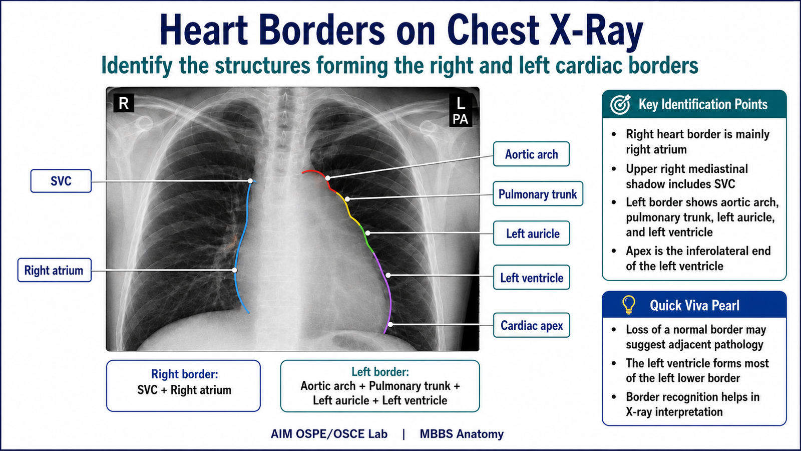
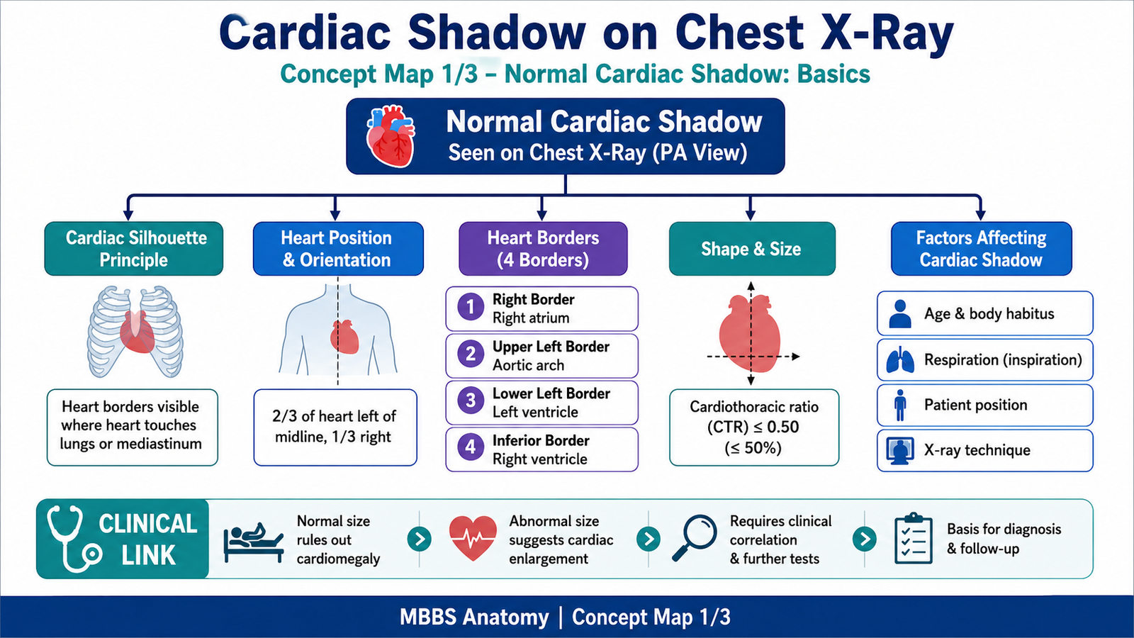
- Cardiac shadow in the lower mediastinum.
- Right heart border mainly formed by the right atrium.
- Right upper border formed by the superior vena cava.
- Left upper border formed by the aortic arch.
- Left middle border formed by the pulmonary trunk / left atrial appendage.
- Left lower border mainly formed by the left ventricle.
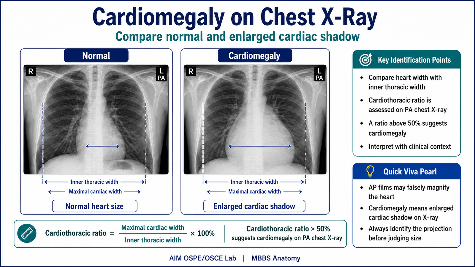
- Cardiothoracic ratio on PA chest X-ray.
- Cardiomegaly when cardiothoracic ratio is more than 50% on a proper PA film.
Result / Interpretation
A normal PA chest X-ray shows a cardiac shadow occupying less than half of the internal thoracic diameter.
Normal cardiothoracic ratio:
Less than 50%
Cardiomegaly:
Cardiothoracic ratio more than 50% on PA chest X-ray.
Clinical significance:
Cardiomegaly may indicate cardiac enlargement due to conditions such as ventricular hypertrophy, dilated cardiomyopathy, heart failure, or pericardial effusion. It should be interpreted with clinical findings.
Viva Questions
1. Which chamber mainly forms the right heart border on chest X-ray?
Right atrium.
2. Which chamber mainly forms the left lower heart border?
Left ventricle.
3. What is the normal cardiothoracic ratio on PA chest X-ray?
Less than 50%.
4. What does cardiomegaly mean on chest X-ray?
Enlargement of the cardiac shadow, usually cardiothoracic ratio more than 50%.
5. Why is PA view preferred for assessing heart size?
Because AP view may falsely magnify the cardiac shadow.
Marking Scheme
Total Marks: 5
| Component | Marks |
|---|---|
| Correct identification / performance | 2 |
| Key observation / procedure steps | 1 |
| Interpretation / principle | 1 |
| Viva answer | 1 |
Checklist detail:
| Checklist Point | Marks |
| Identifies cardiac shadow correctly | 1 |
| Identifies right and left heart borders correctly | 1 |
| Mentions cardiothoracic ratio / assesses cardiac size | 1 |
| Interprets cardiomegaly correctly | 1 |
| Answers viva question correctly | 1 |
Common Student Mistakes
- Confusing right atrium with right ventricle as the right heart border.
- Calling cardiomegaly on an AP film without considering magnification.
- Forgetting that cardiothoracic ratio should be assessed on a proper PA chest X-ray.
- Not identifying the left ventricle as the main left lower border.
AIM Feedback
Revise the cardiac borders as a simple X-ray outline. Remember: right border = right atrium, and left lower border = left ventricle. For cardiomegaly, always check whether the film is PA and then apply the 50% cardiothoracic ratio rule. Do not diagnose cardiomegaly from the X-ray alone without clinical correlation.
Short LMS Caption:
Identify the cardiac shadow, trace the right and left heart borders, and apply the 50% cardiothoracic ratio rule to detect cardiomegaly on PA chest X-ray.
🖼️ Visual / Image Support

🧩 Concept Map / Interpretation Support
🎥 Video Demonstration / Procedure Support