
This chapter follows the supplied KMU learning outcomes and connects anemia physiology, pathology, pharmacotherapy and public-health prevention in one logical sequence. First understand why each anemia develops; then use the morphology, laboratory findings and treatment principles for revision.
Foundations of Anemia, Nutritional & Hypoproliferative Anemias and Their Pharmacotherapy
Blood and Immunology Module
Understand how defective red-cell production, blood loss and nutritional deficiencies cause anemia; recognize iron-deficiency, megaloblastic and aplastic anemia; and connect these disorders with rational hematinic therapy and prevention of nutritional anemia.
Topic Introduction
Anemia is a reduction in the oxygen-carrying capacity of blood, most commonly recognized by a reduction in hemoglobin. It is not a single disease; it is a manifestation of different underlying problems. Red cells may be produced inadequately, lost through bleeding or destroyed prematurely. In this topic, the main focus is on disorders in which red-cell production becomes limited by iron, vitamin B12 or folate deficiency, as well as aplastic anemia caused by bone-marrow failure. You will first connect normal erythropoiesis and red-cell indices with the classification of anemia, then study the major pathological patterns, their diagnostic findings, appropriate hematinic drugs and the public-health prevention of nutritional anemia in Pakistan.
A. Physiological Basis, Erythropoiesis and Classification of Anemia
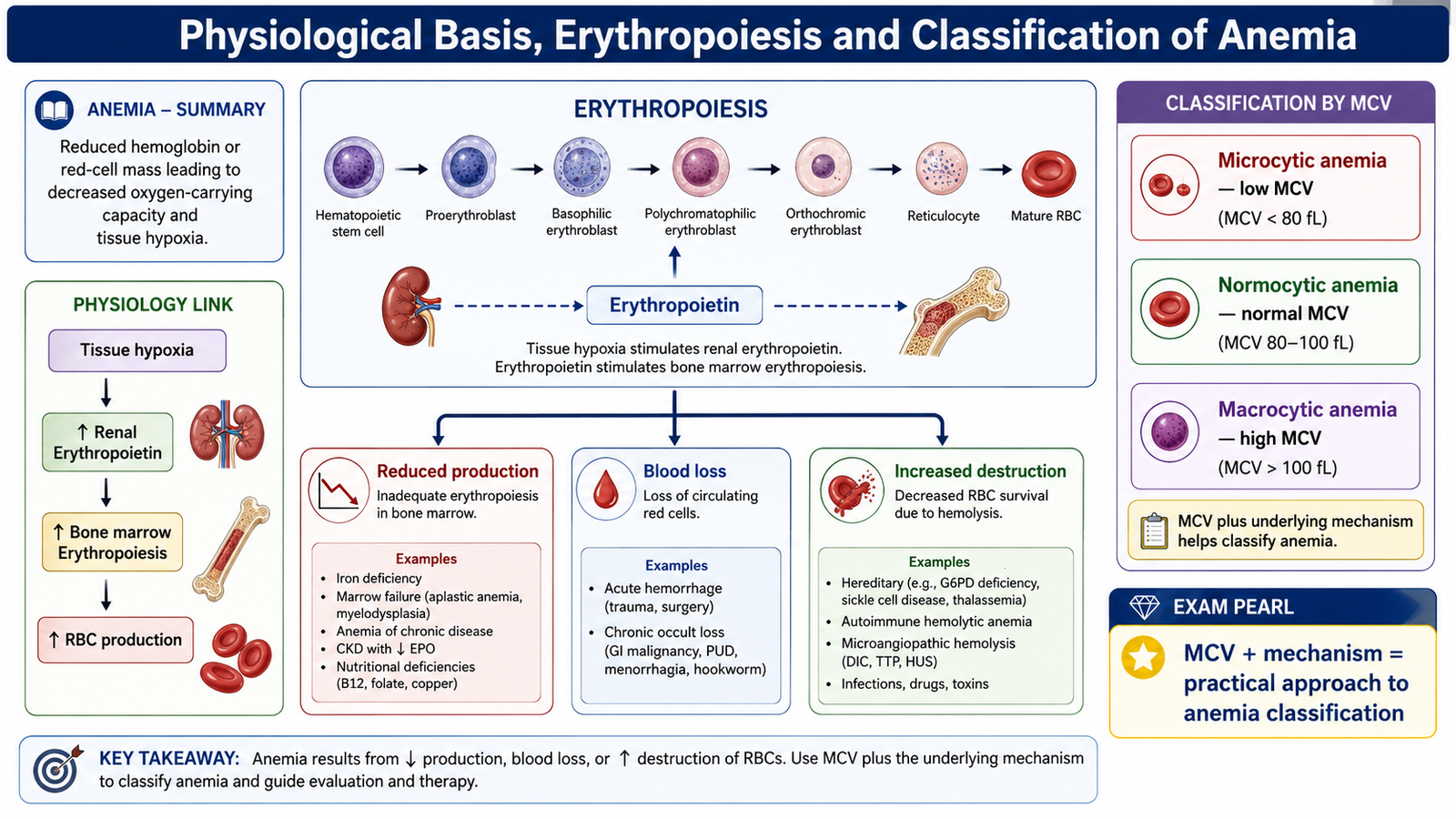
Anemia becomes clinically important because red blood cells contain hemoglobin, which carries oxygen from the lungs to the tissues. When the amount of functional hemoglobin falls, the oxygen-carrying capacity of blood decreases. Tissue oxygen delivery therefore becomes inadequate, particularly during exercise or when anemia is severe.
Physiological basis of anemia
The body attempts to compensate for reduced oxygen delivery. Tissue hypoxia stimulates the kidneys to increase secretion of erythropoietin, which acts on erythroid progenitor cells in the bone marrow and increases red-cell production. Cardiovascular compensation also occurs; heart rate and cardiac output may increase so that more blood reaches the tissues.
These compensatory mechanisms explain why anemia may cause fatigue, weakness, exertional dyspnea, dizziness and palpitations. Symptoms are usually more pronounced when anemia develops rapidly because the body has less time to adapt.
Normal erythropoiesis
Erythropoiesis is the production of red blood cells from hematopoietic stem cells. In adults, it occurs mainly in the bone marrow. Stem cells differentiate through erythroid progenitor and erythroblast stages before becoming reticulocytes and finally mature erythrocytes.
Successful erythropoiesis requires several components working together. Erythropoietin provides an important stimulatory signal. Iron is required for hemoglobin synthesis, while vitamin B12 and folate are required for normal DNA synthesis and cell division. The bone marrow itself must also contain sufficient functional hematopoietic cells.
This immediately explains the major disorders studied in this chapter. Lack of iron impairs hemoglobin formation, vitamin B12 or folate deficiency interferes with DNA synthesis, and aplastic anemia reduces the number of functioning marrow stem and progenitor cells.
Red-cell indices
Red-cell indices describe important characteristics of circulating erythrocytes. They help convert the general finding of “anemia” into a more useful morphological pattern.
| Index | Meaning | Clinical implication |
|---|---|---|
| MCV | Mean corpuscular volume; average red-cell size | Classifies anemia as microcytic, normocytic or macrocytic |
| MCH | Average amount of hemoglobin in each red cell | Falls when hemoglobin production is markedly impaired |
| MCHC | Average concentration of hemoglobin within red cells | Reduction supports a hypochromic pattern |
| Reticulocytes | Recently released immature red cells | Help assess whether the marrow is responding appropriately to anemia |
Classification according to underlying mechanism
A mechanistic classification asks a simple question: Why has the circulating red-cell mass become inadequate? There are three broad possibilities.
- Decreased red-cell production: the marrow cannot produce enough effective red cells. Nutritional deficiencies and aplastic anemia fall mainly into this category.
- Blood loss: red cells leave the circulation through acute or chronic bleeding.
- Increased red-cell destruction: red cells are destroyed prematurely, producing hemolytic anemia.
Morphology provides another useful classification. Microcytic anemia contains abnormally small cells, normocytic anemia contains cells of approximately normal size, and macrocytic anemia contains abnormally large cells. Iron deficiency usually produces a microcytic pattern, whereas megaloblastic anemia due to vitamin B12 or folate deficiency produces a macrocytic pattern.
Pathogenesis of blood-loss anemia
In acute blood loss, red cells and plasma are lost together. The immediate physiological problem is loss of circulating blood volume. If loss is substantial, reduced venous return and cardiac output can produce tissue hypoperfusion. Fluid subsequently moves into the circulation and plasma volume is restored, making the reduction in red-cell concentration more apparent. Renal hypoxia then stimulates erythropoietin and increases marrow red-cell production.
Chronic blood loss behaves differently. Repeated bleeding repeatedly removes iron contained in hemoglobin. Initially, stored iron can be used to support new red-cell production. With continued loss, iron stores are eventually depleted. Hemoglobin synthesis then becomes inadequate, and chronic blood-loss anemia commonly develops the morphological and laboratory features of iron deficiency.

B. Iron-Deficiency Anemia: Mechanism, Morphology and Diagnosis
Iron-deficiency anemia develops when the amount of iron available for erythropoiesis is insufficient to maintain normal hemoglobin production. Iron deficiency may result from inadequate intake, increased requirement, impaired absorption or continuing blood loss. Whatever the initiating cause, the central pathological problem is the same: progressively less iron becomes available to developing erythroid cells.
Pathophysiological mechanism
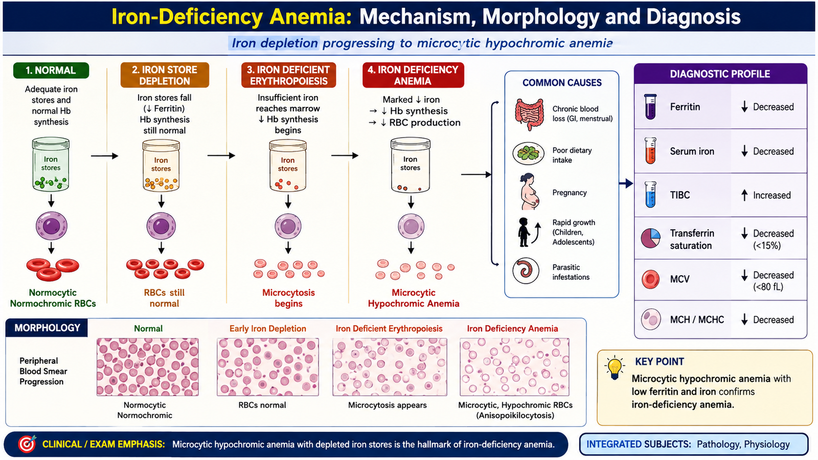
Iron deficiency develops in stages. The body initially uses stored iron to maintain erythropoiesis. As negative iron balance continues, storage iron becomes depleted. Eventually the iron supplied to developing red cells becomes inadequate, so hemoglobin synthesis falls.
Why do the cells become microcytic? Developing erythroblasts continue dividing while attempting to reach an adequate intracellular hemoglobin concentration. When hemoglobin synthesis is deficient, additional cell divisions result in smaller cells.
Why are they hypochromic? Each cell contains less hemoglobin than it should. On a peripheral smear this appears as increased central pallor.
Clinical course
Iron deficiency usually progresses gradually. Depletion of storage iron occurs before overt anemia develops. As iron supply becomes increasingly inadequate, hemoglobin synthesis becomes restricted and the characteristic red-cell abnormalities appear.
The general clinical effects largely reflect tissue hypoxia:
- Fatigue and weakness.
- Pallor.
- Reduced exercise tolerance.
- Exertional breathlessness.
- Palpitations or tachycardia when physiological compensation is significant.
Prolonged iron deficiency may also produce epithelial abnormalities. Glossitis and angular cheilitis may occur because rapidly renewing epithelium is affected. Koilonychia describes thin, spoon-shaped nails and is a classical feature of established deficiency. Pica, an unusual desire to consume non-food substances, may also be associated with iron deficiency.
Morphological changes
The peripheral-blood appearance reflects deficient hemoglobin production. Important findings include:
- Microcytosis — reduced red-cell size.
- Hypochromia — increased central pallor due to reduced hemoglobin content.
- Anisocytosis — increased variation in cell size.
- Poikilocytosis — variation in red-cell shape in more established disease.
- Elongated or pencil-shaped red cells may be present.
Laboratory investigations
A diagnosis should demonstrate both anemia and evidence of depleted iron availability. The results are interpreted as a pattern rather than as isolated values.
| Investigation | Typical pattern | Reason |
|---|---|---|
| Hemoglobin | Reduced | Hemoglobin synthesis is inadequate |
| MCV | Reduced | Red cells become microcytic |
| Peripheral smear | Microcytic, hypochromic pattern | Reduced hemoglobin synthesis |
| Serum ferritin | Reduced | Body iron stores have been depleted |
| Serum iron | Reduced | Less circulating iron is available |
| Total iron-binding capacity | Usually increased | Iron-binding capacity rises as available iron falls |
| Transferrin saturation | Reduced | A smaller proportion of transferrin is carrying iron |
Recognizing iron deficiency is only the first part of diagnosis. The cause of the deficiency must also be identified. The clinical assessment should therefore consider dietary intake, increased physiological requirements, gastrointestinal absorption and possible sources of chronic blood loss.
C. Megaloblastic Anemia: Vitamin B12 and Folic-Acid Deficiency
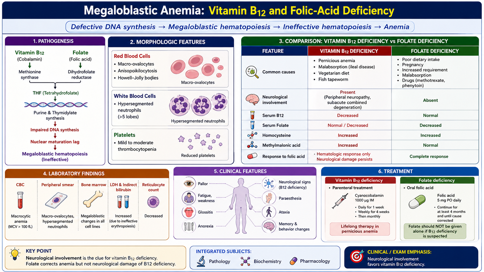
Megaloblastic anemia is a macrocytic anemia caused by defective DNA synthesis in rapidly dividing hematopoietic cells. Vitamin B12 and folate are both essential for normal nucleotide metabolism and DNA synthesis. Deficiency of either vitamin therefore interferes with nuclear maturation and cell division.
Pathogenesis
The essential abnormality is nuclear-cytoplasmic asynchrony. DNA synthesis and nuclear maturation are delayed, while cytoplasmic development continues relatively normally. Developing hematopoietic cells therefore become abnormally large and show immature nuclei relative to their cytoplasm.
Many abnormal precursor cells fail to mature and are destroyed within the marrow. This is called ineffective hematopoiesis. The marrow may therefore contain many developing cells while relatively few normal mature cells reach the peripheral circulation.
Morphological changes in red cells
The peripheral blood characteristically contains large oval red cells called macro-ovalocytes. Because the cells are larger than normal, the MCV is increased. Variation in red-cell size and shape may become prominent as the disorder progresses.
Within the marrow, erythroid precursors are enlarged and demonstrate the characteristic mismatch between delayed nuclear maturation and more advanced cytoplasmic development.
Changes in white cells
Defective DNA synthesis is not restricted to erythroid precursors. Granulocyte precursors are also affected. The classic peripheral-blood manifestation is the hypersegmented neutrophil. In marked disease, effective white-cell production can decrease, producing leukopenia.
Changes in platelets
Megakaryocyte development can also be disturbed. Consequently, significant megaloblastic disease may produce thrombocytopenia. Severe deficiency can therefore affect all three major blood-cell lines rather than producing an isolated anemia.
Determining whether B12 or folate deficiency is responsible
Vitamin B12 and folate deficiency produce very similar hematological morphology, so the blood film alone cannot reliably distinguish them. Serum vitamin levels and the clinical setting are therefore important.
A particularly important distinction is neurological involvement. Vitamin B12 deficiency can cause neurological dysfunction, whereas folate deficiency does not produce the same characteristic neurological syndrome. This difference is clinically important because treatment with folate may improve the anemia of vitamin B12 deficiency while allowing neurological damage to continue.
| Feature | Vitamin B12 deficiency | Folate deficiency |
|---|---|---|
| Anemia | Macrocytic, megaloblastic | Macrocytic, megaloblastic |
| Peripheral smear | Macro-ovalocytes; hypersegmented neutrophils | Macro-ovalocytes; hypersegmented neutrophils |
| Neurological abnormalities | May occur | Characteristic neurological syndrome absent |
| Methylmalonic acid | May be increased | Not characteristically increased |
| Homocysteine | May be increased | May be increased |
Diagnostic approach to the underlying cause
The investigation should proceed from recognition of the megaloblastic pattern to identification of the deficient vitamin and then to determination of why that deficiency occurred.
- A complete blood count identifies macrocytosis and associated cytopenias.
- The peripheral smear can demonstrate macro-ovalocytes and hypersegmented neutrophils.
- Serum vitamin B12 and folate assessment helps identify the deficient nutrient.
- Metabolic markers can assist when the distinction remains uncertain.
- Dietary history may identify inadequate intake.
- Gastrointestinal disease or previous gastrointestinal procedures may suggest malabsorption.
- Loss of normal intrinsic-factor-dependent absorption should be considered in vitamin B12 deficiency.
- Increased physiological requirements are especially relevant to folate deficiency.
Pernicious anemia is an important cause of vitamin B12 deficiency in which effective intrinsic-factor-dependent absorption is lost. Where clinically appropriate, testing directed at this mechanism can support identification of the underlying cause.
D. Aplastic Anemia: Causes, Pathophysiology and Diagnosis
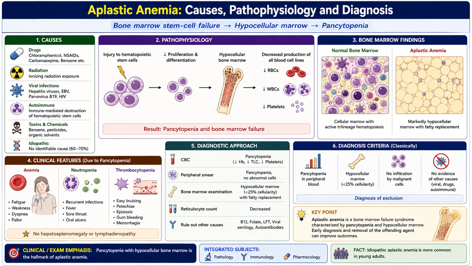
Aplastic anemia is a bone-marrow failure disorder characterized by marked reduction of functional hematopoietic stem and progenitor cells. The marrow therefore cannot produce adequate red cells, white cells or platelets. The resulting reduction in multiple cell lines is called pancytopenia.
Causes
Aplastic anemia may be acquired or inherited. In many acquired cases, a definite external cause cannot be identified. The important concept is that different initiating factors ultimately reduce the population or function of hematopoietic stem cells.
- Idiopathic acquired aplastic anemia — no definite precipitating factor is demonstrated.
- Immune-mediated marrow injury — an important mechanism in acquired disease.
- Drugs and chemicals capable of producing marrow toxicity.
- Ionizing radiation damaging proliferating hematopoietic cells.
- Certain infections associated with marrow failure.
- Inherited marrow-failure disorders, including Fanconi anemia.
Pathophysiology
In many acquired cases, immune mechanisms suppress or destroy hematopoietic stem and progenitor cells. Once the number of functioning precursors falls markedly, normal marrow production cannot be maintained.
Each component of pancytopenia produces a predictable clinical consequence:
- Anemia causes fatigue, pallor and manifestations of reduced oxygen delivery.
- Neutropenia reduces protection against infection.
- Thrombocytopenia causes easy bruising, petechiae and bleeding tendency.
Diagnostic approach
The complete blood count may show pancytopenia. Because red-cell production is impaired, the reticulocyte response is inappropriately low for the degree of anemia.
The central diagnostic finding is obtained by bone-marrow examination. Normal hematopoietic cells are markedly reduced and much of the marrow space is replaced by fat, producing a hypocellular appearance.
The diagnostic process therefore includes:
- Identification of cytopenias on complete blood count.
- Peripheral-blood examination.
- Assessment of the reticulocyte response.
- Bone-marrow examination demonstrating marked hypocellularity.
- History of relevant drug, chemical or radiation exposure.
- Assessment for associated infection or inherited marrow-failure disorders where suggested clinically.
- Exclusion of other causes of pancytopenia.
E. Drugs Used in Anemia and Iron Pharmacotherapy
Pharmacological treatment of deficiency anemia should replace the substance that is limiting effective erythropoiesis. The major hematinic agents in this topic are iron, vitamin B12 and folic acid. Rational therapy therefore begins by identifying which component is deficient rather than treating every anemia with the same drug.
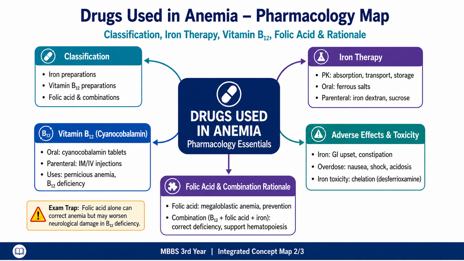
Classification of drugs used in anemia
| Group | Main preparations | Main role |
|---|---|---|
| Iron | Oral and parenteral iron preparations | Restores iron needed for hemoglobin synthesis |
| Vitamin B12 | Oral and parenteral cyanocobalamin | Corrects B12-deficient megaloblastic hematopoiesis |
| Folic acid | Oral folic acid | Corrects folate-deficient DNA synthesis |
Pharmacokinetics of iron
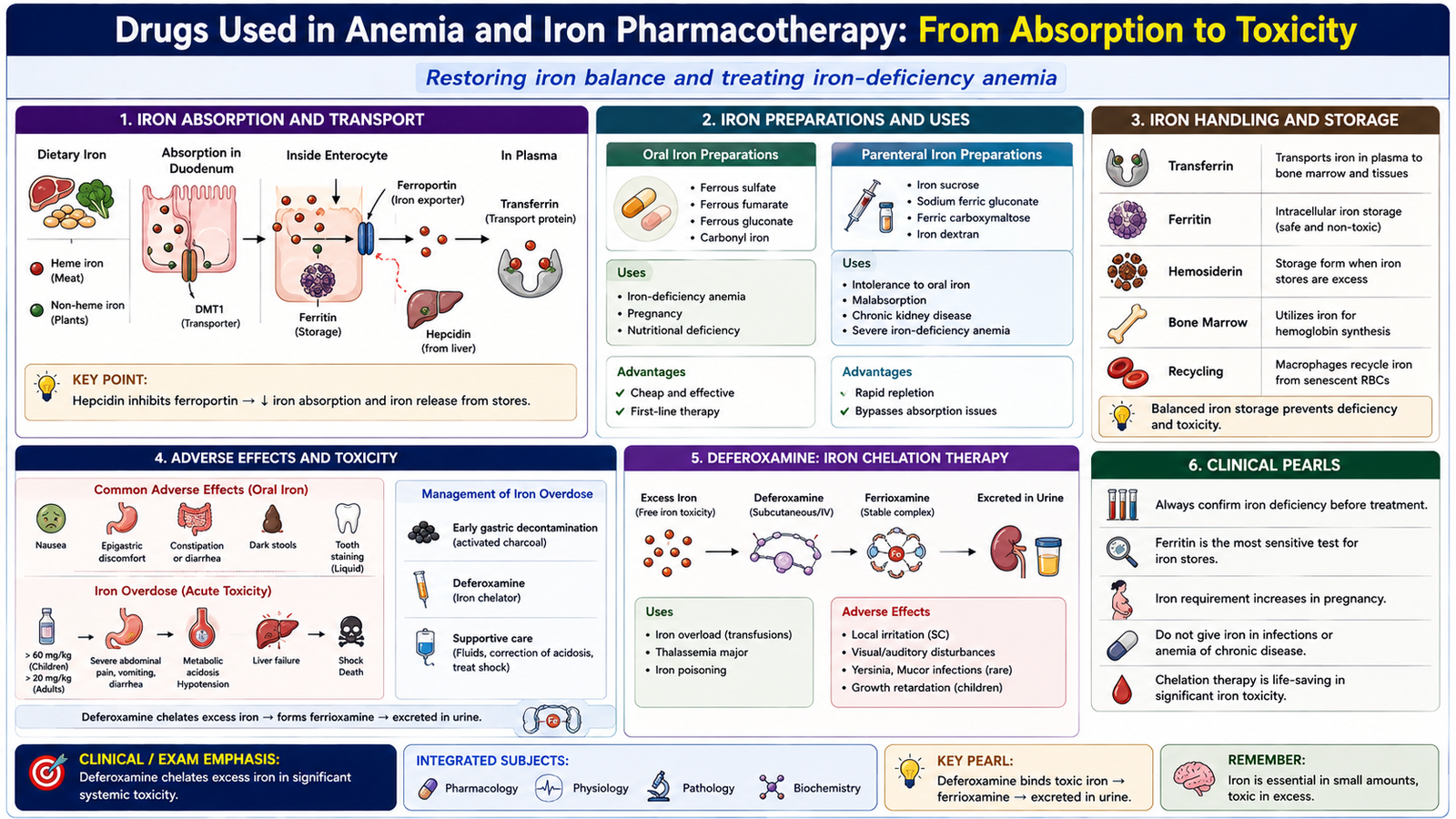
Iron absorption occurs mainly in the duodenum and proximal small intestine. Absorbed iron enters intestinal epithelial cells and is either temporarily stored or transferred into the circulation through the iron-export protein ferroportin.
In plasma, iron binds to transferrin. Transferrin delivers iron to tissues, especially developing erythroid cells in bone marrow, where it is incorporated into heme and ultimately hemoglobin. Iron that is not immediately required is stored mainly as ferritin.
The body has no efficient mechanism for actively excreting large excesses of iron. Iron balance is therefore regulated mainly at the level of absorption and release from storage sites.
Hepcidin, produced by the liver, is a major regulator of systemic iron availability. Increased hepcidin reduces ferroportin-mediated movement of iron into plasma, whereas lower hepcidin activity permits greater iron availability.
Oral iron preparations
Oral therapy is suitable when iron deficiency is present and gastrointestinal absorption is adequate. Common preparations contain ferrous iron.
- Ferrous sulfate.
- Ferrous fumarate.
- Ferrous gluconate.
Food and several substances in the gastrointestinal tract can reduce iron absorption. The clinically important principle is that the amount of iron actually absorbed is influenced by the chemical form of iron, gastrointestinal conditions and existing body iron needs.
Adverse effects of oral iron
Unabsorbed iron can irritate the gastrointestinal tract. This explains why gastrointestinal adverse effects are particularly common with oral therapy.
- Nausea.
- Epigastric discomfort.
- Abdominal pain.
- Constipation or diarrhea.
- Dark stools.
Parenteral iron preparations
Parenteral iron bypasses gastrointestinal absorption. It is useful when adequate iron replacement cannot be achieved satisfactorily through the oral route, such as when absorption is impaired or oral preparations are not tolerated.
Examples include:
- Iron sucrose.
- Ferric carboxymaltose.
- Iron dextran.
Parenteral preparations can cause infusion-related reactions and, less commonly, clinically important hypersensitivity. The possibility and severity of these reactions vary among preparations.
Iron toxicity and its treatment
Excessive iron can produce severe toxicity because free iron damages gastrointestinal mucosa and, after systemic absorption, promotes cellular injury. Significant poisoning may therefore progress from gastrointestinal symptoms to circulatory, metabolic and organ dysfunction.
Management begins with appropriate supportive assessment and stabilization. In significant systemic iron poisoning, deferoxamine is an iron-chelating drug. It binds free iron to form a complex that can be eliminated from the body.
F. Vitamin B12, Folic Acid and Rational Hematinic Combination Therapy
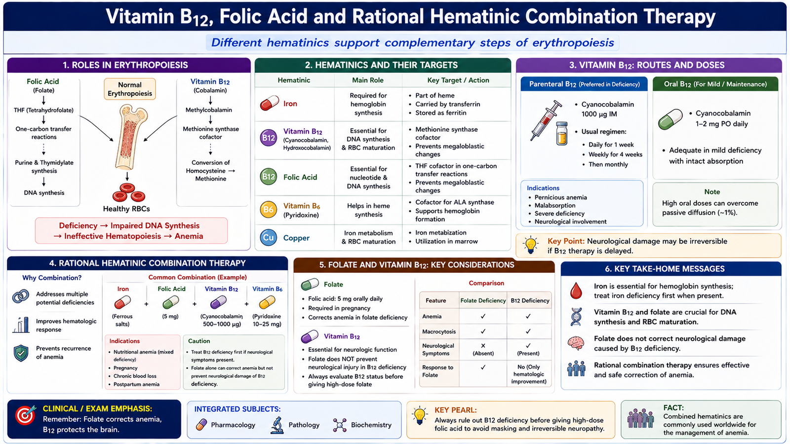
Vitamin B12 and folate are required for normal DNA synthesis. Their pharmacological replacement allows abnormal megaloblastic hematopoiesis to return toward normal when deficiency of the corresponding vitamin is responsible. Because vitamin B12 and folate deficiency can produce very similar blood findings, identifying the deficient nutrient before treatment is particularly important.
Cyanocobalamin preparations
Cyanocobalamin is a preparation of vitamin B12. It can be given through the oral route or by parenteral administration. Selection of the route depends mainly on whether adequate gastrointestinal absorption can be relied upon.
- Oral cyanocobalamin provides vitamin B12 through gastrointestinal absorption.
- Parenteral cyanocobalamin bypasses gastrointestinal absorption and is useful when normal B12 absorption is seriously impaired.
Clinical uses of vitamin B12
Vitamin B12 is used to correct confirmed or appropriately recognized B12 deficiency. Important settings include:
- Nutritional vitamin B12 deficiency.
- Pernicious anemia.
- Vitamin B12 malabsorption.
- Conditions in which normal gastrointestinal absorption of B12 is significantly impaired.
Replacement corrects defective DNA synthesis in hematopoietic tissue and is especially important because untreated B12 deficiency may also produce neurological injury.
Clinical uses of folic acid
Folic acid replacement supplies folate required for DNA synthesis. Its major uses relevant to this topic include:
- Treatment of folate-deficiency megaloblastic anemia.
- Supplementation when folate requirements are increased.
- Prevention of deficiency in appropriately identified high-risk individuals.
- Folate supplementation in relation to pregnancy because adequate folate is required for normal fetal neural-tube development.
Why cyanocobalamin, folic acid and iron may be combined
The pharmacological rationale becomes clear when normal erythropoiesis is considered. These nutrients do not perform the same function.
Vitamin B12 + folate → normal DNA synthesis and maturation of dividing erythroid cells
Adequate supply of all required nutrients → effective erythropoiesis
Combination therapy is therefore rational when more than one deficiency is present or when a clinical setting places the patient at significant risk of combined deficiency. It is not rational simply to give every patient with anemia iron, folate and vitamin B12 without determining the underlying disorder.
This distinction is important because empirical combination therapy can alter the blood picture while an important cause remains unrecognized. Treatment should therefore support, rather than replace, the diagnostic evaluation of anemia.
G. Nutritional Anemia in Pakistan: Vulnerable Populations, Risk Factors and Prevention
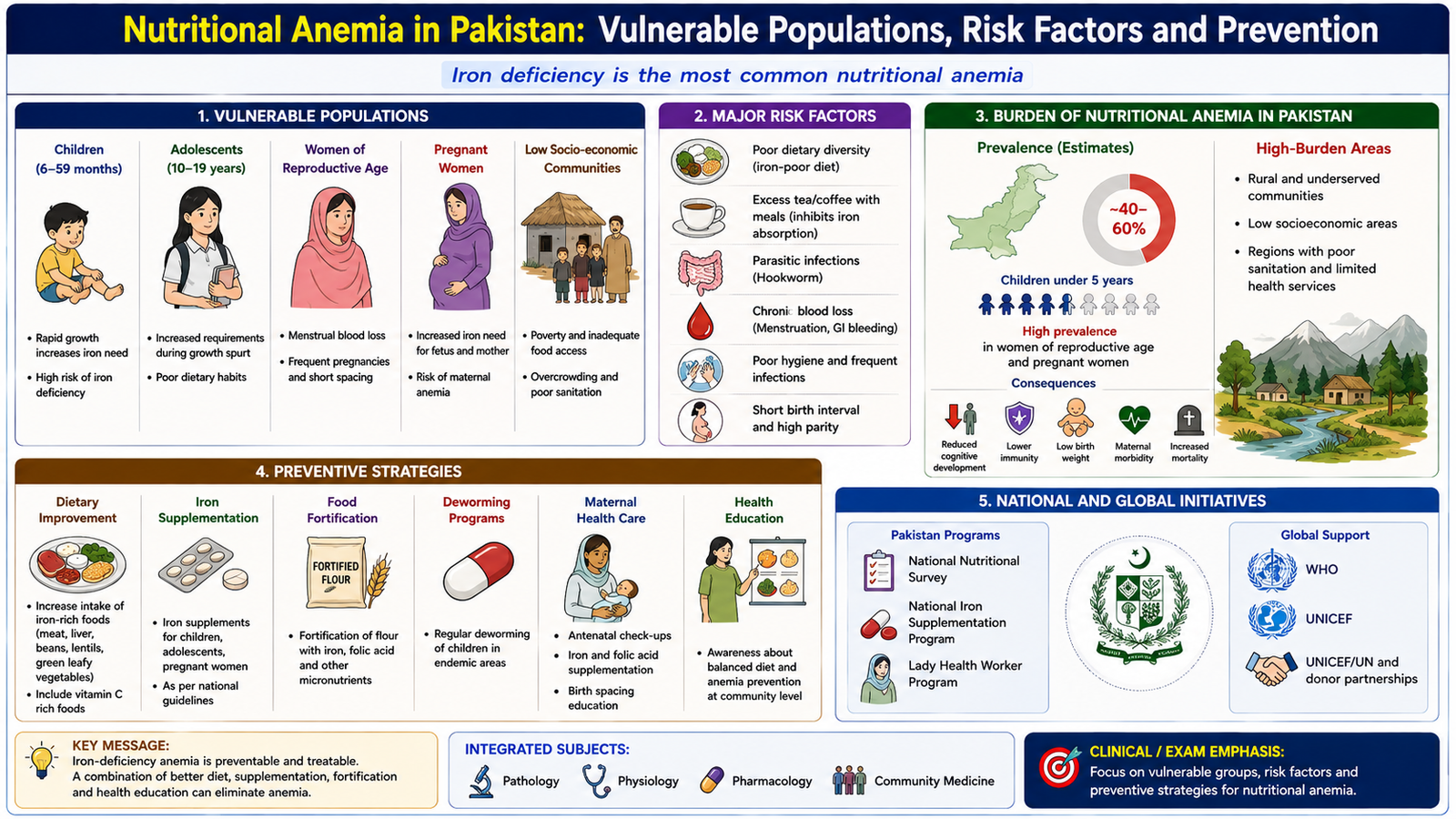
Nutritional anemia develops when one or more nutrients required for normal erythropoiesis are deficient. At population level, prevention requires more than treatment of individual patients. Public-health measures must identify which groups are most vulnerable, why deficiency develops and which interventions can reduce the underlying risk.
Classification of nutritional anemias
- Iron-deficiency anemia — inadequate iron availability limits hemoglobin synthesis.
- Folate-deficiency anemia — inadequate folate impairs DNA synthesis and produces megaloblastic hematopoiesis.
- Vitamin B12-deficiency anemia — B12 deficiency impairs DNA synthesis and may additionally produce neurological abnormalities.
- Mixed nutritional anemia — more than one required nutrient is deficient.
Important nutritional anemia affecting Pakistan
Iron deficiency is a major nutritional cause of anemia in Pakistan and is especially important among women and children. Folate and vitamin B12 deficiencies may also contribute, particularly where dietary supply, physiological demand or absorption is inadequate.
The exact numerical prevalence can vary according to the population studied and the survey used. The important undergraduate public-health principle is therefore to recognize the major vulnerable groups and determinants rather than attach an unsupported single prevalence figure to the entire country.
Vulnerable populations and why they are at risk
Vulnerability usually develops when physiological need exceeds nutrient intake or when nutrients are lost faster than they can be replaced.
- Infants and young children: rapid growth increases the requirement for nutrients needed for erythropoiesis.
- Adolescents: rapid increase in body mass and blood volume increases iron requirements.
- Adolescent girls: growth-related requirements are combined with menstrual iron loss.
- Women of reproductive age: repeated menstrual blood loss increases the risk of negative iron balance.
- Pregnant women: maternal blood-volume expansion and fetal growth substantially increase nutritional requirements.
- Women with closely spaced or repeated pregnancies: depleted nutritional stores may not be fully restored before the next pregnancy.
- People with poorly diversified diets: intake of iron, folate, vitamin B12 or other important nutrients may be inadequate.
Major risk factors
The determinants of nutritional anemia can be understood by grouping them according to how they disturb nutrient balance.
| Mechanism | Important risk factors |
|---|---|
| Inadequate intake | Poor dietary diversity and inadequate intake of micronutrient-rich foods |
| Increased requirement | Rapid childhood growth, adolescence and pregnancy |
| Blood or nutrient loss | Menstrual loss and other chronic blood loss |
| Reduced absorption | Gastrointestinal conditions interfering with nutrient absorption |
| Contributing disease | Infections or parasitic disease when they produce blood loss or nutritional depletion |
| Social determinants | Limited access to a nutritionally adequate and diverse diet |
Public-health strategies for prevention
Effective prevention works at more than one point in the pathway from inadequate nutritional supply to established anemia. Strategies should improve nutrient intake, protect high-risk groups, reduce avoidable losses and detect clinically important anemia early enough for its cause to be treated.
- Dietary diversification: improve access to and use of foods providing iron and other essential micronutrients.
- Nutrition education: improve understanding of nutrient-rich food choices and the needs of vulnerable groups.
- Food fortification: increase micronutrient availability through appropriate commonly consumed foods.
- Targeted supplementation: provide required micronutrients to appropriately identified high-risk groups.
- Maternal and child health measures: protect groups with high physiological nutrient requirements.
- Control of contributing infection or parasitic disease: reduce chronic blood loss and nutritional depletion where these factors are present.
- Early recognition and appropriate treatment: identify anemia and investigate its underlying cause instead of repeatedly replacing nutrients without assessment.

Integrated Mechanism Flow
The major anemias in this topic differ in their initiating defect but eventually converge on inadequate effective red-cell production and reduced oxygen delivery.
↓
2. Defective hemoglobin synthesis, defective DNA synthesis or reduced marrow-cell production
↓
3. Abnormal or inadequate erythropoiesis
↓
4. Reduced effective circulating red-cell mass and hemoglobin
↓
5. Reduced tissue oxygen delivery
↓
6. Clinical manifestations of anemia and compensatory erythropoietin/cardiovascular responses
Important Comparison: Major Hypoproliferative and Nutritional Anemia Patterns
| Feature | Iron deficiency | Megaloblastic anemia | Aplastic anemia |
|---|---|---|---|
| Main defect | Reduced hemoglobin synthesis | Defective DNA synthesis | Bone-marrow stem/progenitor-cell failure |
| Typical red-cell size | Microcytic | Macrocytic | Usually not defined by a distinctive microcytic or megaloblastic pattern |
| Characteristic clue | Microcytosis + hypochromia + depleted iron stores | Macro-ovalocytes + hypersegmented neutrophils | Pancytopenia + hypocellular marrow |
| Other cell lines | Not primarily defined by pancytopenia | WBCs and platelets may fall in severe disease | WBCs and platelets characteristically reduced with RBCs |
| Core diagnostic direction | Iron studies + search for cause of iron deficiency | B12/folate assessment + search for cause | Bone-marrow examination + search for marrow-failure cause |
⭐ AIM High-Yield Review
- Anemia reduces the oxygen-carrying capacity of blood and stimulates compensatory erythropoietin production.
- Mechanistically, anemia results from decreased production, blood loss or increased destruction of red cells.
- MCV separates anemia into microcytic, normocytic and macrocytic patterns.
- Chronic blood loss can deplete iron stores and eventually produce iron-deficiency anemia.
- ⭐ Iron deficiency causes impaired hemoglobin synthesis → microcytosis + hypochromia.
- Reduced ferritin indicates depleted iron stores and is an important clue to iron deficiency.
- Vitamin B12 and folate deficiency impair DNA synthesis and produce megaloblastic hematopoiesis.
- ⭐ Macro-ovalocytes + hypersegmented neutrophils are classic clues to megaloblastic anemia.
- Neurological abnormalities distinguish vitamin B12 deficiency from uncomplicated folate deficiency.
- ⭐ Aplastic anemia produces marrow failure with pancytopenia and hypocellular fatty marrow.
- Oral iron commonly causes gastrointestinal adverse effects; parenteral iron bypasses gastrointestinal absorption.
- Deferoxamine is an iron-chelating drug used in significant systemic iron toxicity.
- Folic acid may improve the anemia of B12 deficiency without correcting B12-related neurological injury.
- Iron supports hemoglobin synthesis, whereas vitamin B12 and folate support normal DNA synthesis and cell maturation.
- Prevention of nutritional anemia requires identification of vulnerable groups, improved nutritional supply and correction of contributing causes rather than nutrient replacement alone.
Types of Anemias — Hematology
Use this video to reinforce anemia classification, iron-deficiency anemia, vitamin B12 and folate deficiency, blood-loss anemia and aplastic anemia after completing the AIM learning material.