
This chapter follows the supplied KMU learning outcomes and builds the topic in a logical sequence. First understand how each lymphoid neoplasm develops and appears morphologically; then use the final high-yield review to consolidate the important examination points.
Topic 4 — Lymphoid Neoplasms, Lymphomas and Plasma Cell Disorders
Blood and Immunology Module • Pathology
A structured introduction to the classification, pathogenesis, morphology and diagnosis of major lymphoid neoplasms, including ALL, CLL, multiple myeloma, Hodgkin lymphoma and major non-Hodgkin lymphomas.
Topic Introduction
Lymphoid neoplasms are clonal disorders produced by malignant transformation of B lymphocytes, T lymphocytes, natural killer cells or their precursors. Because normal lymphocytes develop through several stages of maturation, malignant transformation can occur at different stages and produce very different diseases. Some mainly involve bone marrow and blood and present as leukemia, while others primarily form masses in lymph nodes or extranodal tissues and present as lymphoma. Plasma cell neoplasms arise from terminally differentiated B cells that produce immunoglobulins. Understanding these disorders becomes easier when each disease is connected to its cell of origin, mechanism of growth, characteristic morphology and diagnostic markers. This chapter develops these links systematically.
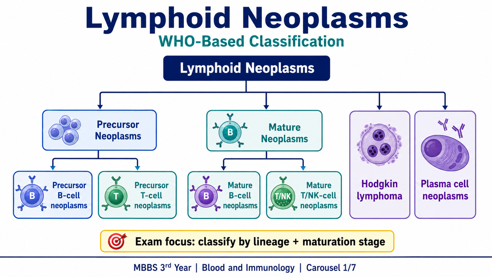
A. Lymphoid Neoplasms: Core Concept and WHO-Based Classification
Lymphoid neoplasms form a large group of malignant clonal proliferations derived from cells of the immune system. A useful way to understand them is to remember that a normal lymphocyte passes through a sequence of precursor development, maturation and antigen-dependent differentiation. A malignant clone generally retains features of the normal lymphoid cell from which it developed. Therefore, morphology, immunophenotype and genetic abnormalities can be used together to determine the lineage and maturation stage of the tumor.
The terms leukemia and lymphoma describe the dominant anatomical pattern of disease rather than two completely separate biological processes. A lymphoid malignancy predominantly involving marrow and peripheral blood tends to be called leukemia, while one forming tissue masses is usually called lymphoma. Some entities can produce either presentation. This is why precursor tumors are commonly named lymphoblastic leukemia/lymphoma and why chronic lymphocytic leukemia and small lymphocytic lymphoma represent closely related manifestations of the same basic disease.
Major groups of lymphoid neoplasms
The WHO approach classifies lymphoid neoplasms primarily according to cell lineage and stage of differentiation, and then uses morphology, immunophenotype and genetic features to define individual entities.
- Precursor B-cell neoplasms: mainly B-lymphoblastic leukemia/lymphoma.
- Precursor T-cell neoplasms: mainly T-lymphoblastic leukemia/lymphoma.
- Mature B-cell neoplasms: include chronic lymphocytic leukemia/small lymphocytic lymphoma, follicular lymphoma, mantle cell lymphoma, marginal zone lymphomas, diffuse large B-cell lymphoma and Burkitt lymphoma.
- Mature T-cell and NK-cell neoplasms: include peripheral T-cell lymphomas, anaplastic large cell lymphoma, adult T-cell leukemia/lymphoma and extranodal NK/T-cell lymphoma.
- Hodgkin lymphoma: includes classical Hodgkin lymphoma and nodular lymphocyte-predominant Hodgkin lymphoma.
- Plasma cell neoplasms: include plasmacytoma and plasma cell myeloma, commonly called multiple myeloma.
Why classification matters
Lymphoid tumors may look similar clinically but arise from biologically different cells. For example, a lymph node enlarged by follicular lymphoma contains neoplastic mature B cells, whereas lymphoblastic lymphoma is composed of immature precursor lymphocytes. Identifying lineage and maturation stage therefore helps establish the diagnosis and explains the expected morphology and biological behavior.
B. Acute Lymphoblastic Leukemia/Lymphoblastic Lymphoma
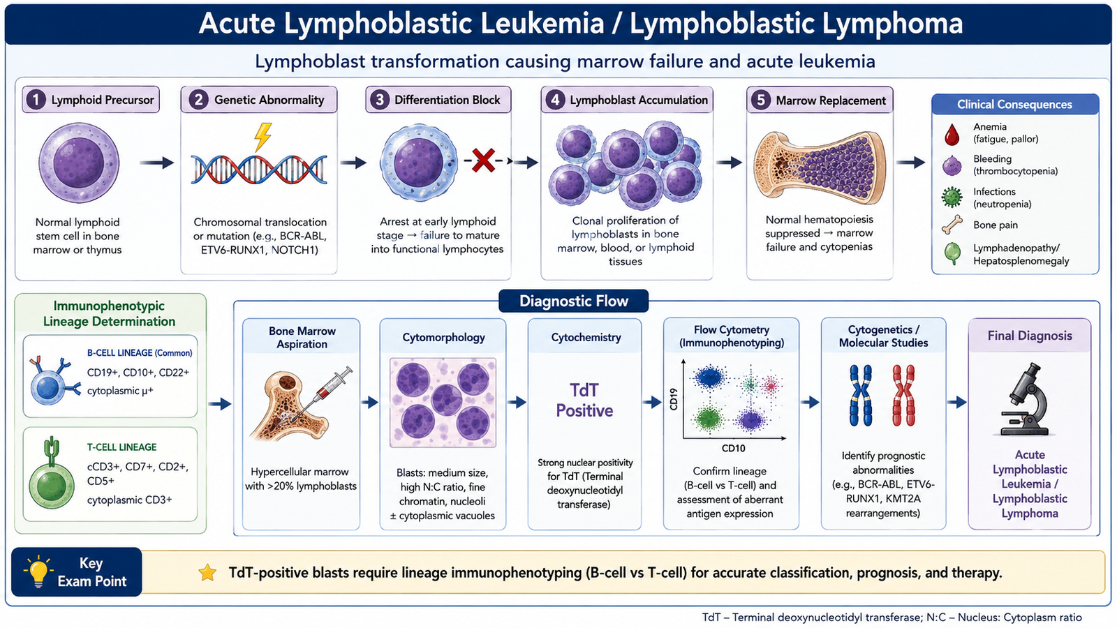
Acute lymphoblastic leukemia (ALL) is a malignant neoplasm of immature lymphoid precursor cells called lymphoblasts. The abnormal clone may belong to either the B-cell or T-cell lineage. When the malignant lymphoblasts predominantly involve bone marrow and blood, the disease presents as leukemia; when tissue masses dominate, the term lymphoblastic lymphoma is used.
Pathophysiology
ALL develops when genetic abnormalities arise in an immature lymphoid precursor and disturb the normal balance between differentiation, proliferation and cell death. The affected blast fails to mature normally but retains the ability to proliferate. Expansion of this clone progressively replaces normal bone marrow cells.
Genetic alteration in a lymphoid precursor → impaired differentiation with excessive survival and proliferation → accumulation of lymphoblasts → replacement of normal marrow → anemia, thrombocytopenia and neutropenia → spread to blood and other tissues.
The consequences of marrow replacement explain many manifestations. Reduced erythropoiesis produces anemia and fatigue. Reduced platelet production causes bruising or bleeding. Loss of normal neutrophils increases susceptibility to infection. Lymphoblasts may also infiltrate lymph nodes, liver, spleen, the central nervous system and other tissues.
B-ALL is the major precursor B-cell neoplasm. Important genetic abnormalities occur in different subgroups. At undergraduate level, useful examples include hyperdiploidy, ETV6-RUNX1 fusion and BCR-ABL1 fusion. These abnormalities demonstrate that ALL is not one genetically uniform disorder.
T-ALL arises from immature T-lineage cells and commonly has biological features related to thymic T-cell development. Because these cells normally develop in the thymus, a mediastinal mass caused by thymic involvement is an important clue to T-lineage lymphoblastic disease.
Morphological features
The peripheral blood and bone marrow contain increased numbers of lymphoblasts. These immature cells usually appear relatively uniform rather than showing the wide range of maturation seen in reactive lymphoid processes.
- Small to medium-sized lymphoblasts.
- High nuclear-to-cytoplasmic ratio.
- Scant, usually basophilic cytoplasm.
- Fine or relatively open nuclear chromatin.
- Nucleoli may be inconspicuous or visible.
- Bone marrow becomes increasingly replaced by blasts.
Unlike myeloblasts in acute myeloid leukemia, lymphoblasts do not characteristically contain Auer rods. This is a useful morphological distinction.
Diagnostic approach
Diagnosis requires more than recognizing immature cells. The central diagnostic task is to establish that the cells are lymphoblasts, determine whether they are B- or T-lineage, and identify important genetic abnormalities.
- Complete blood count: may demonstrate anemia, thrombocytopenia and abnormalities in the leukocyte count.
- Peripheral blood smear: may show circulating lymphoblasts.
- Bone-marrow examination: demonstrates marrow involvement by lymphoblasts and allows detailed morphological assessment.
- Flow cytometry or immunophenotyping: establishes precursor phenotype and lineage.
- Genetic and molecular testing: identifies disease-defining or prognostically important abnormalities.
Immature lymphoid cells characteristically express terminal deoxynucleotidyl transferase (TdT). B-lineage blasts commonly express B-cell-associated markers such as CD19 and CD79a, while T-lineage blasts express T-cell markers such as cytoplasmic or surface CD3 and other T-cell-associated antigens.
C. Chronic Lymphocytic Leukemia/Small Lymphocytic Lymphoma
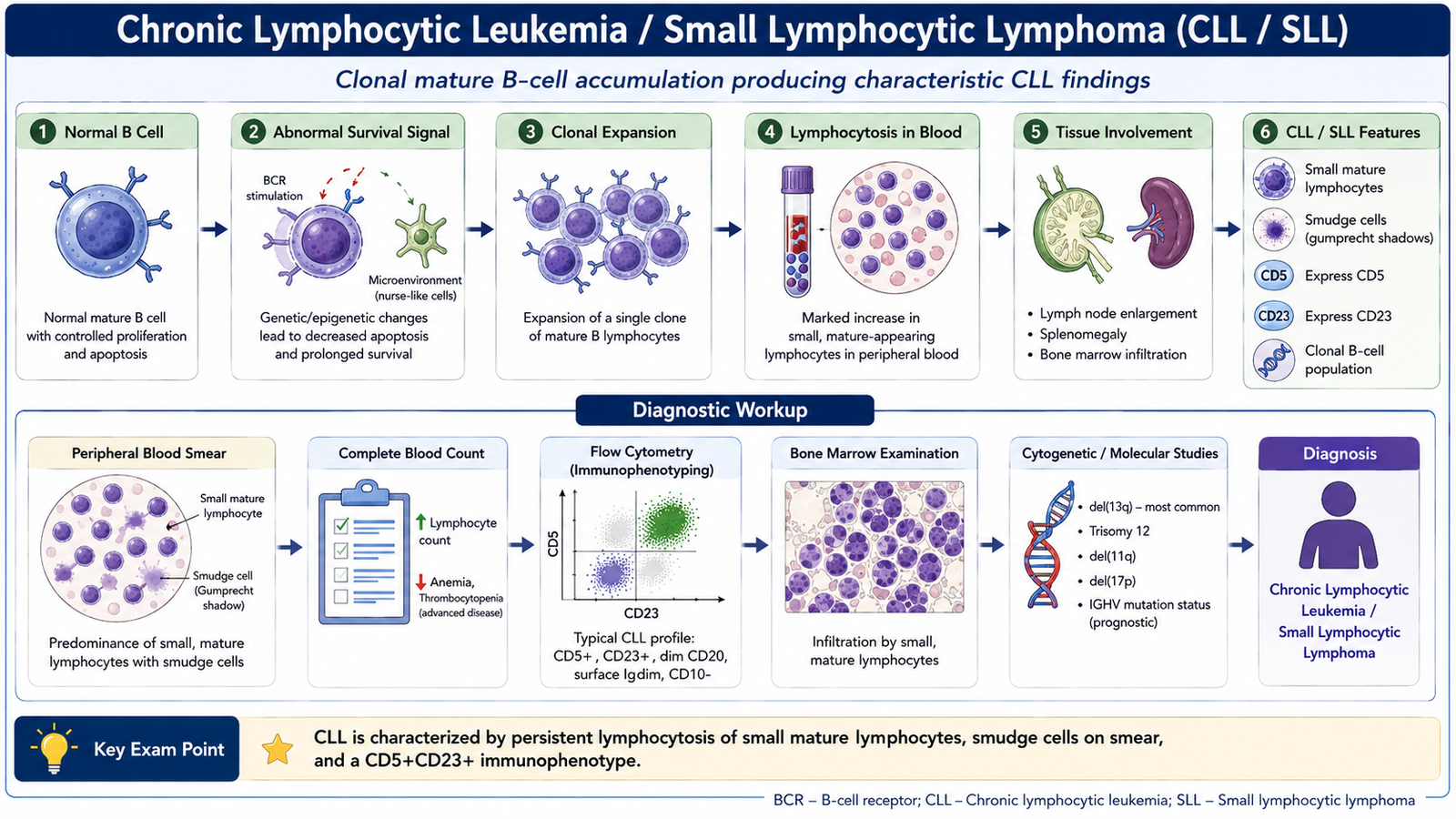
Chronic lymphocytic leukemia (CLL) is a mature B-cell neoplasm characterized by accumulation of small, relatively mature-appearing clonal B lymphocytes in blood, bone marrow and lymphoid tissues. Small lymphocytic lymphoma (SLL) represents the closely related tissue-based form in which lymph nodes or other lymphoid tissues are predominantly involved.
Pathophysiology
The malignant cells in CLL are not rapidly dividing blasts. Instead, the disorder is characterized largely by abnormal survival and gradual accumulation of a clonal population of B lymphocytes. Alterations affecting pathways that control apoptosis, B-cell receptor signaling and cell survival allow these cells to persist longer than normal.
Several chromosomal abnormalities can occur. Commonly recognized examples include deletion of chromosome 13q, trisomy 12, deletion of 11q and deletion of 17p involving the TP53 pathway. These abnormalities help demonstrate the molecular heterogeneity of CLL and may influence disease behavior.
Although the malignant lymphocytes accumulate, they are immunologically dysfunctional. Therefore, patients may eventually develop impaired humoral immunity and increased susceptibility to infection. Extensive marrow replacement can also produce anemia or thrombocytopenia.
Distinguishing morphological features
The characteristic peripheral smear shows a large population of small mature-appearing lymphocytes. Their nuclei contain dense, coarse chromatin, while the cytoplasm is scant.
- Small, relatively uniform lymphocytes.
- Round nuclei with densely clumped chromatin.
- Scant cytoplasm.
- Smudge cells are frequently seen on peripheral blood films.
Smudge cells form because CLL lymphocytes are fragile and may rupture while the blood film is being prepared. They support the morphological impression but are not, by themselves, sufficient for diagnosis.
In involved lymph nodes, normal architecture is replaced by small lymphocytes. Areas containing larger proliferating cells may form pale structures known as proliferation centers, which are characteristic of CLL/SLL tissue involvement.
Diagnostic workup
The diagnosis combines persistent clonal lymphocytosis with characteristic immunophenotypic features. The workup therefore moves from recognition of lymphocytosis to confirmation that the expanded cells constitute a single abnormal B-cell population.
- Complete blood count: identifies lymphocytosis and associated cytopenias when present.
- Peripheral smear: demonstrates small mature lymphocytes and smudge cells.
- Flow cytometry: confirms clonality and the characteristic immunophenotype.
- Cytogenetic or molecular studies: may identify abnormalities important for biological characterization.
- Lymph-node examination or biopsy: is relevant when tissue-based disease predominates.
CLL cells typically express the B-cell marker CD19 together with CD5 and CD23. CD20 and surface immunoglobulin expression are generally weaker than in many other mature B-cell neoplasms. The unusual co-expression of a B-cell phenotype with CD5 is therefore a useful diagnostic feature.
D. Multiple Myeloma and Plasma Cell Neoplasia
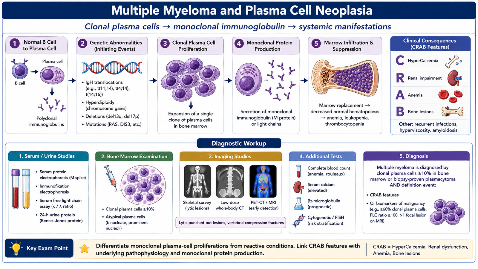
Multiple myeloma, also called plasma cell myeloma, is a malignant neoplasm of clonal plasma cells that usually involves multiple sites within bone marrow. Plasma cells are terminally differentiated B lymphocytes specialized for immunoglobulin production. Consequently, the malignant clone commonly produces a single type of immunoglobulin or immunoglobulin component known as a monoclonal protein.
Pathogenesis
Multiple myeloma develops through progressive genetic alterations in a plasma cell clone. Early abnormalities commonly involve changes affecting immunoglobulin gene regions and genes controlling plasma-cell growth. Additional abnormalities accumulate as the clone expands and becomes more biologically aggressive.
Because malignant plasma cells depend strongly on the bone-marrow environment, interaction with marrow stromal cells and local cytokines helps support their survival. The expanding clone also alters normal bone remodeling. Osteoclast activity is increased while normal bone formation is relatively suppressed, producing focal bone destruction.
Important molecular genetic abnormalities
The molecular abnormalities in myeloma are heterogeneous rather than identical in every patient. Important undergraduate examples include:
- Translocations involving the immunoglobulin heavy-chain locus on chromosome 14.
- Abnormal activation of genes controlling the cell cycle, including cyclin-related pathways.
- Hyperdiploidy in an important subgroup of tumors.
- Secondary abnormalities involving pathways such as RAS and MYC during progression.
- Loss or disruption of TP53 may be associated with more aggressive biological behavior.
The purpose of learning these abnormalities is to understand that myeloma results from a genetically altered plasma-cell clone, rather than from a simple reactive increase in normal plasma cells.
Types according to monoclonal protein
Multiple myeloma may be grouped according to the immunoglobulin produced by the neoplastic plasma cells.
- IgG myeloma: a common immunoglobulin-producing form.
- IgA myeloma: another important secretory form.
- Light-chain myeloma: produces predominantly free immunoglobulin light chains.
- Less common immunoglobulin types: other heavy-chain classes may occasionally be produced.
- Nonsecretory myeloma: little or no detectable monoclonal protein is released into blood or urine despite the presence of a plasma-cell neoplasm.
Morphology
Bone marrow shows increased clonal plasma cells. A typical plasma cell has an eccentric nucleus with coarse chromatin arranged in a characteristic clock-face pattern. The cytoplasm is basophilic because plasma cells contain abundant rough endoplasmic reticulum for immunoglobulin synthesis. A pale perinuclear zone, or hof, represents the Golgi region.
The peripheral blood may show rouleaux formation, in which red cells appear stacked like coins. This occurs because increased circulating proteins reduce the normal repulsive forces between red cells.
Clinical features and their pathological basis
Osteoclast activation produces focal bone destruction, weakening the skeleton.
Plasma-cell infiltration of marrow interferes with normal red-cell production.
Bone destruction releases calcium into the circulation.
Filtered monoclonal light chains and other disease-related factors can damage the kidneys.
The malignant clone suppresses effective normal immunoglobulin production.
A single plasma-cell clone produces a restricted immunoglobulin or light-chain product.
When multiple myeloma is suspected, the clonal protein can be investigated by serum or urine protein studies and immunofixation, while bone-marrow examination demonstrates the plasma-cell proliferation. Free light chains may appear in the urine and are traditionally called Bence Jones proteins. Imaging helps demonstrate the skeletal damage produced by the disease.
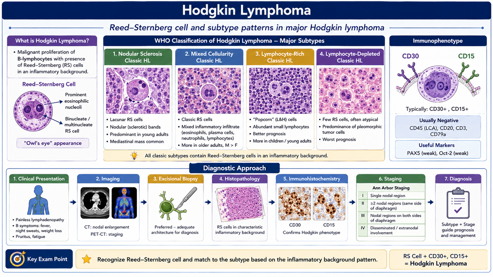
E. Hodgkin Lymphoma
Hodgkin lymphoma is a malignant lymphoid neoplasm in which the characteristic neoplastic cells are present within a much larger background of non-neoplastic inflammatory and immune cells. In classical Hodgkin lymphoma, the characteristic malignant cells include Reed-Sternberg cells and their variants. Although these cells are usually derived from B lymphocytes, they often lose many features of normal mature B cells.
Classification
The principal pathological division is between classical Hodgkin lymphoma and nodular lymphocyte-predominant Hodgkin lymphoma.
Classical Hodgkin lymphoma is divided into four major morphological subtypes:
- Nodular sclerosis classical Hodgkin lymphoma.
- Mixed cellularity classical Hodgkin lymphoma.
- Lymphocyte-rich classical Hodgkin lymphoma.
- Lymphocyte-depleted classical Hodgkin lymphoma.
Nodular lymphocyte-predominant Hodgkin lymphoma is biologically distinct from classical Hodgkin lymphoma and contains characteristic lymphocyte-predominant cells rather than typical classical Reed-Sternberg cells.
Etiology and pathogenesis
The malignant cells of classical Hodgkin lymphoma arise from germinal-center or post-germinal-center B cells. Genetic and signaling abnormalities allow these abnormal cells to survive despite losing normal B-cell functions. The malignant cells release cytokines and chemokines that attract large numbers of reactive lymphocytes, macrophages, eosinophils and other inflammatory cells.
This explains an important pathological feature: the tumor cells may represent only a small fraction of the total cellular population in an involved lymph node, while most of the mass consists of reactive cells recruited into the tumor microenvironment.
Epstein-Barr virus (EBV) is associated with a proportion of classical Hodgkin lymphomas. Viral proteins can provide survival signals to infected B cells, but EBV is not present in every case and therefore is not the sole cause of Hodgkin lymphoma.
Morphological changes
The classical Reed-Sternberg cell is large and commonly has two nuclear lobes or two nuclei, each containing a prominent eosinophilic nucleolus. The appearance of prominent nucleoli surrounded by clear chromatin produces the traditional description of an “owl-eye” appearance.
Classical Reed-Sternberg cells typically express CD30 and commonly CD15. Their B-cell marker expression is much weaker than that of ordinary mature B-cell lymphomas.
Morphological pattern of the major subtypes
- Nodular sclerosis: broad collagen bands divide the lymph node into nodules. Lacunar-type Reed-Sternberg variants are characteristic.
- Mixed cellularity: numerous classical Reed-Sternberg cells are found within a mixed inflammatory background containing lymphocytes, eosinophils, plasma cells and histiocytes.
- Lymphocyte-rich: the background contains many lymphocytes with relatively fewer Reed-Sternberg cells.
- Lymphocyte-depleted: relatively few normal lymphocytes are present and atypical tumor cells are more prominent.
- Nodular lymphocyte-predominant form: contains lymphocyte-predominant cells with multilobed nuclei, traditionally called “popcorn cells,” in a predominantly lymphocytic background.
Clinical course
Hodgkin lymphoma commonly presents as painless lymph-node enlargement. Cervical and mediastinal lymph nodes are frequently involved. A characteristic feature is a tendency to spread from one lymph-node group to an anatomically adjacent group in a relatively orderly fashion.
Some patients develop systemic symptoms such as fever, night sweats and weight loss. These manifestations reflect the biological effects of cytokines released by the tumor and its surrounding inflammatory cells.
F. Non-Hodgkin Lymphomas: Major Types and Predisposing Factors
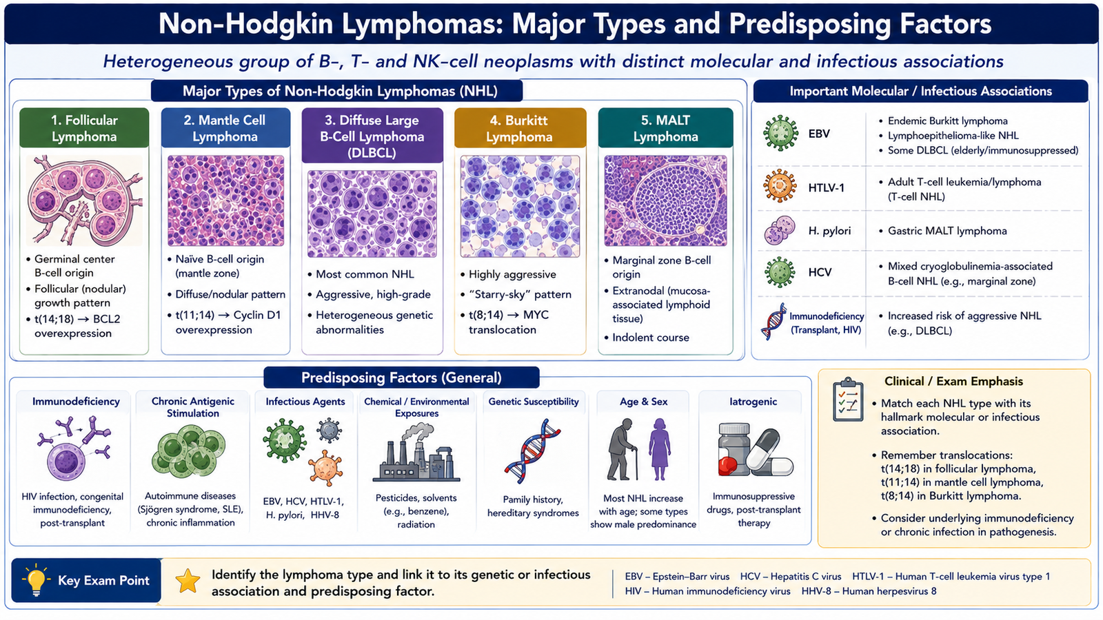
Non-Hodgkin lymphoma (NHL) is a collective term for a diverse group of lymphoid malignancies other than Hodgkin lymphoma. Most arise from mature B cells, but tumors of precursor lymphocytes, mature T cells and NK cells also occur. The WHO approach therefore classifies these diseases according to lineage, maturation stage, morphology, immunophenotype and characteristic molecular abnormalities rather than treating NHL as a single disease.
Basic pathological classification
For undergraduate learning, the major pathological groups can be organized as follows:
- Precursor B-cell neoplasms: B-lymphoblastic leukemia/lymphoma.
- Precursor T-cell neoplasms: T-lymphoblastic leukemia/lymphoma.
- Mature B-cell lymphomas: CLL/SLL, follicular lymphoma, mantle cell lymphoma, marginal zone lymphoma, diffuse large B-cell lymphoma and Burkitt lymphoma.
- Mature T-cell and NK-cell neoplasms: include peripheral T-cell lymphoma, anaplastic large cell lymphoma, adult T-cell leukemia/lymphoma and extranodal NK/T-cell lymphoma.
Major mature B-cell lymphomas
Diffuse large B-cell lymphoma (DLBCL) is composed of large neoplastic B cells growing in a diffuse pattern that effaces normal tissue architecture. It is an aggressive lymphoma and may arise in lymph nodes or extranodal sites.
Follicular lymphoma is a mature B-cell neoplasm that commonly forms a nodular or follicular growth pattern resembling normal germinal centers. A characteristic molecular abnormality is the t(14;18) translocation, which leads to increased expression of the anti-apoptotic protein BCL2. The important biological result is prolonged survival of abnormal B cells.
Mantle cell lymphoma is a mature B-cell lymphoma associated with dysregulation of cyclin D1, commonly through t(11;14). Increased cyclin D1 promotes progression through the cell cycle and contributes to abnormal proliferation.
Marginal zone lymphoma arises from mature B cells resembling marginal-zone cells. Some extranodal forms develop in mucosa-associated lymphoid tissue after prolonged immune stimulation.
Burkitt lymphoma is a highly proliferative mature B-cell lymphoma characterized by dysregulation of the MYC oncogene, classically involving chromosome 8 and an immunoglobulin gene locus. Microscopically, the rapid tumor-cell turnover produces numerous apoptotic cells that are removed by macrophages, creating the classic starry-sky appearance.
Important mature T-cell and NK-cell neoplasms
T-cell and NK-cell lymphomas are less common than mature B-cell lymphomas but form an important part of the classification. Major examples include peripheral T-cell lymphoma, adult T-cell leukemia/lymphoma, anaplastic large cell lymphoma and extranodal NK/T-cell lymphoma.
Predisposing factors and infectious associations
Some lymphomas arise in settings of chronic antigenic stimulation, immunodeficiency or infection. The infectious organism does not always transform the lymphocyte directly; in some diseases, chronic immune stimulation creates an environment in which abnormal clones can emerge.
| Infectious agent or setting | Associated lymphoid neoplasm | Key relationship |
|---|---|---|
| Epstein-Barr virus | Burkitt lymphoma and several other lymphoid proliferations | Promotes B-cell survival and proliferation in susceptible settings. |
| Helicobacter pylori | Gastric MALT lymphoma | Chronic antigenic stimulation promotes prolonged B-cell proliferation. |
| HTLV-1 | Adult T-cell leukemia/lymphoma | Viral effects promote abnormal proliferation and survival of T cells. |
| HHV-8 | Primary effusion lymphoma | Viral oncogenic signaling contributes to lymphoid transformation. |
| EBV | Extranodal NK/T-cell lymphoma | Strong association exists between the virus and this lymphoma. |
Immunodeficiency is another important predisposing setting. When immune surveillance is reduced, particularly against oncogenic virus-infected lymphocytes, abnormal lymphoid clones have a greater opportunity to expand.
G. Non-Hodgkin Lymphoma: Lymph-Node Morphology and Diagnostic Investigation
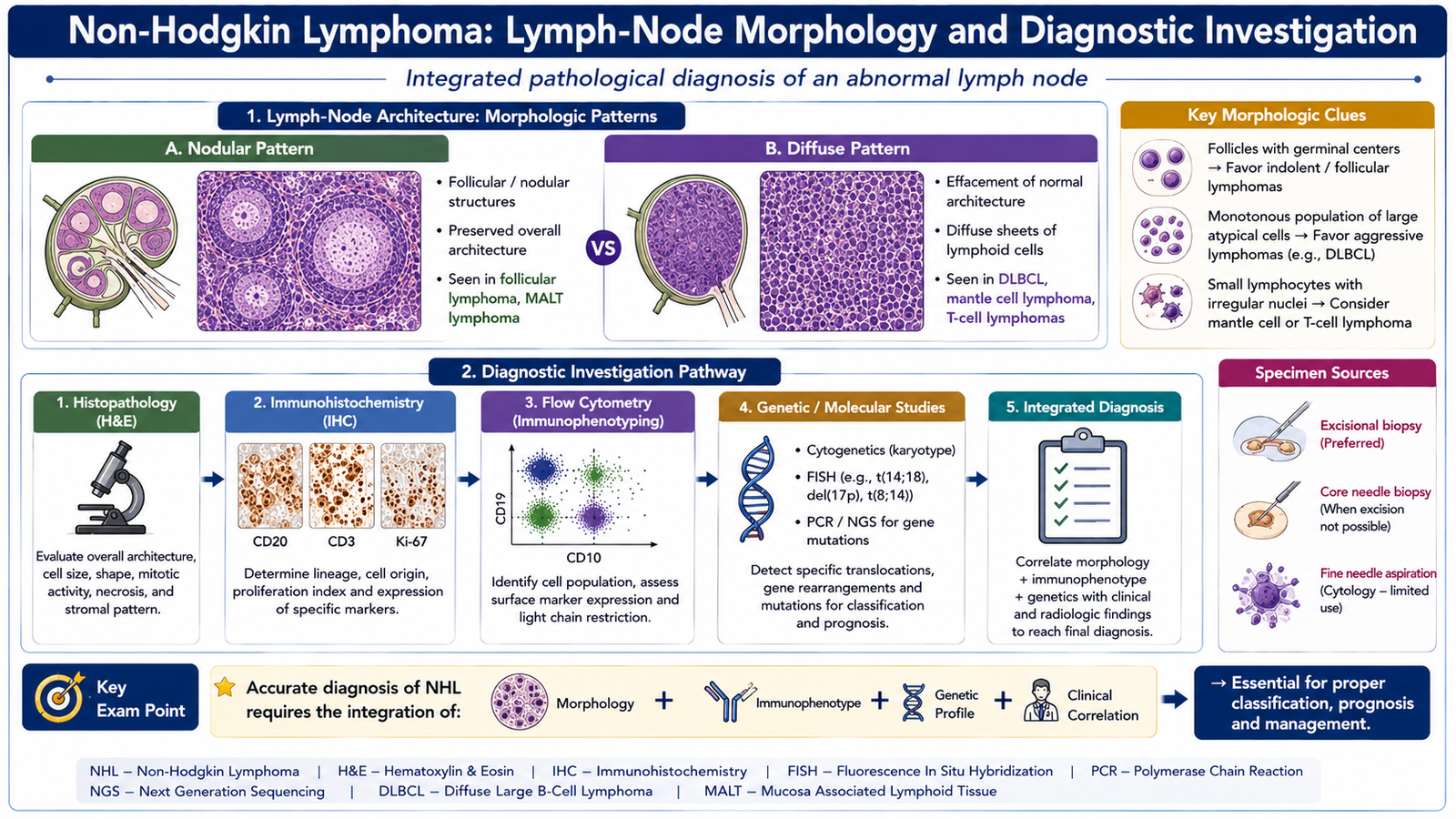
The diagnosis of non-Hodgkin lymphoma cannot usually be made from lymph-node enlargement alone because reactive lymphoid hyperplasia and many different lymphomas can produce similar clinical findings. Pathological diagnosis therefore requires assessment of the architecture of the node, the morphology of the abnormal cells and the immunophenotypic and genetic characteristics of the lymphoid population.
Morphological changes in an involved lymph node
A normal lymph node contains a highly organized arrangement of follicles, paracortex, sinuses and medullary structures. Lymphoma tends to disturb or replace this normal architecture.
- Partial or complete architectural effacement: normal nodal compartments become replaced by neoplastic lymphoid cells.
- Nodular or follicular pattern: tumor cells may grow as abnormal follicles or nodules, as in follicular lymphoma.
- Diffuse pattern: neoplastic cells may grow in sheets and completely erase normal architecture, as commonly seen in DLBCL.
- Monomorphic population: many lymphomas show a relatively uniform abnormal lymphoid population compared with the mixture of cell types normally present in reactive nodes.
- Cell size and nuclear features: small-cell and large-cell lymphomas differ in nuclear size, chromatin pattern, nucleoli and mitotic activity.
- Special morphological patterns: individual lymphomas may show distinctive findings, such as a starry-sky pattern in Burkitt lymphoma.
Morphology provides the first major clue, but morphology alone is often insufficient because several lymphomas can produce overlapping appearances.
Investigations required for diagnosis
A properly handled tissue specimen is central to lymphoma diagnosis because preservation of tissue architecture is important. An excisional lymph-node biopsy, when appropriate and feasible, allows the pathologist to evaluate both the cells and their arrangement within the node.
- Histopathological examination: determines whether normal architecture is preserved or effaced and identifies the growth pattern and tumor-cell morphology.
- Immunohistochemistry: identifies lineage and differentiation markers within tissue and helps distinguish individual lymphoma entities.
- Flow cytometry: can identify abnormal clonal lymphocyte populations and determine B-cell or T-cell immunophenotype.
- Cytogenetic and fluorescence in situ hybridization studies: identify characteristic chromosomal rearrangements when relevant.
- Molecular testing: may demonstrate clonality or disease-associated genetic abnormalities.
- Blood and bone-marrow assessment: is used when circulating or marrow involvement is suspected and helps determine the extent of disease.
Why immunophenotyping is essential
The appearance of a lymphoid cell does not always identify its lineage reliably. Immunophenotyping detects proteins characteristic of B cells, T cells, precursor cells or plasma cells. For example, CD20 supports mature B-cell differentiation, CD3 supports T-cell lineage and TdT supports an immature lymphoid precursor. A panel of markers is interpreted as a pattern; a single marker should not usually be interpreted in isolation.
Integrated Mechanism Flow
Acquired genetic or molecular abnormality
Abnormal survival, impaired differentiation and/or uncontrolled proliferation
Expansion of a single lymphoid clone
Bone-marrow, blood, lymph-node or extranodal involvement
Characteristic morphology, immunophenotype and clinical manifestations
Diagnosis by integrated morphological, immunophenotypic and genetic assessment
Important Comparisons
ALL versus CLL
| Feature | ALL | CLL |
|---|---|---|
| Cell type | Immature lymphoid precursor | Mature clonal B lymphocyte |
| Typical morphology | Lymphoblasts with high N:C ratio | Small mature lymphocytes with clumped chromatin |
| Characteristic clue | TdT-positive precursor phenotype | Smudge cells with CD5/CD23-positive B-cell phenotype |
| Major mechanism | Blocked differentiation with blast proliferation | Progressive accumulation of long-lived abnormal B cells |
Hodgkin versus Non-Hodgkin Lymphoma
| Feature | Hodgkin lymphoma | Non-Hodgkin lymphoma |
|---|---|---|
| Characteristic tumor cell | Reed-Sternberg cell in classical disease | Depends on specific lymphoma subtype |
| Tumor background | Prominent reactive inflammatory background | Often dominated by the neoplastic lymphoid population |
| Pattern of nodal spread | Often orderly and contiguous | May be less predictable and noncontiguous |
| Extranodal involvement | Less prominent as an initial pattern | Common in several NHL types |
| Classification | Classical subtypes and nodular lymphocyte-predominant form | Multiple B-cell, T-cell, NK-cell and precursor entities |
⭐ AIM High-Yield Review
- Lymphoid neoplasms are classified mainly according to cell lineage and stage of differentiation, supported by morphology, immunophenotype and genetics.
- ALL is a precursor lymphoid neoplasm in which lymphoblast accumulation replaces normal marrow and produces cytopenias.
- ⭐ TdT is an important marker of immature lymphoid cells; lineage-specific markers distinguish B-ALL from T-ALL.
- CLL consists of small mature-appearing clonal B cells; smudge cells are a classic peripheral smear clue.
- ⭐ CLL characteristically shows a B-cell phenotype with abnormal co-expression of CD5 and CD23.
- Multiple myeloma is a clonal plasma-cell neoplasm associated with monoclonal immunoglobulin production and destructive bone disease.
- Bone destruction in myeloma explains bone pain, pathological fractures and hypercalcemia, while marrow replacement contributes to anemia.
- ⭐ Typical plasma cells show an eccentric nucleus, clock-face chromatin and a perinuclear hof; increased serum proteins can produce rouleaux formation.
- Reed-Sternberg cells are the characteristic neoplastic cells of classical Hodgkin lymphoma and typically express CD30 with frequent CD15 expression.
- Classical Hodgkin lymphoma includes nodular sclerosis, mixed cellularity, lymphocyte-rich and lymphocyte-depleted subtypes.
- ⭐ Follicular lymphoma → BCL2 dysregulation; mantle cell lymphoma → cyclin D1 dysregulation; Burkitt lymphoma → MYC dysregulation.
- Important infection associations include H. pylori with gastric MALT lymphoma, HTLV-1 with adult T-cell leukemia/lymphoma, and EBV with several lymphoid neoplasms including Burkitt and extranodal NK/T-cell lymphoma.
- Lymphoma can partially or completely efface normal lymph-node architecture and may grow in nodular or diffuse patterns.
- ⭐ A definitive lymphoma diagnosis integrates histological architecture, cell morphology, immunophenotyping and selected genetic studies.
Lymphoid Neoplasms, Lymphomas and Plasma Cell Disorders
Watch these focused pathology videos after completing the AIM Learning Material. Together they reinforce the classification, pathogenesis, morphology and diagnostic features of the major lymphoid neoplasms included in this topic.
Lymphoid Neoplasms — Classification, Features & Key Principles
Use this first to build the overall framework: leukemia versus lymphoma, classification, morphology, immunophenotyping, clonality and major Hodgkin/non-Hodgkin principles.
Acute Lymphoblastic Leukemia — ALL
Reinforces ALL pathogenesis, important genetic abnormalities, lymphoblast morphology and the major clinicopathological features of B-ALL and T-ALL.
Chronic Lymphocytic Leukemia — CLL
Reviews CLL/SLL pathogenesis, important molecular abnormalities, characteristic small lymphocytes, smudge cells and immunophenotypic diagnosis.
Multiple Myeloma — Plasma Cell Neoplasm
Focuses on plasma-cell biology, pathogenesis of multiple myeloma and the mechanisms producing its important clinical manifestations.
Hodgkin Lymphoma
Reinforces Hodgkin lymphoma clinical and pathological concepts after students have learned its classification, Reed-Sternberg morphology and major subtypes from the AIM chapter.
Begin with the lymphoid-neoplasm overview, then review ALL, CLL, multiple myeloma and Hodgkin lymphoma. While watching, concentrate on the same four links used throughout AIM: pathogenesis → morphology → clinical finding → diagnosis.