
Foundations of Immunity, Immune Responses, Antibodies and Complement
A connected introduction to innate and adaptive immunity, B- and T-cell responses, antibodies, immunological memory and the complement system.
Topic Introduction
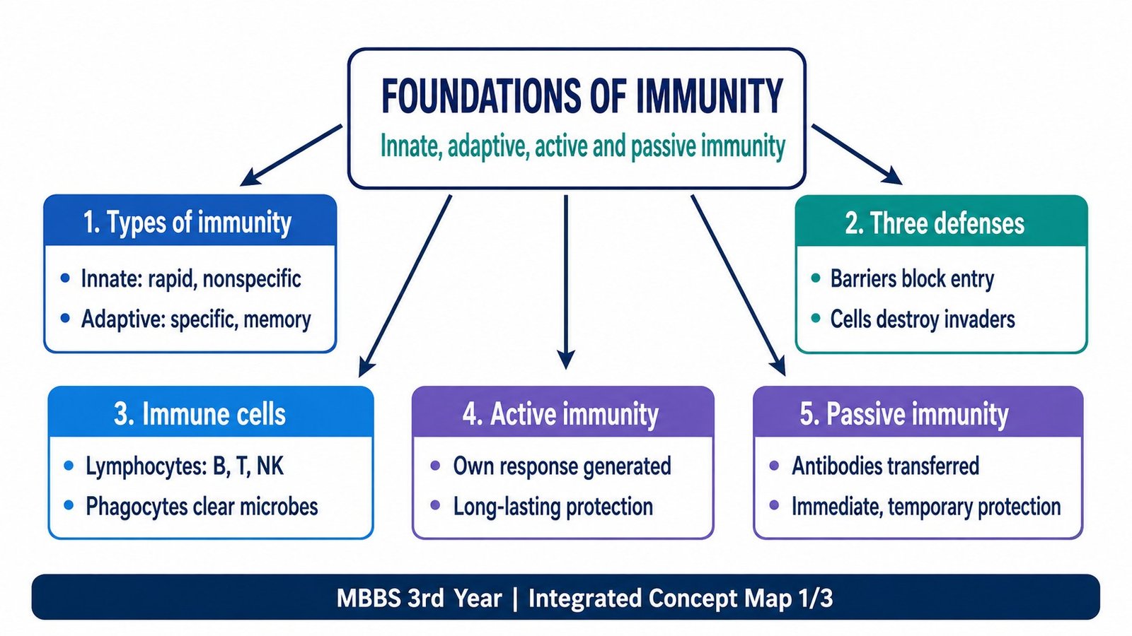
The immune system protects the body through several layers of defense that work together rather than independently. Innate immunity provides rapid protection through barriers, phagocytes, natural killer cells and soluble proteins. Adaptive immunity develops highly specific responses through B and T lymphocytes and can remember previous exposure to an antigen. B cells produce antibodies that act mainly against extracellular microbes and toxins, whereas T cells are central to cell-mediated immunity. The complement system supports both innate and antibody-mediated defense by promoting inflammation, phagocytosis and microbial killing. Understanding these connections makes later immunological diseases much easier to interpret.
A. Organization of the Immune System: Types, Lines of Defense and Immune Cells
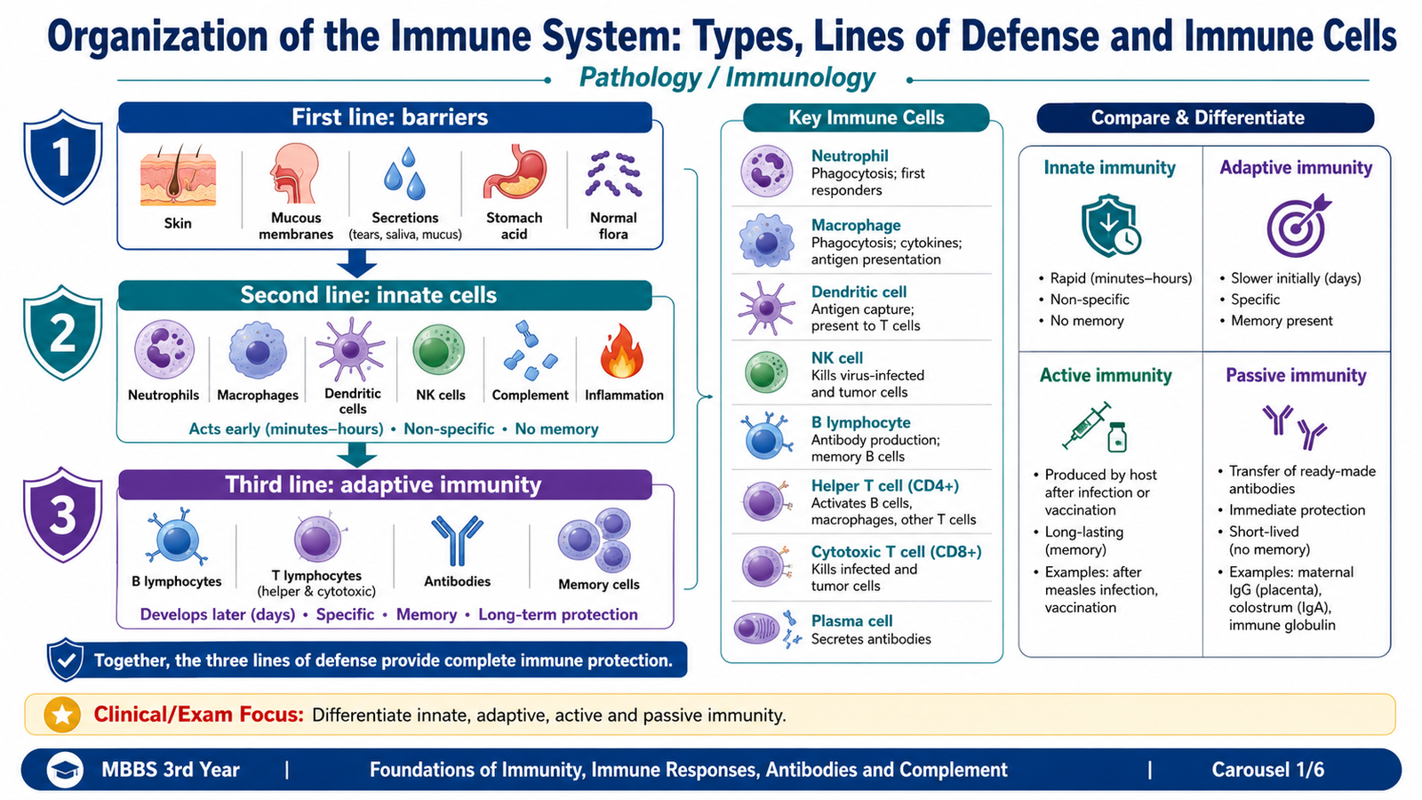
Immunity is the body’s ability to recognize and defend itself against harmful foreign material, especially microorganisms and their products. Protection begins before an organism enters the tissues and continues through increasingly specialized responses if the first defenses are overcome. For learning purposes, immunity is divided into innate immunity and adaptive or acquired immunity, but these systems normally cooperate closely.
Functions and main types of immunity
The immune system performs several connected functions. It prevents entry of microbes, recognizes organisms that cross surface barriers, contains and eliminates infection, removes infected or abnormal cells and develops memory after many adaptive immune responses.
- Innate immunity is present before exposure to a particular antigen and responds rapidly.
- Adaptive immunity develops after antigen recognition and is characterized by high specificity and memory.
- Humoral immunity is the antibody-mediated arm of adaptive immunity.
- Cell-mediated immunity is the T-lymphocyte-mediated arm of adaptive immunity.
Three lines of defense
The body’s protective mechanisms can be viewed as three lines. The first prevents entry, the second reacts rapidly if a barrier is crossed, and the third develops a highly specific adaptive response.
| Line | Main components | Important property |
|---|---|---|
| First line | Skin, mucosal epithelium and antimicrobial substances in secretions | Acts as a physical and chemical barrier that prevents microbial entry. |
| Second line | Phagocytes, dendritic cells, NK cells, inflammatory mediators and complement | Responds rapidly after microbes enter tissues and is mainly innate. |
| Third line | B lymphocytes, T lymphocytes and antibodies | Provides antigen-specific adaptive immunity and develops memory. |
Origin and functions of important immune cells
Cells of the immune system ultimately arise from hematopoietic stem cells in the bone marrow. These stem cells produce myeloid and lymphoid lineages. The resulting cells differ in their structure, location and function, but they cooperate during immune responses.
- Neutrophils: short-lived circulating phagocytes that are rapidly recruited to sites of infection. They are particularly important in early defense against many bacteria.
- Monocytes and macrophages: monocytes circulate in blood and can enter tissues, where cells of the monocyte-macrophage system perform phagocytosis, remove damaged material, produce inflammatory mediators and participate in antigen presentation.
- Dendritic cells: specialized antigen-presenting cells that capture antigens in tissues and are particularly effective at activating naïve T lymphocytes.
- Natural killer cells: lymphoid cells of innate immunity that can kill infected or abnormal cells without using the antigen-specific receptors of T lymphocytes.
- B lymphocytes: lymphocytes of adaptive immunity that recognize antigen and can differentiate into antibody-secreting plasma cells and memory B cells.
- Plasma cells: terminally differentiated B cells specialized for producing and secreting large quantities of antibody.
- T lymphocytes: adaptive immune cells responsible for cell-mediated immunity and for regulation of many immune responses. Major functional groups include helper and cytotoxic T cells.
Innate and adaptive immunity compared
Innate immunity recognizes broad patterns that are shared by groups of microbes. Adaptive immunity uses highly diverse antigen receptors on individual lymphocyte clones, allowing much greater specificity.
| Feature | Innate immunity | Adaptive immunity |
|---|---|---|
| Onset | Rapid | Slower during first exposure |
| Recognition | Shared microbial patterns | Highly specific antigens |
| Main cells | Phagocytes, dendritic cells, NK cells | B and T lymphocytes |
| Memory | No classical antigen-specific memory | Characteristic immunological memory |
Active and passive immunity
Active immunity develops when a person’s own adaptive immune system responds to an antigen. Because lymphocytes are activated and memory cells can develop, protection may be prolonged. Passive immunity results from transfer of preformed antibodies. It provides immediate protection but is temporary because the recipient has not generated the antibody response or immunological memory personally.
| Feature | Active immunity | Passive immunity |
|---|---|---|
| Antibody production | Produced by the person’s own immune system | Received from another source |
| Onset | Takes time to develop | Immediate |
| Memory | Present | Absent |
| Natural example | Response after natural infection | Maternal antibody transferred to the fetus or infant |
B. Specificity of Adaptive Immunity and Coordination of B and T Lymphocytes
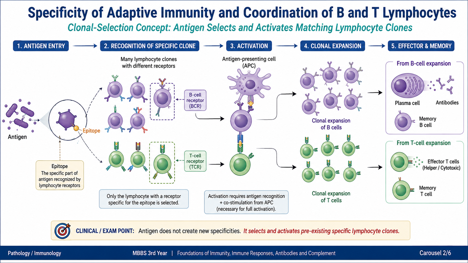
The major strength of adaptive immunity is its ability to distinguish between different antigens. This specificity depends on large populations of B and T lymphocytes, with each lymphocyte clone carrying receptors that recognize a particular antigenic structure. When the matching antigen is encountered, the appropriate clone is selected, activated and expanded.
Antigen recognition and clonal selection
An antigen is a substance that can be specifically recognized by antibodies or lymphocyte antigen receptors. Many antigens contain several smaller antigenic regions called epitopes. Different lymphocyte clones can therefore recognize different epitopes on the same antigen.
The body produces a very large diversity of lymphocytes before a particular infection occurs. Entry of an antigen selects the lymphocytes that already possess receptors capable of recognizing it. The selected cells then proliferate. This is called clonal selection and expansion.
→ specific antigen enters
→ matching clone recognizes antigen
→ activation and clonal expansion
→ effector cells + memory cells
This mechanism explains why adaptive immunity is both specific and capable of memory. Only the matching lymphocyte population expands substantially, and some descendants remain as long-lived memory cells.
Different recognition by B and T lymphocytes
B and T cells both provide antigen specificity, but they recognize antigen in different ways.
- B lymphocytes can recognize many antigens directly in their native form through membrane immunoglobulin that acts as the B-cell receptor.
- T lymphocytes generally recognize peptide fragments presented on cell surfaces by major histocompatibility complex (MHC) molecules.
- Antigen-presenting cells, especially dendritic cells, process protein antigens and display peptide fragments to T cells.
Roles of B and T lymphocytes
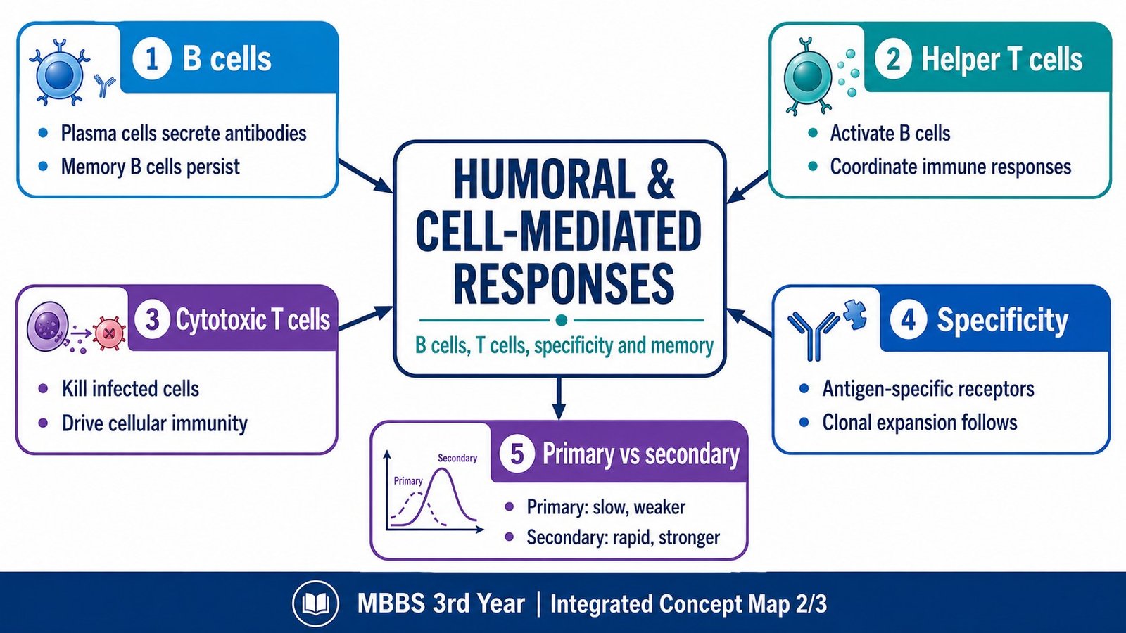
B and T lymphocytes form complementary arms of adaptive immunity. B cells ultimately produce antibodies and are therefore central to humoral immunity. T cells do not normally secrete antibodies. Instead, helper T cells regulate other immune cells, while cytotoxic T cells can directly eliminate selected infected or abnormal cells.
- B cells: antigen recognition → activation → plasma cells → antibody secretion.
- Helper T cells: recognize antigen presented by other cells and provide signals that regulate B cells, macrophages and other leukocytes.
- Cytotoxic T cells: recognize relevant antigen on target cells and induce death of those cells.
- Memory B and T cells: persist after the primary response and allow more efficient responses on later exposure.
How helper T cells regulate immune responses
Helper T cells act as coordinators. Once activated by antigen, they provide signals through cell-surface molecules and cytokines. These signals can promote B-cell proliferation and antibody production, enhance macrophage antimicrobial activity and help support cytotoxic T-cell responses. Thus, helper T cells allow different components of adaptive immunity to respond in a coordinated way rather than as isolated cell populations.
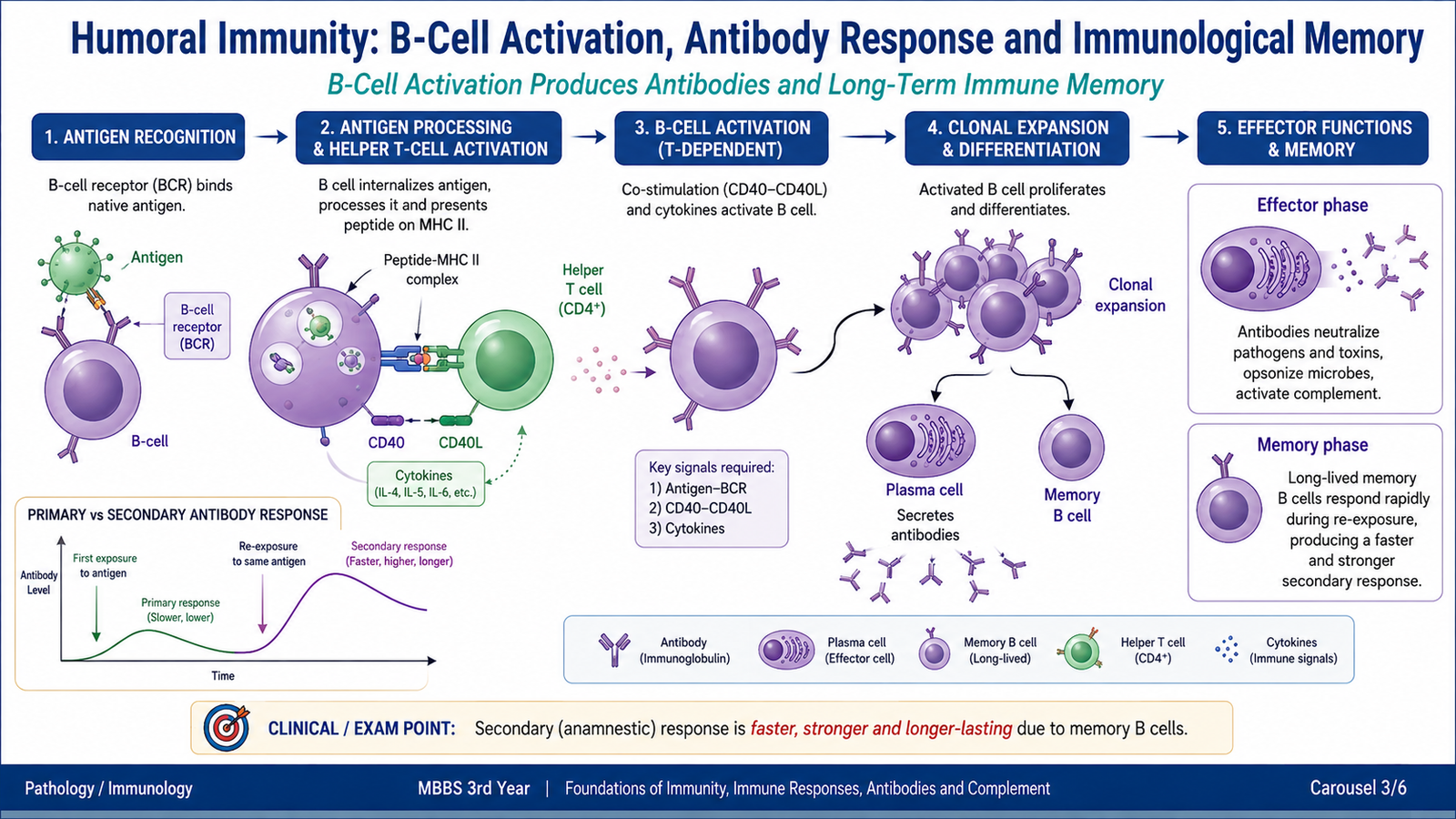
C. Humoral Immunity: B-Cell Activation, Antibody Response and Immunological Memory
Humoral immunity is the arm of adaptive immunity mediated mainly by antibodies. It is particularly effective against microbes and microbial products located outside cells. B lymphocytes provide antigen specificity, while plasma cells formed from activated B cells secrete the antibodies that perform the effector functions.
Role of B lymphocytes in humoral immunity
Each mature B cell expresses antigen receptors on its surface. When an antigen binds to the receptor of a matching B-cell clone, the antigen-specific cell is selected for activation. For many protein antigens, efficient B-cell activation also requires signals from helper T lymphocytes.
The B cell can internalize the bound antigen, process it and display antigenic peptide on MHC class II. A helper T cell that recognizes the presented peptide then provides additional signals. An important interaction occurs between CD40 on the B cell and CD40 ligand on the activated helper T cell, together with cytokines produced by the helper T cell.
→ antigen processing and presentation
→ helper T-cell recognition and support
→ B-cell proliferation
→ plasma cells + memory B cells
→ antibody production
Plasma cells and memory B cells
Activated B cells undergo clonal expansion. Some differentiate into plasma cells, which are specialized for antibody production. Others become memory B cells. Plasma cells provide the immediate antibody response, whereas memory B cells help produce a more rapid and effective response if the antigen is encountered again.
Primary immune response
The response after the first exposure to an antigen is called the primary immune response. Initially, only a small number of lymphocytes recognize that antigen. These cells must first become activated, multiply and differentiate, so there is a delay before a strong response develops.
IgM is an important early antibody in a primary response. As the response develops, activated B cells may undergo class switching and other changes that improve the effectiveness of the antibody response.
Secondary immune response
On later exposure to the same antigen, memory lymphocytes respond more efficiently. The secondary immune response therefore begins more rapidly, usually reaches a greater magnitude and commonly produces antibodies with higher affinity for the antigen.
| Feature | Primary response | Secondary response |
|---|---|---|
| Exposure | First encounter | Repeat encounter |
| Responding cells | Mainly naïve antigen-specific lymphocytes | Memory lymphocytes |
| Speed | Slower | Faster |
| Magnitude | Usually smaller | Usually greater |
| Antibody affinity | Generally lower early in the response | Generally higher |
D. Cell-Mediated Immunity: T-Cell Activation, Diversity and Effector Functions
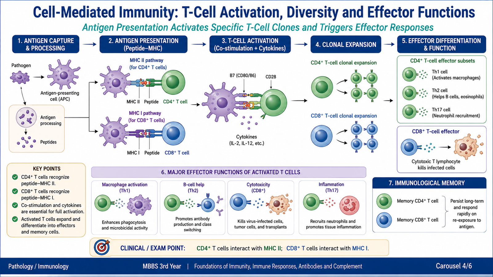
Cell-mediated immunity (CMI) is the adaptive immune response in which T lymphocytes are the major effector and regulatory cells. It is especially important when the relevant antigen is associated with cells, such as in intracellular infection, because antibodies in extracellular fluid cannot directly eliminate microorganisms that are hidden inside infected cells.
Cellular components involved in CMI
Several cell types cooperate in cell-mediated responses. Dendritic cells initiate many T-cell responses, helper T cells coordinate immune activity, cytotoxic T cells kill selected target cells and macrophages can become more effective antimicrobial cells after receiving appropriate T-cell signals.
- Dendritic cells: capture antigen and present it to naïve T lymphocytes.
- CD4+ helper T cells: regulate immune responses mainly through cytokines and cell-to-cell signals.
- CD8+ cytotoxic T cells: kill cells displaying the appropriate antigen.
- Macrophages: phagocytose microbes and may be activated by T-cell signals to increase microbial killing.
How T cells recognize antigen
T cells generally do not recognize intact free protein in the same way as B cells. Protein antigens are processed into peptides and displayed by MHC molecules.
- CD4+ T cells mainly recognize peptides presented by MHC class II.
- CD8+ T cells mainly recognize peptides presented by MHC class I.
This arrangement helps direct different T-cell populations toward appropriate functions. CD4+ cells primarily regulate other immune cells, whereas CD8+ cells can identify and kill cells displaying relevant intracellular antigens.
T-cell activation
Recognition of peptide-MHC by the T-cell receptor provides antigen specificity, but full activation of a naïve T cell requires additional signals. This prevents a strong adaptive response from being triggered simply by antigen recognition in an inappropriate setting.
After activation, the selected T-cell clone proliferates. Some cells become short-lived effector cells and others persist as memory T cells.
T-cell diversity
The immune system must be able to respond to a very large variety of antigens. During lymphocyte development, rearrangement of antigen-receptor gene segments produces a large number of T cells with different receptor specificities. The essential concept is that receptor diversity is generated before exposure to a particular antigen. The antigen then selects and expands the matching clone.
→ peptide-MHC recognition
→ co-stimulation
→ clonal expansion
→ effector T cells + memory T cells
Effector actions of T cells
Helper T cells increase or direct the activity of other immune cells through cytokines and cell-surface interactions. Cytotoxic T cells recognize target cells carrying the appropriate peptide-MHC class I complex and induce controlled cell death. Important cytotoxic mechanisms include release of perforin and granzymes and activation of death-receptor pathways.
Humoral and cell-mediated immunity compared
| Feature | Humoral immunity | Cell-mediated immunity |
|---|---|---|
| Main lymphocyte | B lymphocyte | T lymphocyte |
| Main effector | Antibodies | Activated T cells and cells activated by T-cell signals |
| Major target | Extracellular microbes and toxins | Intracellular microbes and infected cells |
| Memory | Memory B cells | Memory T cells |
E. Antigens and Antibodies: Immunoglobulin Structure, Classes and Functions
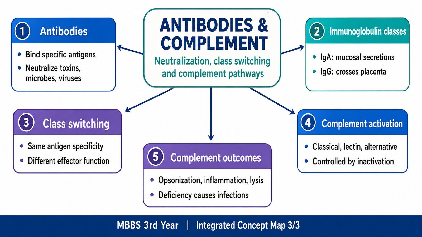
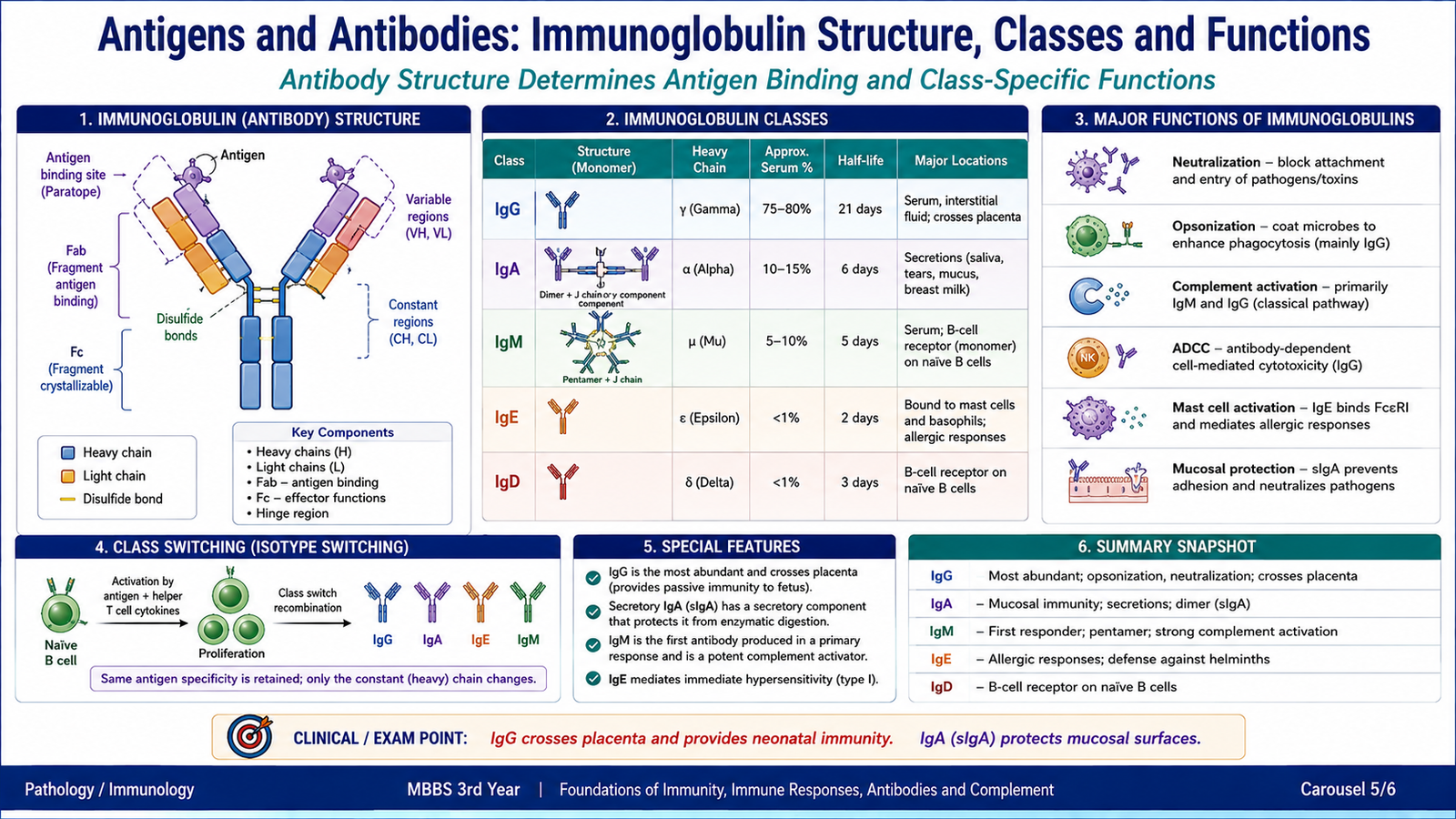
Antigens provide the targets for adaptive immune recognition, while antibodies are specialized proteins produced by plasma cells that bind those antigens. Antibody molecules combine a highly specific antigen-binding region with a constant region that determines many of their biological effects. Differences in the heavy-chain constant region produce the major immunoglobulin classes.
Antigens and antibodies
An antigen is a substance specifically recognized by antibodies or lymphocyte receptors. An antibody, also called an immunoglobulin, is an antigen-binding protein produced in secreted form by plasma cells. The antibody binds to a particular epitope through its variable region.
Basic immunoglobulin structure
A typical antibody contains two identical heavy chains and two identical light chains linked together. Each chain has variable and constant regions.
- Variable regions: form the antigen-binding sites and determine specificity.
- Fab regions: contain the antigen-binding portions of the molecule.
- Fc region: is formed by constant portions of heavy chains and is responsible for many effector functions.
- Hinge region: provides flexibility in immunoglobulin classes that contain a hinge.
Classification of immunoglobulins
Immunoglobulins are divided into five major classes according to the type of heavy chain: IgG, IgA, IgM, IgE and IgD. Each class is suited to particular immune functions.
| Immunoglobulin | Important form/location | Major functions |
|---|---|---|
| IgG | Monomer; major antibody in blood and extracellular fluid | Neutralization, opsonization, complement activation and placental transfer |
| IgA | Monomer in serum; mainly dimeric in mucosal secretions | Mucosal protection and neutralization in secretions |
| IgM | Pentamer when secreted; monomer as B-cell receptor | Important early antibody response and strong activation of classical complement |
| IgE | Binds to mast cells and basophils | Immediate hypersensitivity and defense against certain parasites |
| IgD | Mainly membrane-bound on naïve B cells | Functions as a B-cell antigen receptor |
Structure of immunoglobulin A
IgA is particularly important at mucosal surfaces. Serum IgA is mainly monomeric, whereas secretory IgA is mainly a dimer. Two IgA molecules are linked by a J chain. During transport across mucosal epithelial cells, a portion of the epithelial receptor remains attached as the secretory component.
The secretory component helps protect IgA against enzymatic degradation. This makes secretory IgA well suited for protection in saliva, tears, intestinal secretions and breast milk.
→ J chain joins the molecules
→ epithelial transport
→ secretory component retained
→ secretory IgA at mucosal surfaces
Important functions of antibodies
Antibodies often protect the host by binding an antigen and then preventing its harmful action or recruiting other immune mechanisms. The important principle is that antigen recognition by the antibody is converted into an effective defensive response.
- Neutralization: antibody blocks microbial attachment, viral entry or toxin binding to host cells.
- Opsonization: antibody coats a microbe and makes it easier for phagocytes to recognize and ingest it.
- Complement activation: antigen-bound antibodies can activate the classical complement pathway.
- Antibody-dependent cellular cytotoxicity: immune cells can recognize and damage antibody-coated target cells.
Antibody-mediated neutralization
Neutralizing antibodies bind the part of a toxin, microbe or virus that is required for interaction with a host cell. Once this binding site is covered, the harmful agent cannot interact normally with its cellular receptor.
→ host-cell attachment or receptor binding is blocked
→ entry or toxic action is prevented
→ immune clearance becomes easier
Thus, antibodies may protect against viruses by preventing attachment or entry, against microbes by reducing attachment to host tissues, and against toxins by preventing the toxin from reaching its cellular target.
Immunoglobulin class switching
Activated B cells can change the class of antibody they produce while retaining recognition of the same antigen. This process is called class switching or isotype switching.
The variable antigen-binding region remains essentially unchanged, while the heavy-chain constant region changes. Therefore, antigen specificity remains the same but the biological function of the antibody changes. Helper T-cell signals are important in class switching during responses to many protein antigens.
Maternal immunity to the fetus and breast-fed infant
Maternal antibody transfer is a form of natural passive immunity. IgG crosses the placenta and provides systemic antibody protection to the fetus and newborn. The infant receives ready-made maternal antibody, so this protection is temporary and does not itself create memory in the infant.
After birth, secretory IgA in breast milk provides protection mainly at mucosal surfaces, especially in the gastrointestinal tract. It binds microorganisms and helps prevent their attachment to mucosal epithelial cells.
Monoclonal and polyclonal antibodies
A complex antigen usually carries several epitopes and can activate several B-cell clones. The resulting mixture of antibodies is called a polyclonal antibody response. A monoclonal antibody population arises from a single B-cell clone and therefore has one defined antigen specificity.
| Feature | Monoclonal | Polyclonal |
|---|---|---|
| Origin | Single B-cell clone | Multiple B-cell clones |
| Epitope recognition | One defined epitope | Multiple epitopes |
| Antibody population | Uniform | Mixture |
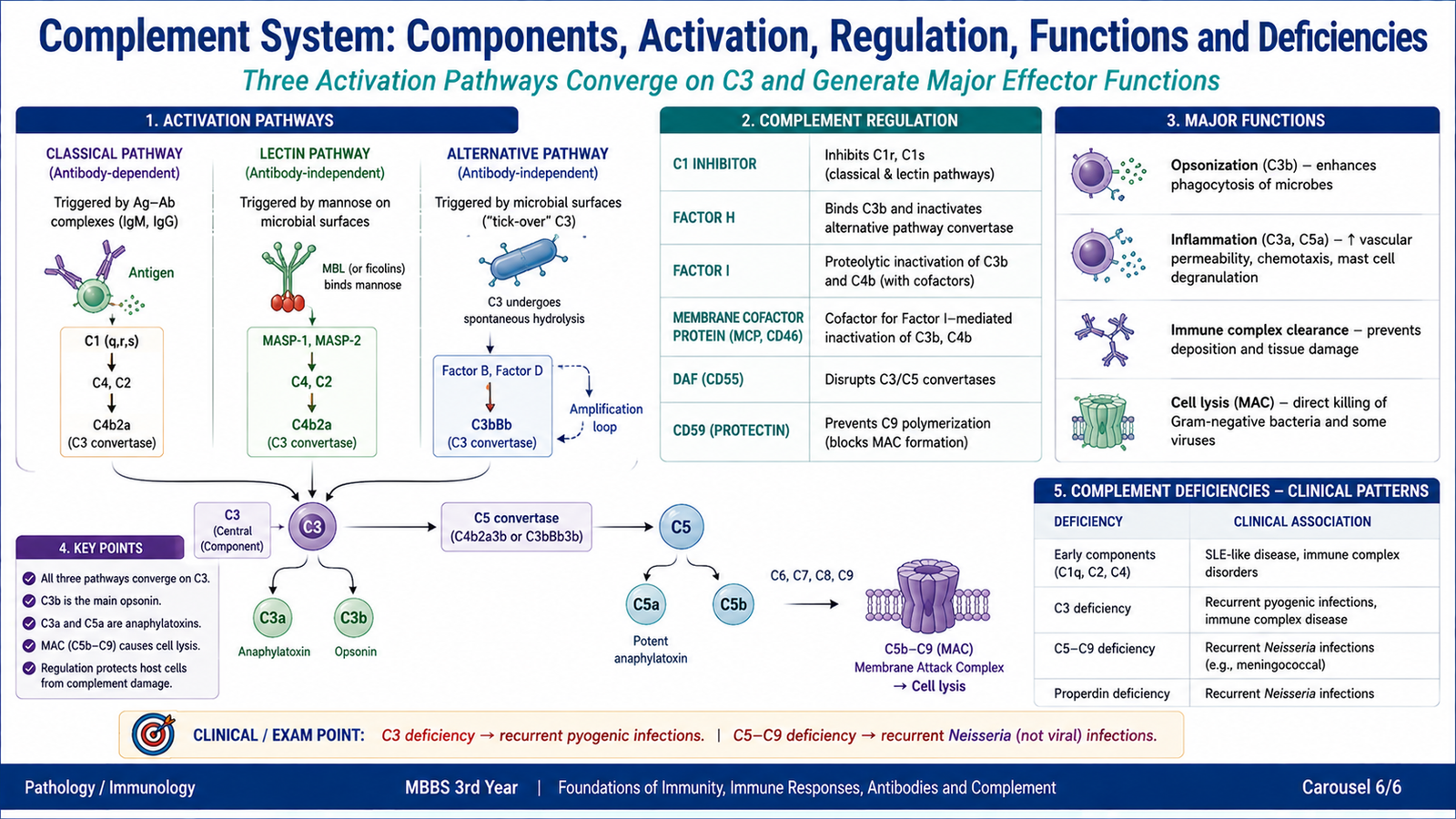
F. Complement System: Components, Activation, Regulation, Functions and Deficiencies
The complement system is a group of soluble and membrane-associated proteins that enhance immune defense. Most circulate in inactive forms and become activated through an enzyme cascade. Complement participates in innate immunity and also works with antibodies, linking innate and adaptive immune mechanisms.
Complement components and synthesis
Major complement proteins are traditionally designated C1 to C9, together with additional factors and regulatory proteins. During activation, several complement proteins are split into fragments with different functions. For example, cleavage of C3 produces C3a and C3b.
The liver is the major source of many circulating complement proteins. Monocytes, macrophages and some other cells can also produce complement components locally. Complement therefore functions both as a circulating plasma defense system and as part of local immune responses.
Complement activation pathways
Complement can be activated by three major pathways. Their initiating events differ, but all ultimately generate a C3 convertase and converge on cleavage of C3.
| Pathway | How it begins | Important principle |
|---|---|---|
| Classical pathway | Usually begins when C1 interacts with antibody bound to antigen | Links antibody responses to complement |
| Lectin pathway | Mannose-binding lectin recognizes characteristic microbial carbohydrates | Does not require antibody |
| Alternative pathway | Complement activation and amplification occur on suitable microbial surfaces | Important innate immune pathway |
Common pathway after C3 activation
Each activation route produces a C3 convertase. This enzyme cleaves C3 into C3a and C3b. C3b contributes to formation of a C5 convertase, which begins the terminal complement sequence.
↓
C3 convertase
↓
C3 → C3a + C3b
↓
C5 convertase
↓
C5b → C6 → C7 → C8 → C9
↓
Membrane attack complex
C5b combines with C6, C7, C8 and C9 to produce the membrane attack complex (MAC). MAC forms membrane pores in susceptible target cells and may cause cell lysis.
Inactivation and regulation of complement
Complement activation must be controlled because the same mechanisms that damage microbes can also injure host tissue. Regulatory proteins therefore interrupt the cascade at several levels and protect normal cell membranes.
- C1 inhibitor: limits early activation involving C1 and related components of the lectin pathway.
- Factors H and I: help control and inactivate C3b, especially on host surfaces.
- Decay-accelerating factor (CD55): accelerates breakdown of complement convertase complexes on host cells.
- CD59: prevents completion of the membrane attack complex on host-cell membranes.
Complement activity is therefore determined by a balance between activation on a target and inhibition on protected host surfaces.
Important functions of complement
Different fragments of the complement cascade perform different defensive functions. This is why deficiency of one component does not produce exactly the same clinical effect as deficiency of another.
Opsonization
C3b becomes deposited on microbial surfaces. Phagocytes possess receptors that recognize C3b, so coating a microbe with C3b increases attachment, ingestion and destruction.
Inflammation and leukocyte recruitment
C3a and C5a promote inflammatory reactions. C5a is also a powerful chemotactic and activating signal for leukocytes, helping recruit them toward sites where complement has been activated.
Microbial lysis
The terminal components C5b-C9 form the membrane attack complex. This damages susceptible microbial membranes through pore formation.
Immune-complex clearance
Complement helps bind and remove antigen-antibody complexes. If this function is impaired, immune complexes may persist and contribute to tissue inflammation.
Diseases associated with complement-protein deficiencies
The pattern of disease reflects the normal function of the missing component. Defects in early classical components interfere particularly with immune-complex handling, C3 deficiency causes a broad loss of complement efficiency, and terminal pathway defects reduce MAC formation.
| Deficiency | Functional consequence | Important association |
|---|---|---|
| Early classical components such as C1, C2 or C4 | Reduced immune-complex clearance | Increased tendency to immune-complex and lupus-like disease |
| C3 | Marked impairment of opsonization and several downstream complement functions | Recurrent severe pyogenic bacterial infections and immune-complex disease |
| C5-C9 | Defective MAC formation | Recurrent Neisseria infections |
| C1 inhibitor | Failure to adequately control specific plasma-protein cascades | Hereditary angioedema |
Integrated Mechanism Flow
The major immune processes in this topic can be connected as one sequence:
↓
Innate immune cells and complement provide rapid early defense
↓
Antigen is recognized and presented to specific lymphocyte clones
↓
B- and T-cell clonal activation and expansion occur
↓
Antibodies, activated T cells and complement eliminate the threat
↓
Memory lymphocytes support a faster secondary adaptive response
⭐ AIM High-Yield Review
- Innate immunity is rapid and recognizes shared microbial patterns; adaptive immunity is highly specific and develops memory.
- The first two lines of defense are mainly innate; the third line is antigen-specific adaptive immunity.
- Dendritic cells are important antigen-presenting cells for activation of naïve T lymphocytes.
- Active immunity is produced by the person’s own immune system and can generate memory; passive immunity transfers preformed antibody and is temporary.
- B lymphocytes mediate humoral immunity by differentiating into plasma cells that secrete antibodies.
- Helper T cells regulate immune responses through cell-surface interactions and cytokines.
- Secondary immune responses are faster and stronger because memory lymphocytes already exist.
- CD4+ T cells mainly recognize antigen with MHC class II; CD8+ T cells mainly recognize antigen with MHC class I.
- Humoral immunity is especially important against extracellular microbes and toxins; cell-mediated immunity is especially important against intracellular infection.
- ⭐ IgG crosses the placenta, whereas secretory IgA provides important mucosal protection to the breast-fed infant.
- Class switching changes the immunoglobulin heavy-chain constant region and therefore effector function, while antigen specificity is retained.
- All three complement activation pathways converge on C3 activation.
- C3b is a major opsonin, C5a is strongly chemotactic and inflammatory, and C5b-C9 form the membrane attack complex.
- ⭐ C3 deficiency predisposes to severe recurrent pyogenic infection; C5-C9 deficiencies particularly predispose to recurrent Neisseria infection.
Foundations of Immunity, Immune Responses, Antibodies and Complement
Use these videos after reading the AIM Learning Material to strengthen the major concepts of innate and adaptive immunity, humoral and cell-mediated responses, antibodies and complement.
Innate vs Adaptive Immunity and Humoral vs Cell-Mediated Immunity
Focus on the major types of immunity, lines of defense and the distinction between humoral and cell-mediated adaptive responses.
B Cells and Antibodies
Focus on B-cell responses, plasma cells, antibody production, immunoglobulin classes and the major functions of antibodies.
Helper T Cells and Regulation of the Immune Response
Focus on helper T-cell activation and how helper T cells coordinate B-cell and other immune responses.
Cytotoxic T Cells and MHC Class I
Focus on antigen presentation through MHC class I, CD8+ cytotoxic T-cell recognition and destruction of target cells.
Classical, Lectin and Alternative Complement Pathways
Focus on the three complement activation pathways, C3 activation, opsonization, inflammatory effects and formation of the membrane attack complex.