
This chapter follows the supplied KMU learning outcomes and builds the topic in a logical sequence. First understand how each myeloid disorder changes bone-marrow function, then use the high-yield section for revision.
Topic 3 — Myeloid Neoplasms, Myelodysplastic Syndromes and Myeloproliferative Disorders
Blood and Immunology Module
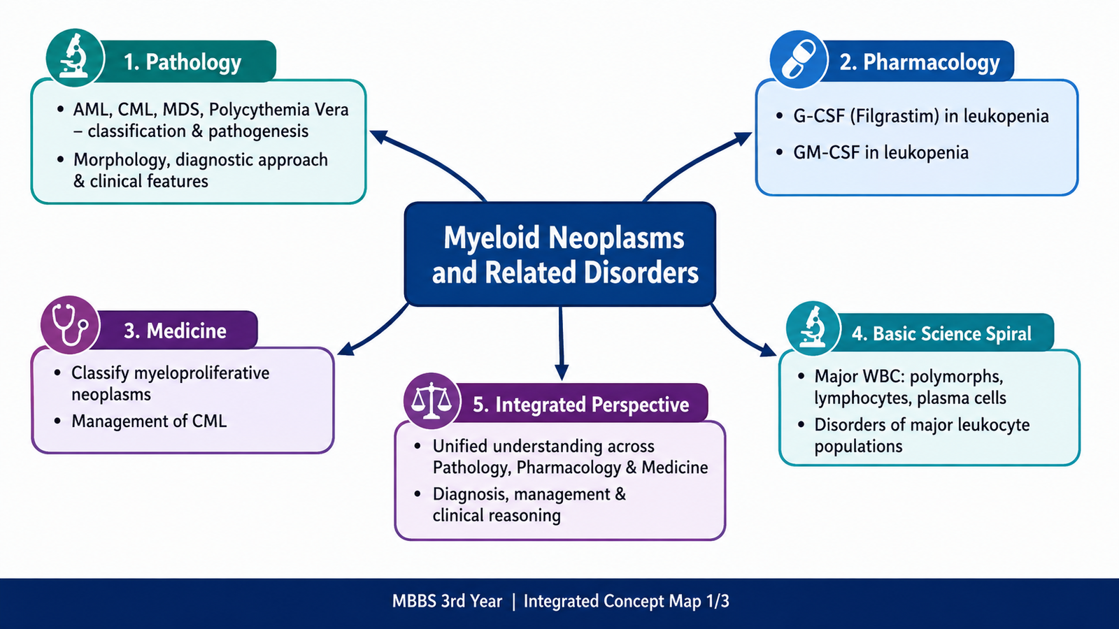
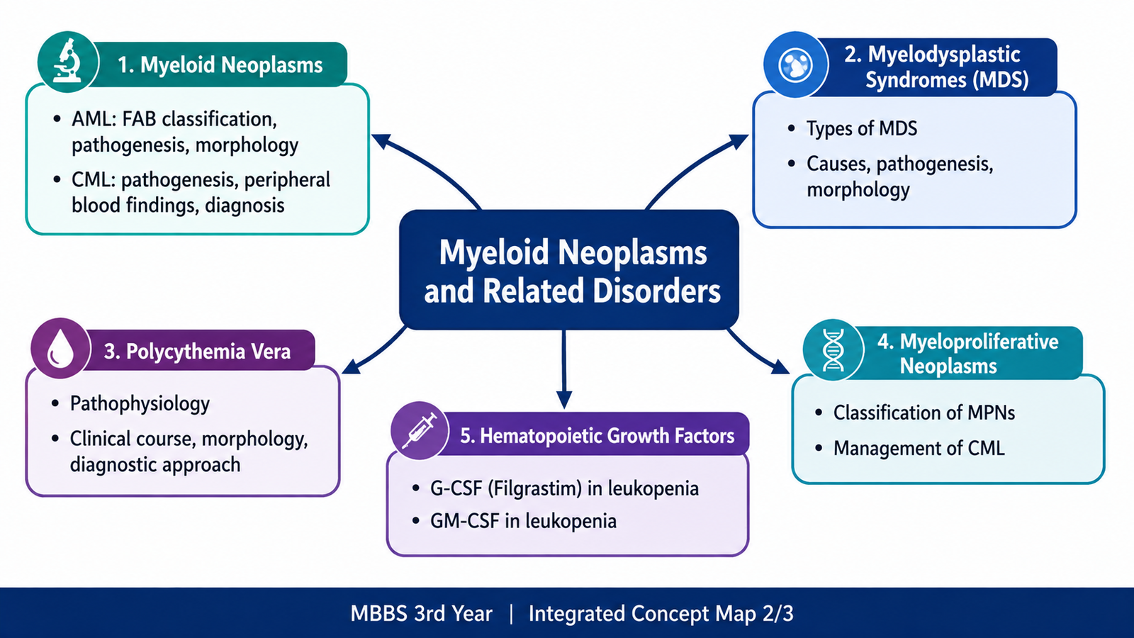
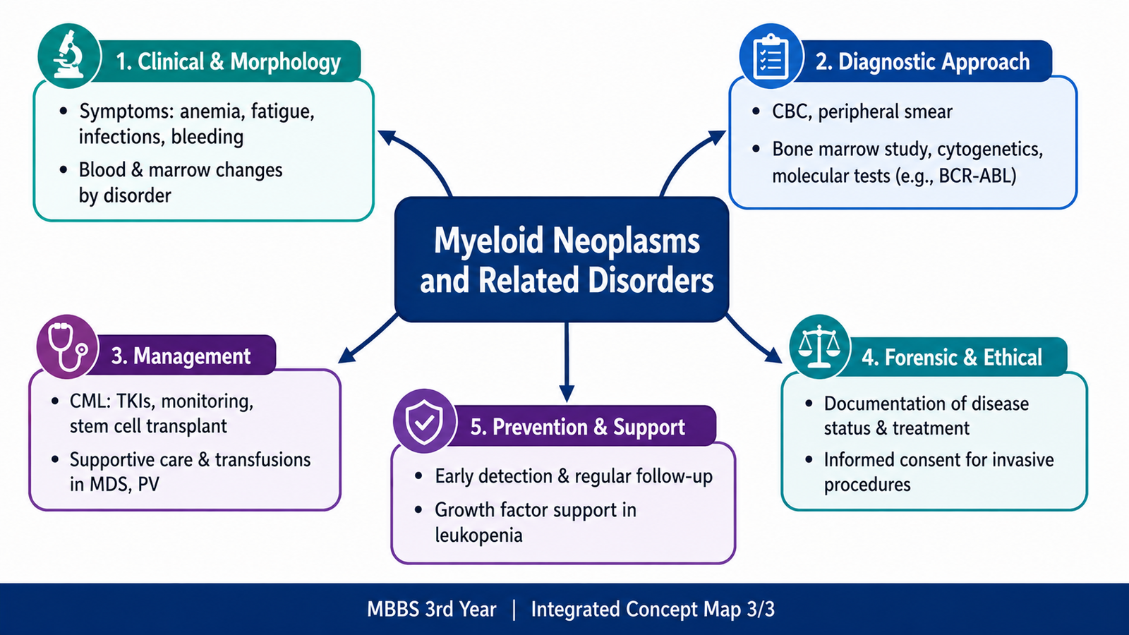
An integrated introduction to AML, CML, myelodysplastic syndromes, polycythemia vera, hematopoietic growth factors and the major white-blood-cell populations.
Topic Introduction
Myeloid disorders arise when cells belonging to the myeloid side of hematopoiesis develop abnormalities in proliferation, maturation or both. Some disorders, such as acute myeloid leukemia, are dominated by accumulation of immature blasts. Others, such as chronic myeloid leukemia and polycythemia vera, produce excessive numbers of relatively mature blood cells. Myelodysplastic syndromes are different again: the marrow may be active and even hypercellular, but blood-cell production is ineffective and dysplastic. Understanding these differences makes the blood count, peripheral smear, bone-marrow findings and diagnostic tests much easier to interpret. This chapter also connects these disorders with normal white-blood-cell populations and the pharmacological role of hematopoietic growth factors in leukopenia.
A. White Blood Cells and the Myeloid Neoplasm Framework
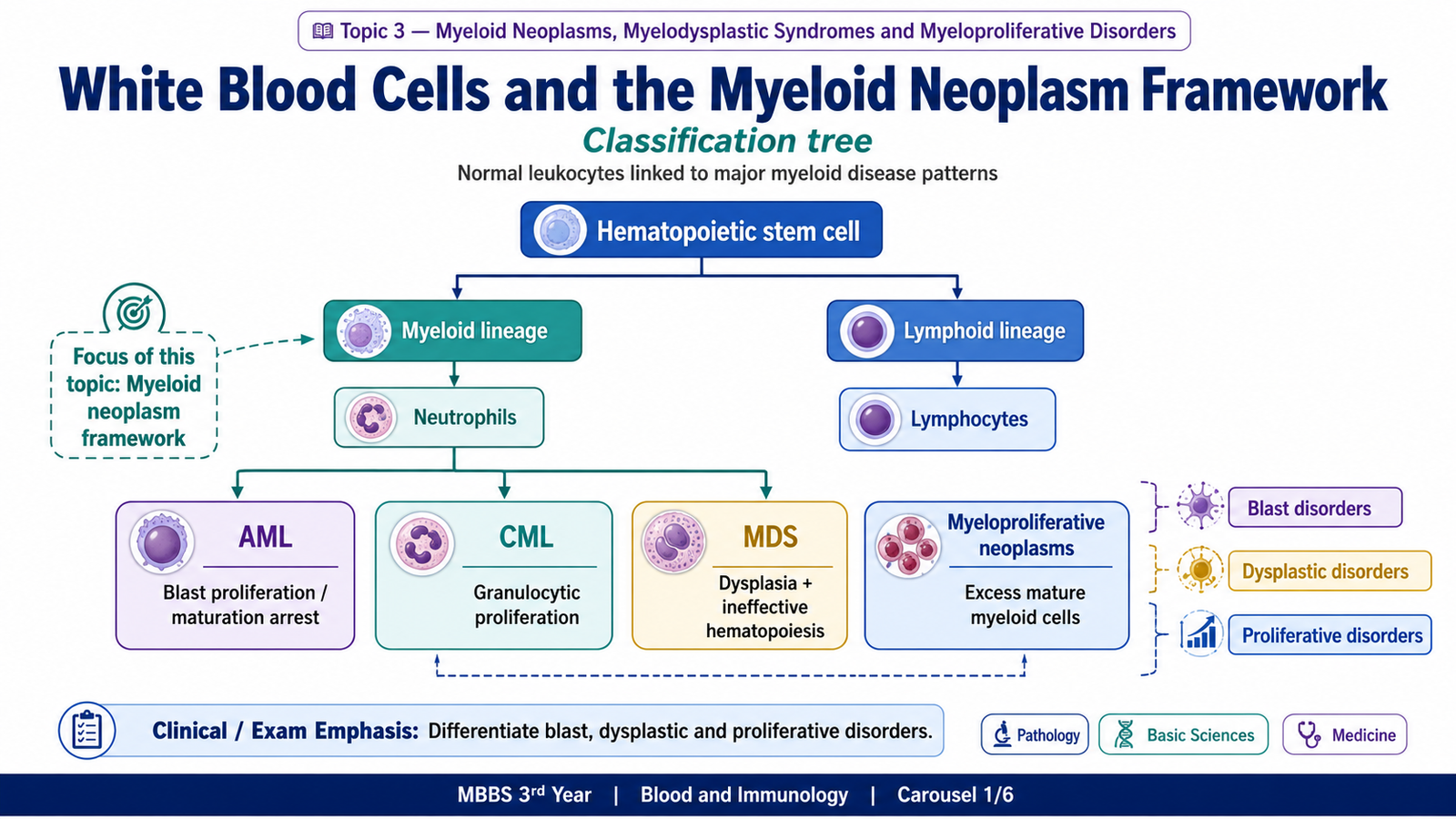
Normal bone marrow continuously produces several types of white blood cells. These cells arise from hematopoietic stem cells and then differentiate along myeloid or lymphoid pathways. Knowing the normal cell populations is important because myeloid neoplasms represent clonal abnormalities of cells that normally arise from the myeloid lineage.
Major White-Blood-Cell Populations
The major leukocyte populations can be understood according to their morphology and function.
- Neutrophils: polymorphonuclear leukocytes that form the major circulating phagocytic defense against many bacterial infections.
- Eosinophils: granulocytes particularly associated with responses to parasites and allergic disease.
- Basophils: granulocytes containing mediators such as histamine; marked basophilia is an important clue in CML.
- Monocytes: circulating cells that enter tissues and may differentiate into macrophages.
- Lymphocytes: include B cells, T cells and natural killer cells and are central to adaptive and cellular immune responses.
- Plasma cells: terminally differentiated B cells specialized for antibody secretion.
Disorders Affecting Leukocyte Populations
Leukocyte abnormalities may involve an increase, a decrease or a neoplastic expansion of a particular population. Examples include neutrophilia during inflammation, neutropenia following marrow suppression, eosinophilia in allergic or parasitic conditions, lymphocytosis in some infections and clonal proliferation in hematological neoplasms.
How Myeloid Disorders Differ
Myeloid neoplasms begin from an abnormal hematopoietic clone, but the biological behavior of that clone differs among diseases.
Abnormal hematopoietic stem/progenitor cell → clonal expansion → altered maturation or proliferation → abnormal marrow and peripheral blood → cytopenias, excessive blood-cell counts or both
Acute myeloid leukemia (AML) is dominated by accumulation of immature myeloid blasts. Chronic myeloid leukemia (CML) produces excessive granulocytic cells at many stages of maturation. Myelodysplastic syndromes (MDS) show ineffective and morphologically abnormal blood-cell production. Polycythemia vera (PV) is a myeloproliferative neoplasm characterized mainly by increased red-cell production, usually with proliferation of other myeloid lineages as well.
Major Myeloproliferative Neoplasms
The major classic myeloproliferative neoplasms relevant at undergraduate level are:
- Chronic myeloid leukemia
- Polycythemia vera
- Essential thrombocythemia
- Primary myelofibrosis
B. Acute Myeloid Leukemia
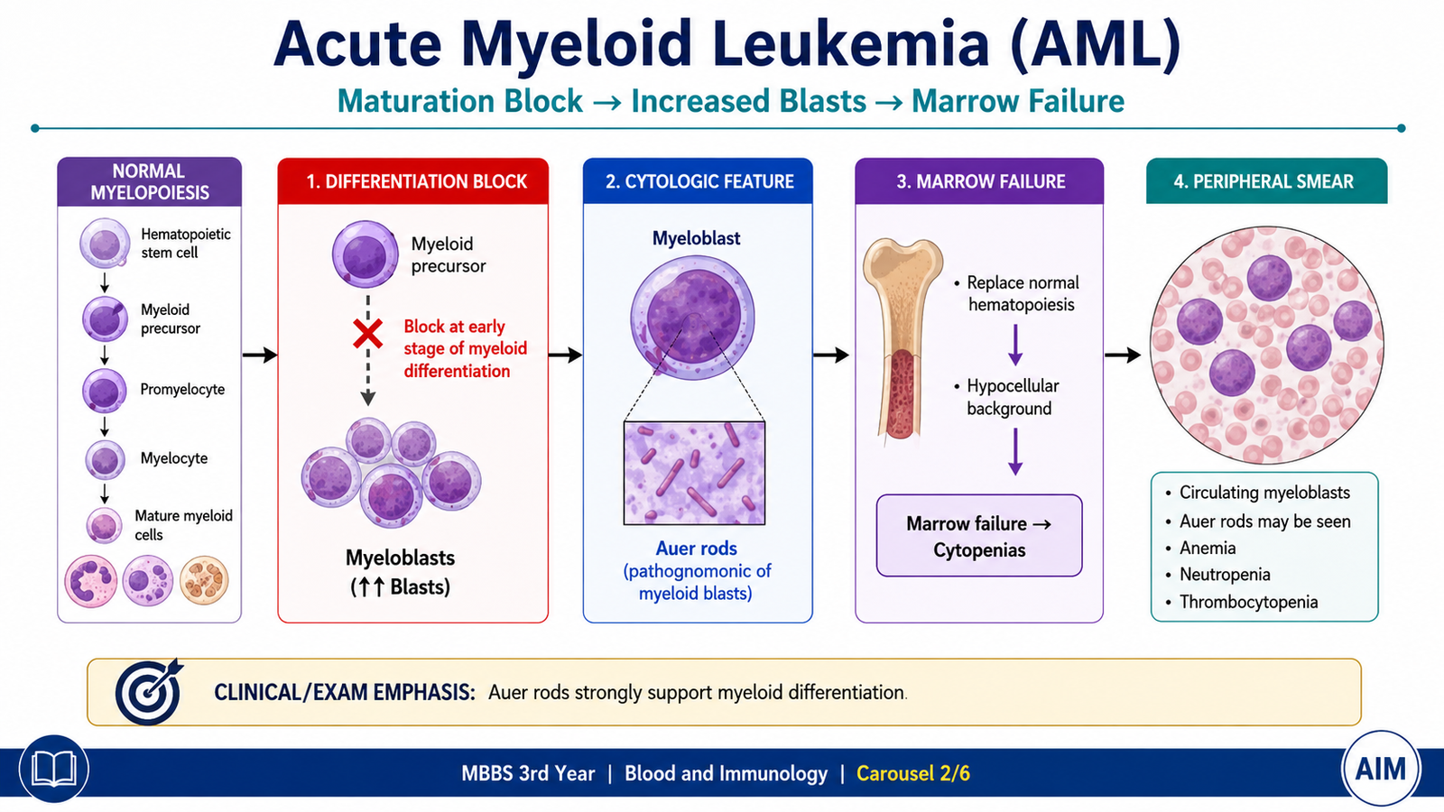
Acute myeloid leukemia (AML) is a malignant clonal disorder in which myeloid precursor cells acquire abnormalities that promote proliferation and interfere with normal differentiation. Immature cells therefore accumulate in the bone marrow and may appear in peripheral blood. As these leukemic blasts progressively replace normal marrow, normal production of red cells, platelets and functional leukocytes falls.
FAB Classification
The FAB classification divides AML mainly according to morphology and the degree or direction of myeloid differentiation. Although modern diagnostic systems make greater use of genetic abnormalities, the FAB system remains useful for understanding and undergraduate examination.
| FAB Type | Description |
|---|---|
| M0 | Minimally differentiated AML |
| M1 | AML without maturation |
| M2 | AML with maturation |
| M3 | Acute promyelocytic leukemia |
| M4 | Acute myelomonocytic leukemia |
| M5 | Acute monocytic/monoblastic leukemia |
| M6 | Acute erythroid leukemia |
| M7 | Acute megakaryoblastic leukemia |
Pathophysiology
AML develops through acquired genetic and molecular abnormalities in a hematopoietic stem or progenitor cell. These abnormalities give the leukemic clone a growth advantage and, importantly, interfere with normal maturation.
The circulating white-cell count in AML may be high, normal or low. A high leukocyte count does not mean that immunity is effective because many circulating cells are immature leukemic blasts rather than normally functioning mature leukocytes.
Acute promyelocytic leukemia, corresponding to FAB M3, has a characteristic abnormality involving chromosomes 15 and 17 and the PML::RARA fusion. The abnormal promyelocytes contain abundant cytoplasmic granules, and this subtype has a particularly important association with coagulation abnormalities.
Morphological Features
Peripheral blood
- Anemia is commonly present because normal erythropoiesis is suppressed.
- Platelets are often reduced because megakaryocytic production is impaired.
- Myeloblasts may circulate in peripheral blood.
- The total leukocyte count may be increased, normal or reduced.
Myeloblast morphology
Myeloblasts are relatively large immature cells with a high nuclear-to-cytoplasmic ratio, fine chromatin and visible nucleoli. Their cytoplasm may contain azurophilic granules.
Bone marrow
The marrow is typically replaced to a substantial degree by leukemic blasts, with suppression of normal hematopoietic elements. In most AML, the diagnosis requires a major increase in myeloid blasts; certain genetically defined forms can be recognized by their characteristic abnormalities even when the blast percentage is lower.
Diagnostic Approach
Diagnosis is not based on the peripheral smear alone. The purpose of investigation is first to demonstrate acute leukemia, then establish myeloid lineage and finally define biologically important abnormalities.
- Complete blood count: demonstrates anemia, platelet abnormalities and the leukocyte pattern.
- Peripheral blood smear: identifies circulating blasts and may reveal Auer rods.
- Bone-marrow examination: demonstrates blast accumulation and assesses marrow morphology.
- Cytochemical or immunophenotypic assessment: establishes myeloid differentiation. Myeloperoxidase is a useful marker of myeloid lineage.
- Flow cytometry: characterizes the immunophenotype of the abnormal cells.
- Cytogenetic and molecular tests: identify important chromosomal and molecular abnormalities and help define disease subtype.
C. Chronic Myeloid Leukemia
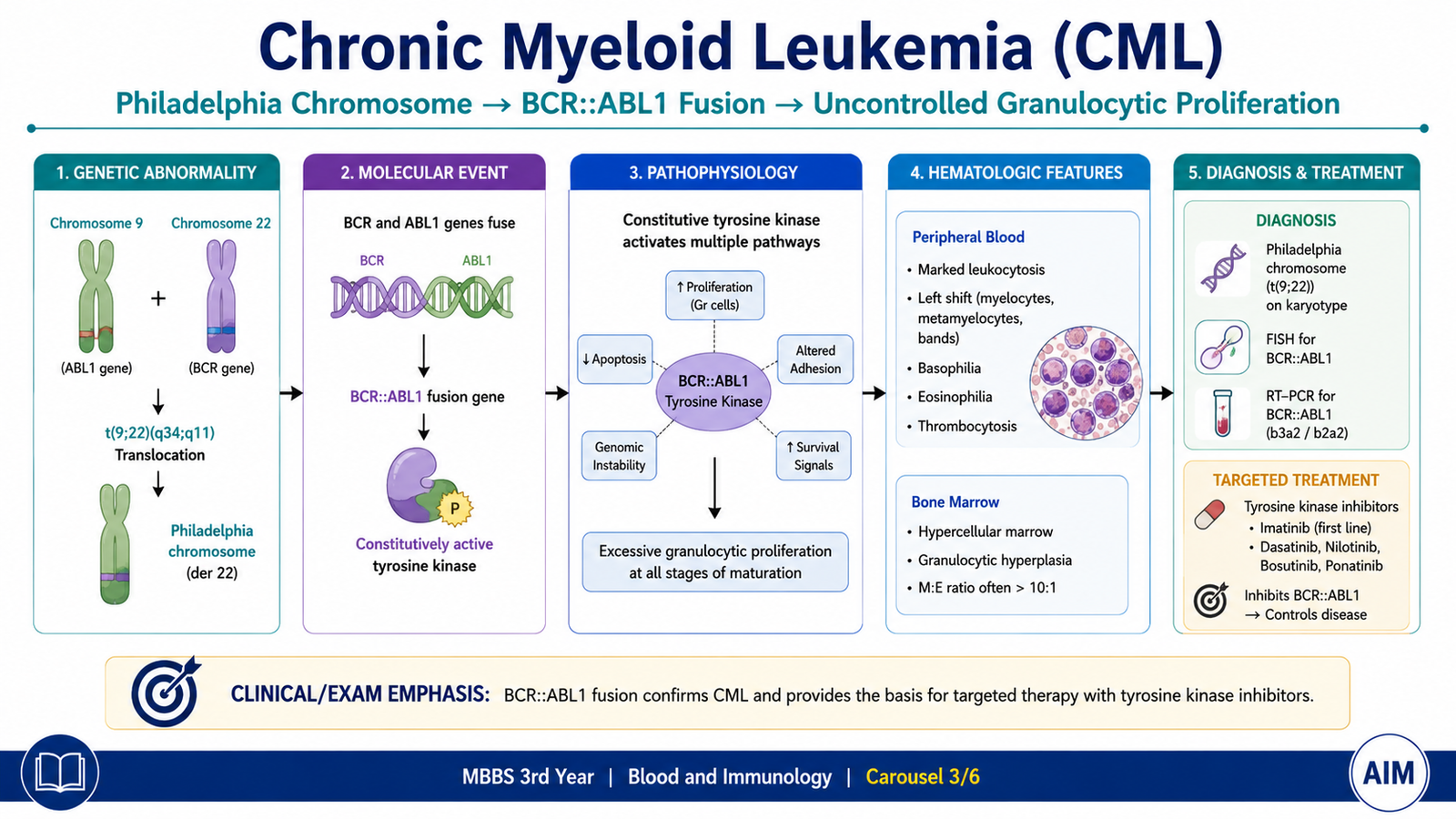
Chronic myeloid leukemia (CML) is a clonal myeloproliferative neoplasm characterized by excessive production of granulocytic cells. Unlike AML, differentiation is largely preserved during the chronic phase. Therefore, peripheral blood contains granulocytes at several different stages of maturation rather than being dominated only by blasts.
Pathophysiology
The central molecular abnormality in CML is formation of the BCR::ABL1 fusion gene. It usually results from reciprocal translocation between chromosomes 9 and 22, written as t(9;22). The shortened abnormal chromosome 22 is called the Philadelphia chromosome.
The abnormal tyrosine kinase provides persistent growth and survival signaling. Because the leukemic cells can still mature in the chronic phase, neutrophils and their precursors appear together in peripheral blood.
Peripheral-Blood Findings
- Marked leukocytosis caused mainly by expansion of the granulocytic series.
- Cells at multiple stages of granulocytic maturation, including myelocytes, metamyelocytes and mature neutrophils.
- Basophilia, an especially useful diagnostic clue.
- Eosinophilia may also occur.
- Platelet count may be increased.
- Mild to moderate anemia may develop.
- Blasts are relatively few during the chronic phase compared with acute leukemia.
The bone marrow is markedly hypercellular with prominent granulocytic proliferation. Splenomegaly is common because the enlarged spleen participates in sequestration of blood cells and may also become a site of extramedullary hematopoiesis.
Clinical Course
Patients may present with fatigue, weight loss, abdominal fullness due to splenomegaly or may be discovered incidentally after a blood count shows marked leukocytosis. The disease usually begins in a relatively stable chronic phase. Without adequate disease control, the abnormal clone may acquire additional abnormalities and progress toward more aggressive disease, eventually resembling acute leukemia in blast phase.
Diagnostic Approach
The blood film can strongly suggest CML, but definitive diagnosis requires demonstration of the characteristic BCR::ABL1 abnormality.
- CBC: confirms leukocytosis and associated abnormalities.
- Peripheral smear: demonstrates the full spectrum of granulocytic maturation and basophilia.
- Bone marrow: shows marked myeloid hyperplasia when examination is required.
- Cytogenetic analysis: may demonstrate the Philadelphia chromosome.
- FISH or molecular testing: demonstrates BCR::ABL1.
- Quantitative molecular testing: is also important for assessing the molecular response after treatment begins.
Management of CML
CML provides a classic example of treatment directed at a disease-producing molecular abnormality. Because BCR::ABL1 functions as an abnormal tyrosine kinase, BCR::ABL1 tyrosine-kinase inhibitors form the central treatment approach.
BCR::ABL1 drives abnormal proliferation → tyrosine-kinase inhibitor suppresses BCR::ABL1 signaling → leukemic-cell growth falls → hematologic and molecular disease burden decreases.
Imatinib is the classic prototype BCR::ABL1 tyrosine-kinase inhibitor. Other agents from the same therapeutic group may be used according to disease response, resistance, tolerance and clinical circumstances. Treatment response is assessed using hematological and molecular parameters.
Stem-cell transplantation has an important role in selected patients, particularly when disease is resistant to appropriate targeted therapy or has progressed to advanced phases. Detailed transplant protocols are beyond the required undergraduate scope.
D. Myelodysplastic Syndromes
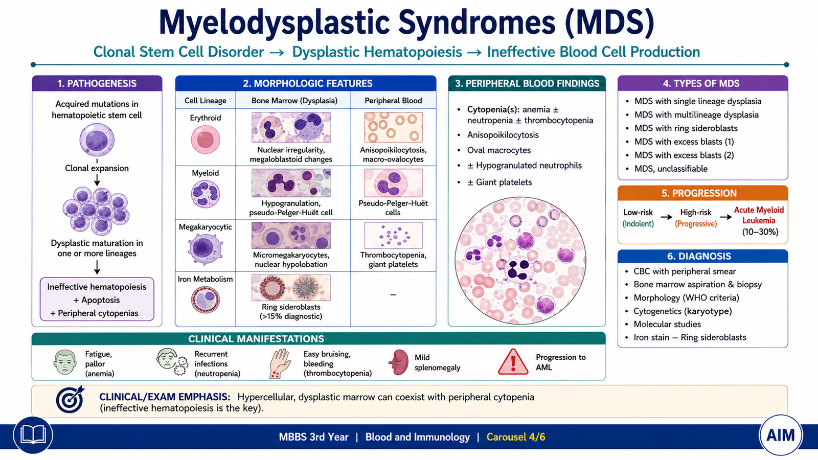
Myelodysplastic syndromes (MDS) are clonal disorders of hematopoietic stem cells characterized by ineffective blood-cell production, morphological dysplasia and persistent cytopenias. A major feature of MDS is the apparent contradiction between the marrow and peripheral blood: the bone marrow is often cellular or hypercellular, yet one or more circulating blood-cell populations are reduced.
This occurs because abnormal precursor cells develop poorly and many undergo death within the marrow before they can become functional circulating cells.
Types of MDS
Classification terminology has evolved as molecular information has increased. At undergraduate level, the important morphological patterns include:
- MDS predominantly involving a single dysplastic lineage
- MDS with multilineage dysplasia
- MDS with ring sideroblasts
- MDS with excess blasts
- MDS associated with an isolated deletion involving chromosome 5q
- MDS that does not fit neatly into the major defined categories
The exact terminology may change between classification systems, but the core concept remains the same: clonal ineffective hematopoiesis with dysplasia and a risk of progression to AML.
Causes and Predisposing Factors
MDS may arise without an identifiable preceding exposure or may occur after injury to hematopoietic stem cells. Important settings include:
- Increasing age
- Previous cytotoxic chemotherapy
- Previous radiation exposure
- Other acquired damage affecting hematopoietic stem-cell DNA
Pathogenesis
A hematopoietic stem cell acquires genetic and epigenetic abnormalities that produce a clonal population. These abnormal progenitors can proliferate in the marrow but do not mature normally. Dysplastic cells are therefore formed, and many precursors die before reaching the circulation.
Peripheral-Blood Morphology
The findings depend on which hematopoietic lineages are involved.
- Red cells: anemia is common; red cells may be macrocytic and show variation in size and shape.
- Neutrophils: may be reduced in number and may show hypogranulation or abnormal nuclear segmentation.
- Pseudo-Pelger-Huët cells: neutrophils with abnormal hyposegmented nuclei are an important example of granulocytic dysplasia.
- Platelets: may be reduced and can show abnormal size or granulation.
- Blasts: may increase, particularly in higher-grade disease, but the disorder remains below the level required for classification as overt AML unless transformation occurs.
Bone-Marrow Changes
The bone marrow is commonly hypercellular despite cytopenias in peripheral blood. Dysplastic abnormalities may involve erythroid, granulocytic and megakaryocytic lineages.
- Abnormal erythroid maturation may be present.
- Ring sideroblasts may occur in defined forms and represent erythroid precursors with abnormal mitochondrial iron accumulation around the nucleus.
- Granulocytic precursors may show abnormal maturation and granulation.
- Megakaryocytes may be abnormal in size and nuclear morphology.
- The blast proportion may be increased but is monitored carefully because further increase can indicate progression toward AML.
Symptoms
Symptoms usually result from the affected blood-cell line rather than from a single characteristic symptom of MDS.
- Anemia → fatigue, weakness and exertional symptoms.
- Neutropenia → increased susceptibility to infections.
- Thrombocytopenia → bruising, petechiae or bleeding.
Diagnostic Strategy
The diagnosis requires more than simply finding a low blood count. Other causes of cytopenia and dysplastic-looking blood cells must be considered and excluded where appropriate.
- Demonstrate persistent cytopenia on the complete blood count.
- Inspect the peripheral smear for dysplastic changes.
- Examine bone-marrow aspirate and biopsy for cellularity, dysplasia and blast proportion.
- Use appropriate marrow staining when abnormalities such as ring sideroblasts are suspected.
- Perform cytogenetic and molecular evaluation where required to identify clonal abnormalities and assist classification.
- Exclude important non-neoplastic causes that can produce cytopenia or abnormal maturation.
E. Polycythemia Vera
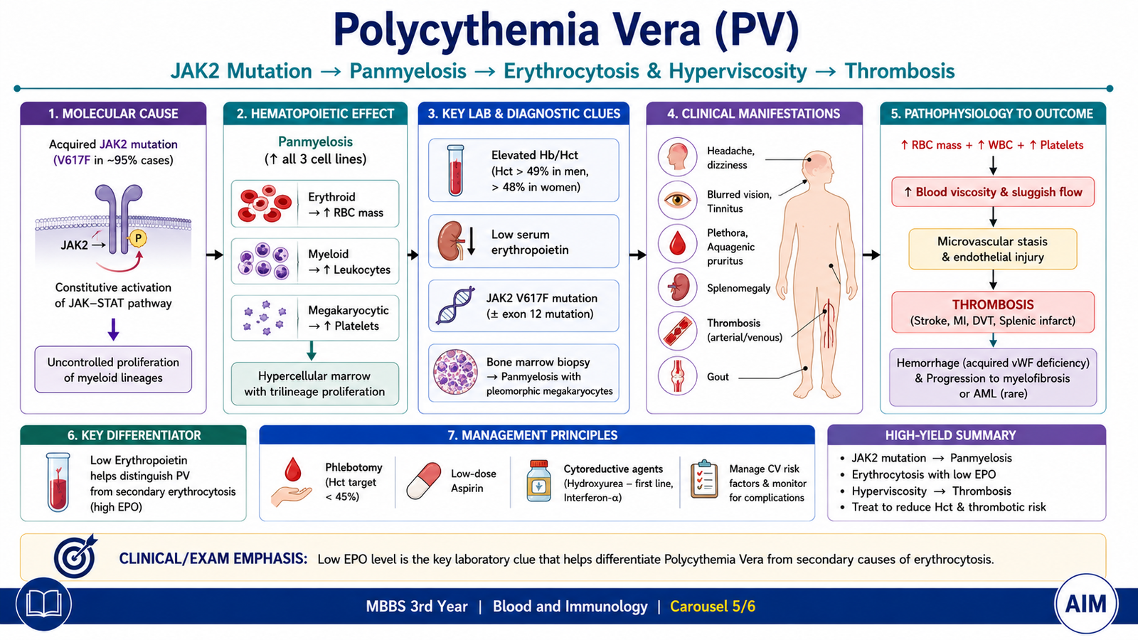
Polycythemia vera (PV) is a chronic clonal myeloproliferative neoplasm in which hematopoietic cells proliferate independently of the normal physiological drive for red-cell production. The most obvious effect is erythrocytosis, but PV is actually a disorder of the myeloid stem-cell compartment and may also increase granulocytes and platelets.
Pathophysiology
Most patients have an activating abnormality involving JAK2, an intracellular signaling protein normally used by hematopoietic growth-factor receptors. Persistent JAK-STAT signaling allows erythroid progenitors to proliferate even when the normal erythropoietin drive is reduced.
Because red-cell production is autonomous, the kidneys normally reduce erythropoietin production. Therefore, a low serum erythropoietin level supports PV and helps distinguish it from many forms of secondary erythrocytosis.
Clinical Features and Course
The expanded red-cell mass increases blood viscosity. Slow and abnormal blood flow can produce headache, dizziness, visual disturbance and vascular complications. Patients may appear plethoric because of the increased circulating red-cell mass.
- Headache and dizziness
- Plethora
- Visual or other microvascular symptoms
- Splenomegaly
- Thrombotic complications
- Bleeding may occur despite the increased cell counts because platelet function can be abnormal.
- Pruritus, especially after contact with warm water, is a characteristic clinical clue.
Increased turnover of blood cells may increase uric-acid production. Over time, some patients develop increasing marrow fibrosis, while a smaller proportion may undergo transformation to an acute leukemia.
Morphology
Peripheral blood
- Marked erythrocytosis is the principal finding.
- Leukocytes may also be increased.
- Platelets may be increased.
Bone marrow
The marrow is hypercellular and demonstrates panmyelosis, meaning proliferation of multiple myeloid lineages, particularly erythroid, granulocytic and megakaryocytic elements. This finding helps show that PV is not simply a physiological increase in red cells.
Diagnostic Approach
The diagnostic problem is to establish true persistent erythrocytosis and then determine whether it is caused by an autonomous myeloproliferative process or by an appropriate increase in erythropoietin.
- Complete blood count: demonstrates persistent erythrocytosis and may show leukocytosis or thrombocytosis.
- Clinical assessment: looks for splenomegaly, hyperviscosity manifestations and alternative causes of erythrocytosis.
- Serum erythropoietin: is usually reduced in PV because excessive red-cell production suppresses normal renal erythropoietin release.
- JAK2 testing: provides strong evidence of a clonal myeloproliferative process when an appropriate activating abnormality is found.
- Bone-marrow examination: can demonstrate characteristic panmyelosis.
| Feature | Polycythemia Vera | Secondary Erythrocytosis |
|---|---|---|
| Basic mechanism | Clonal autonomous marrow proliferation | Erythropoietin-driven red-cell production |
| Erythropoietin | Usually low | Often increased when driven by hypoxia or inappropriate EPO production |
| JAK2 abnormality | Common and diagnostically important | Not the usual mechanism |
| Other myeloid cells | May also be increased | Usually not increased as part of a clonal panmyelosis |
F. Hematopoietic Growth Factors in Leukopenia
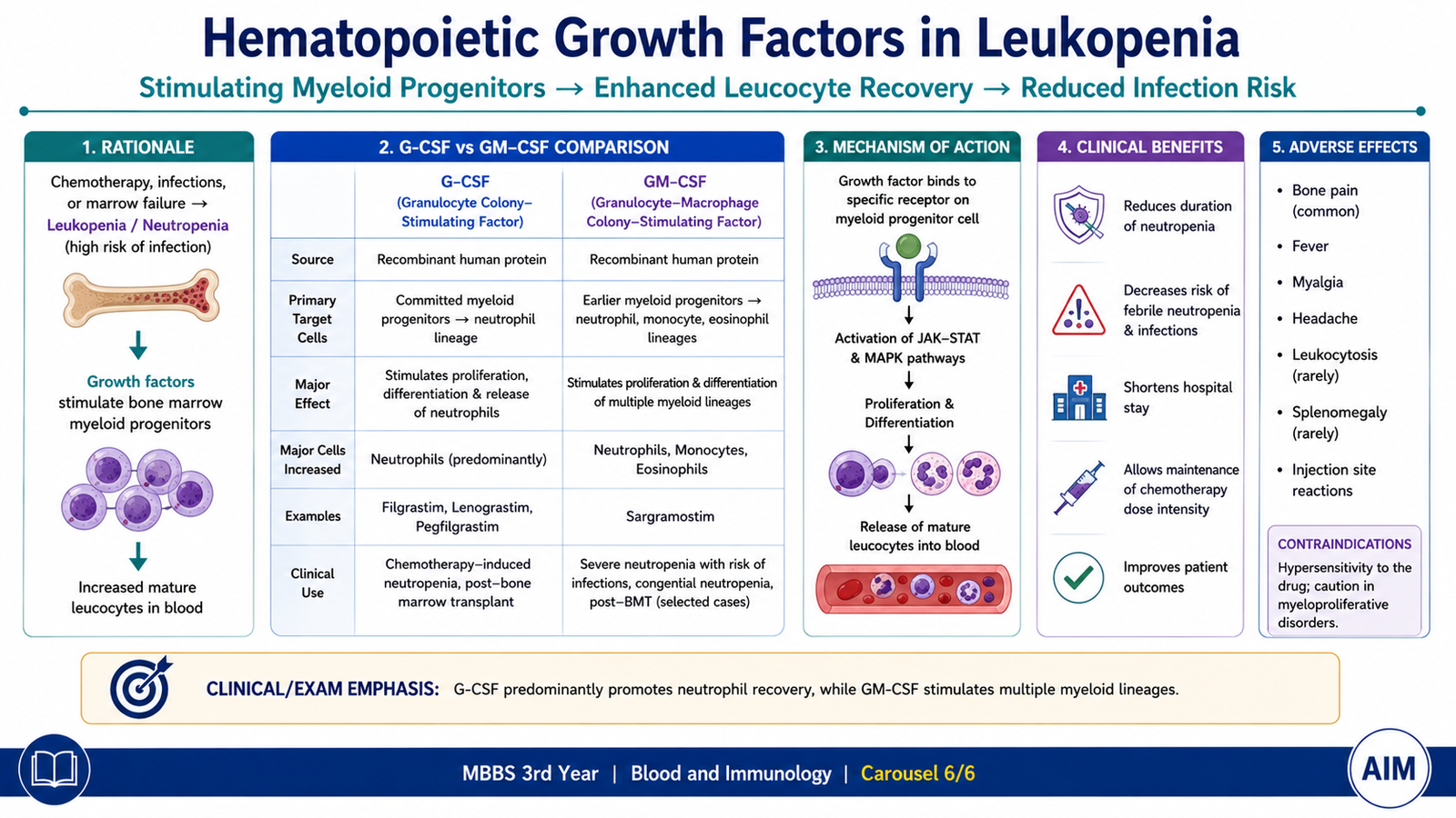
Hematopoietic growth factors are signaling proteins that regulate proliferation, differentiation and survival of bone-marrow precursor cells. Pharmacological forms of these growth factors can be used when the marrow needs stimulation to restore circulating leukocytes, particularly neutrophils. The two important groups in this topic are granulocyte colony-stimulating factor (G-CSF) and granulocyte-monocyte colony-stimulating factor (GM-CSF).
Granulocyte Colony-Stimulating Factor
Filgrastim is a recombinant form of G-CSF. It acts on G-CSF receptors expressed mainly on neutrophil precursors and promotes their proliferation, differentiation and functional maturation.
Role in Leukopenia
G-CSF is particularly useful when leukopenia is mainly due to neutropenia. It is commonly used to accelerate neutrophil recovery after myelosuppressive treatment and in other clinical situations where stimulation of neutrophil production is required.
A characteristic adverse effect of G-CSF therapy is bone pain. This is related to increased marrow activity and expansion of hematopoietic tissue. Excessive leukocyte elevation can also occur if stimulation is marked.
Granulocyte-Monocyte Colony-Stimulating Factor
GM-CSF acts on a broader range of myeloid progenitor cells than G-CSF. It stimulates production and activity of both granulocytic and monocyte/macrophage lineages. Sargramostim is a recombinant GM-CSF preparation.
G-CSF versus GM-CSF
The key distinction is the breadth of their action. G-CSF has a more selective effect on the neutrophil lineage, whereas GM-CSF stimulates granulocyte and monocyte progenitors more broadly.
| Feature | G-CSF | GM-CSF |
|---|---|---|
| Prototype | Filgrastim | Sargramostim |
| Main lineage effect | Predominantly neutrophils | Granulocytes and monocytes |
| Main principle | Accelerates neutrophil recovery | Broader stimulation of myeloid recovery |
Integrated Mechanism Flow
Important Comparison
| Feature | AML | CML | MDS | Polycythemia Vera |
|---|---|---|---|---|
| Dominant abnormality | Accumulation of myeloid blasts | Excess granulocytic proliferation | Ineffective dysplastic hematopoiesis | Autonomous erythrocytosis with panmyelosis |
| Cell maturation | Markedly impaired | Largely preserved in chronic phase | Abnormal and ineffective | Mature cells overproduced |
| Key blood clue | Blasts ± Auer rods | Full granulocytic maturation spectrum with basophilia | Cytopenias with dysplastic cells | Persistent erythrocytosis |
| Important molecular clue | Varies by subtype; PML::RARA is characteristic of APL | BCR::ABL1 | Clonal cytogenetic/molecular abnormalities may be demonstrated | JAK2 pathway abnormality |
| Major clinical consequence | Marrow failure | Marked leukocytosis and splenomegaly | Anemia, infection and bleeding from cytopenias | Hyperviscosity and thrombosis |
| Key diagnostic principle | Blast identification plus confirmation of myeloid lineage | Demonstrate BCR::ABL1 | Persistent cytopenia + marrow dysplasia | Erythrocytosis + clonal/JAK2 evidence + typically low EPO |
⭐ AIM High-Yield Review
- AML is characterized by clonal accumulation of immature myeloid blasts and suppression of normal hematopoiesis.
- The FAB classification divides AML from M0 to M7; M3 corresponds to acute promyelocytic leukemia.
- Auer rods are a highly useful morphological clue to myeloid differentiation.
- Anemia, infection and bleeding in AML largely result from replacement and suppression of normal bone-marrow hematopoiesis.
- CML is driven by the BCR::ABL1 fusion, usually produced by t(9;22), forming the Philadelphia chromosome.
- CML typically shows marked leukocytosis with multiple stages of granulocytic maturation and basophilia.
- BCR::ABL1 tyrosine-kinase inhibitors such as imatinib directly target the central molecular driver of CML.
- MDS causes ineffective and dysplastic hematopoiesis; the marrow may be hypercellular despite peripheral cytopenias.
- Pseudo-Pelger-Huët neutrophils and ring sideroblasts are important examples of dysplastic morphology in appropriate MDS patterns.
- MDS carries a clinically important risk of progression to AML.
- Polycythemia vera is a clonal myeloproliferative neoplasm usually associated with JAK2-pathway activation and marrow panmyelosis.
- PV should be distinguished from secondary erythrocytosis: serum erythropoietin is usually low in PV.
- Hyperviscosity and thrombosis are major consequences of the increased cell mass in PV.
- Filgrastim is G-CSF and mainly promotes neutrophil production; GM-CSF stimulates a broader range of granulocyte-monocyte progenitors.
- Across these disorders, the examination pattern is: blood count → peripheral smear → bone marrow → lineage/molecular confirmation.
Myeloid Neoplasms, MDS & Myeloproliferative Disorders
Use these videos after completing the AIM Learning Material to reinforce the major pathological mechanisms, morphology, diagnostic approach and relevant pharmacology.
Acute Myeloid Leukemia — Classification & Pathogenesis
Reinforces AML classification, pathogenesis and the major concepts underlying myeloid blast proliferation.
Chronic Myeloid Leukemia — BCR::ABL1, Diagnosis & Treatment
Covers the Philadelphia chromosome, BCR::ABL1 pathogenesis, peripheral-blood findings, diagnosis and tyrosine-kinase inhibitor therapy.
Myelodysplastic Syndromes — Pathogenesis, Features & Diagnosis
Reviews ineffective hematopoiesis, cytopenias, abnormal marrow maturation, clinical manifestations and the diagnostic approach to MDS.
Polycythemia Vera — JAK2, Morphology & Diagnosis
Reinforces JAK2-driven pathogenesis, panmyelosis, clinical manifestations, morphology and diagnostic principles of polycythemia vera.
▶ Watch Polycythemia Vera Video
Hematopoietic Growth Factors — G-CSF & GM-CSF
Reviews hematopoietic growth factors with emphasis on G-CSF, filgrastim, GM-CSF and their role in neutropenia and leukopenia.
Complete the AIM Learning Material first → watch the disease-specific videos → revise the AIM High-Yield Review → attempt the Post-Test.