
This chapter follows the supplied KMU learning outcomes and builds the concepts in a logical sequence. First understand how abnormal immune responses develop, then revise the high-yield mechanisms, disease associations and drug actions at the end.
Hypersensitivity, Immune Tolerance, Autoimmunity, Immunodeficiency and Immunomodulatory Drugs
Blood and Immunology — understanding harmful immune responses, loss of self-tolerance, immune failure, transplantation reactions and the drugs used to suppress or stimulate immunity.
Topic Introduction
The immune system normally protects the body while remaining tolerant to its own tissues. Disease develops when this balance is disturbed. An exaggerated immune response against a harmless or foreign antigen produces hypersensitivity, whereas loss of tolerance to self-antigens produces autoimmunity. In contrast, failure of one or more immune components causes immunodeficiency. Similar immune mechanisms are important in transplantation because the recipient may recognize a graft as foreign, while immune cells in a donor graft may sometimes attack the recipient. These responses can be modified therapeutically. Immunosuppressive drugs reduce unwanted immune activity in transplantation and autoimmune disease, while selected immunostimulants and monoclonal antibodies can strengthen or redirect immunity, particularly in certain malignancies.
A. Hypersensitivity: Core Mechanisms and Types
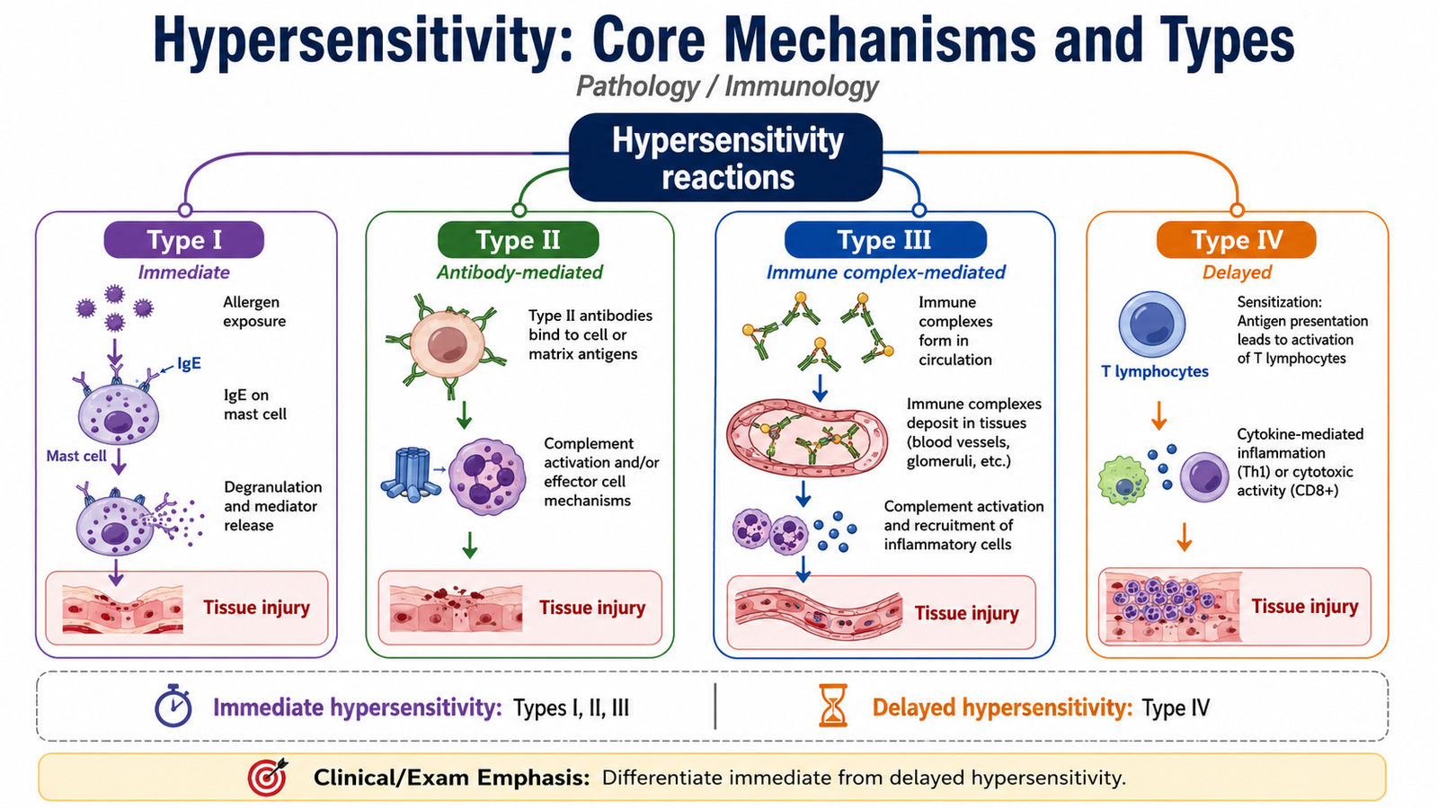
Hypersensitivity is tissue injury produced by an immune response that is excessive, inappropriate or directed against an antigen in a way that damages the host. The antigen may be environmental, microbial or a self-antigen. The important principle is that the same immune mechanisms that normally protect the body can cause disease when they are misdirected or excessively activated.
Allergy usually refers to hypersensitivity against normally harmless environmental antigens, particularly IgE-mediated type I reactions such as allergic rhinitis, some forms of bronchial asthma and anaphylaxis.
Type I — Immediate Hypersensitivity
Type I hypersensitivity is mediated mainly by IgE antibodies, mast cells and Th2 cells. It develops in two main stages: sensitization during the first exposure and mast-cell activation after later exposure.
Allergen exposure → activation of Th2 cells → IL-4 and IL-13 promote IgE production → IgE binds high-affinity receptors on mast cells → re-exposure cross-links mast-cell-bound IgE → mast-cell degranulation → vascular, bronchial and inflammatory effects.
Preformed mast-cell mediators such as histamine rapidly produce vasodilation, increased vascular permeability and smooth-muscle effects. Newly synthesized mediators such as leukotrienes and prostaglandins further contribute to bronchoconstriction and inflammation. Cytokines recruit inflammatory cells, especially eosinophils, producing a later inflammatory phase.
Important examples include:
- Systemic anaphylaxis — widespread mast-cell activation can cause hypotension, edema and bronchospasm.
- Allergic rhinitis — local mast-cell activation produces nasal congestion and secretion.
- Atopic bronchial asthma — IgE-mediated reactions contribute to bronchoconstriction and airway inflammation.
- Food and drug allergies — manifestations may range from localized urticaria to systemic reactions.
Type II — Antibody-Mediated Hypersensitivity
Type II reactions are caused mainly by IgG or IgM antibodies directed against antigens present on cells or in extracellular tissues. Tissue injury can occur by several mechanisms.
- Opsonization and cell destruction: antibody-coated cells are removed by phagocytes or complement. Autoimmune hemolytic anemia is an example.
- Inflammation: antibodies against tissue antigens activate complement and leukocytes. Goodpasture syndrome is a classic example.
- Abnormal receptor function: antibodies may stimulate or block receptors without destroying the cell. Graves disease results from stimulation of the TSH receptor, whereas myasthenia gravis involves interference with acetylcholine receptor function.
Type III — Immune Complex-Mediated Hypersensitivity
In type III hypersensitivity, soluble antigens combine with antibodies and form immune complexes. When these complexes are not adequately cleared, they may deposit in blood vessels, glomeruli, joints or other tissues. Complement activation then recruits neutrophils, whose enzymes and reactive products damage the surrounding tissue.
Examples include systemic lupus erythematosus, serum sickness and the localized Arthus reaction.
Type IV — T-Cell-Mediated Hypersensitivity
Type IV hypersensitivity is mediated by T lymphocytes rather than antibodies. Sensitized CD4+ T cells release cytokines that recruit and activate inflammatory cells, while CD8+ cytotoxic T cells may directly destroy target cells. Because cellular recruitment and activation require time, these reactions usually develop more slowly than immediate IgE-mediated reactions.
Examples include contact dermatitis, the tuberculin-type reaction and T-cell-mediated tissue injury in several autoimmune diseases.
| Feature | Immediate Hypersensitivity | Delayed Hypersensitivity |
|---|---|---|
| Main type | Type I | Type IV |
| Main mediator | IgE and mast cells | Sensitized T lymphocytes |
| Onset | Rapid, usually within minutes after re-exposure | Usually develops over many hours |
| Typical effector mechanism | Mast-cell mediators | Cytokine-mediated inflammation or direct T-cell cytotoxicity |
| Example | Anaphylaxis, allergic rhinitis | Contact dermatitis, tuberculin reaction |

B. Immune Tolerance and Unresponsiveness to Self
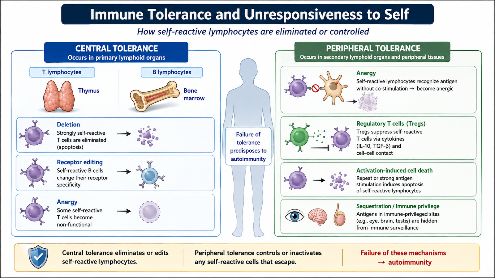
Immune tolerance is a state in which lymphocytes remain unresponsive to a particular antigen. The most important form is self-tolerance, which prevents the immune system from attacking the body’s own tissues. Self-reactive lymphocytes can arise during normal lymphocyte development, so the body requires mechanisms that either eliminate them or prevent them from becoming harmful.
The two major types are central tolerance and peripheral tolerance.
Central Tolerance
Central tolerance occurs while lymphocytes are developing in the primary lymphoid organs. T lymphocytes mature in the thymus, while B lymphocytes mature mainly in the bone marrow.
If an immature lymphocyte recognizes a self-antigen strongly, several outcomes are possible:
- Deletion: strongly self-reactive lymphocytes undergo apoptosis.
- Development of regulatory T cells: some self-reactive CD4+ T cells become regulatory T cells rather than harmful effector cells.
- Receptor editing in B cells: some immature B cells alter their antigen receptor and may lose self-reactivity.
Central tolerance is highly important but not perfect. Some potentially self-reactive lymphocytes therefore enter the peripheral circulation.
Peripheral Tolerance
Peripheral tolerance controls self-reactive lymphocytes that escape central tolerance. It uses several complementary mechanisms.
- Anergy: a lymphocyte recognizes an antigen but does not receive the additional signals required for full activation. It becomes functionally unresponsive.
- Suppression by regulatory T cells: regulatory T cells restrain immune responses through inhibitory signals and anti-inflammatory cytokines.
- Deletion: repeated recognition of self-antigens may activate apoptotic pathways and remove self-reactive cells.
- Limited exposure to some self-antigens: certain antigens are normally relatively inaccessible to immune cells, which helps reduce inappropriate activation.
Self-reactive lymphocyte → deletion, inactivation or regulation → absence of damaging response to self → maintenance of immune tolerance.
Failure of these mechanisms allows self-reactive T or B lymphocytes to survive and become activated. This provides the immunological basis for autoimmune disease.
C. Transplantation Immunology: Graft Rejection and Graft-Versus-Host Disease
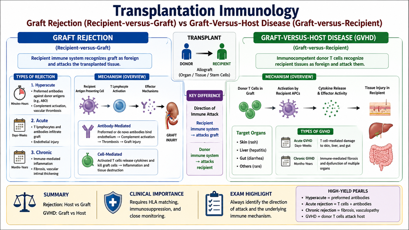
Transplantation introduces cells or tissues carrying antigens that may differ from those of the recipient. An allograft is a graft transferred between genetically different individuals of the same species. The major immunological problem is that recipient lymphocytes may recognize donor antigens, especially donor major histocompatibility complex molecules, as foreign.
Recognition of the Graft
Recipient T cells can recognize donor antigens by two main routes. In direct allorecognition, recipient T cells recognize donor MHC molecules displayed by donor antigen-presenting cells. In indirect allorecognition, donor proteins are processed by recipient antigen-presenting cells and presented to recipient T cells.
These responses activate cytotoxic T cells, helper T cells, macrophages and sometimes antibodies. The resulting injury may affect graft cells, blood vessels or both.
Hyperacute Rejection
Hyperacute rejection is caused by pre-existing recipient antibodies against antigens in the graft. Antibodies bind to graft endothelium soon after blood flow is established, activate complement and promote thrombosis.
Acute Rejection
Acute rejection develops mainly through newly activated T-cell responses and/or antibodies against the graft. T cells may directly damage graft cells or produce cytokines that promote inflammation. Antibody-mediated acute rejection mainly injures graft vessels.
Acute rejection may occur relatively early after transplantation but can also appear later if immunosuppression becomes inadequate.
Chronic Rejection
Chronic rejection is a progressive process characterized particularly by vascular narrowing and interstitial fibrosis. Persistent immune-mediated vascular injury stimulates smooth-muscle proliferation and fibrosis. The graft therefore receives progressively less blood and gradually loses function.
Graft-Versus-Host Disease
Graft-versus-host disease (GVHD) is different from ordinary graft rejection. Here, immunologically competent donor T lymphocytes attack recipient tissues. It is especially important after transplantation of hematopoietic stem cells because the graft itself contains many immune cells.
GVHD is favored when:
- the graft contains immunocompetent T cells;
- the recipient expresses tissue antigens that the donor cells recognize as foreign; and
- the recipient cannot eliminate the attacking donor lymphocytes effectively.
Activated donor T cells recognize recipient antigens, proliferate and produce cytokine-mediated and cytotoxic tissue injury. The skin, gastrointestinal tract and liver are important target organs.
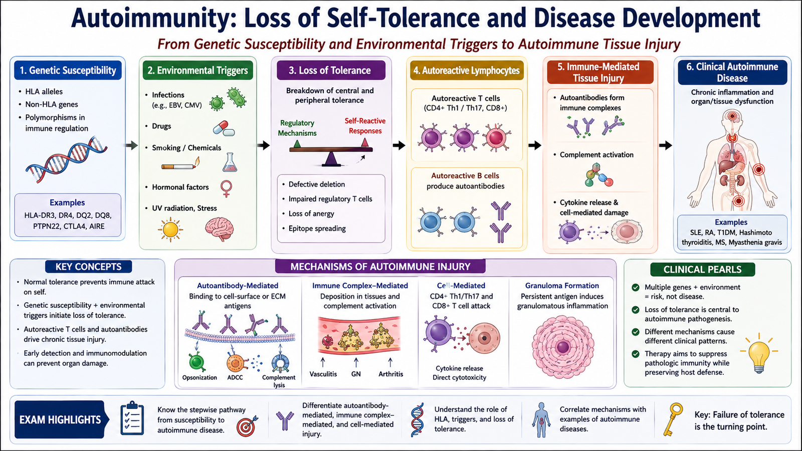
D. Autoimmunity: Loss of Self-Tolerance and Disease Development
Autoimmunity is an immune response directed against the body’s own antigens. Autoimmune disease develops when this response becomes sufficiently strong or persistent to produce tissue injury or abnormal organ function. The central abnormality is therefore a breakdown of self-tolerance.
Autoimmune diseases may predominantly affect one organ, such as autoimmune thyroid disease or type 1 diabetes mellitus, or they may be systemic, as in systemic lupus erythematosus.
How Autoimmune Disease Develops
Autoimmune disease usually results from a combination of inherited susceptibility and environmental influences rather than from a single abnormality.
Genetic Factors
Genes regulating antigen recognition and lymphocyte activation influence susceptibility to autoimmunity. Particular HLA alleles are associated with several autoimmune diseases because HLA molecules determine which peptides are presented to T cells. Abnormalities affecting lymphocyte deletion, regulatory pathways or immune signaling may also impair tolerance.
Failure of Tolerance Mechanisms
Self-reactive lymphocytes may survive when central deletion is incomplete or when peripheral mechanisms such as anergy and regulatory T-cell suppression fail. Once activated, these cells can expand and maintain a continuing immune response against self-antigens.
Environmental and Tissue Factors
- Infections: microbial antigens may resemble self-antigens, allowing cross-reactive immune responses. This is called molecular mimicry.
- Tissue injury: damage may release or alter self-antigens and increase inflammatory signals, making activation of autoreactive lymphocytes more likely.
- Drugs or other environmental exposures: selected exposures can alter immune regulation or modify self-antigens in susceptible individuals.
Mechanisms of Tissue Injury
Autoimmune diseases use the same effector mechanisms seen in hypersensitivity reactions. Autoantibodies may destroy cells, activate complement, form immune complexes or alter receptor function. Autoreactive T lymphocytes may produce cytokine-mediated inflammation or directly kill target cells.
Examples include:
- Autoimmune hemolytic anemia: antibodies promote destruction of red blood cells.
- Graves disease: antibodies stimulate the TSH receptor and increase thyroid function.
- Myasthenia gravis: antibodies interfere with neuromuscular transmission.
- Systemic lupus erythematosus: autoantibodies contribute to immune-complex-mediated tissue injury.
- Type 1 diabetes mellitus: T-cell-mediated destruction damages pancreatic beta cells.
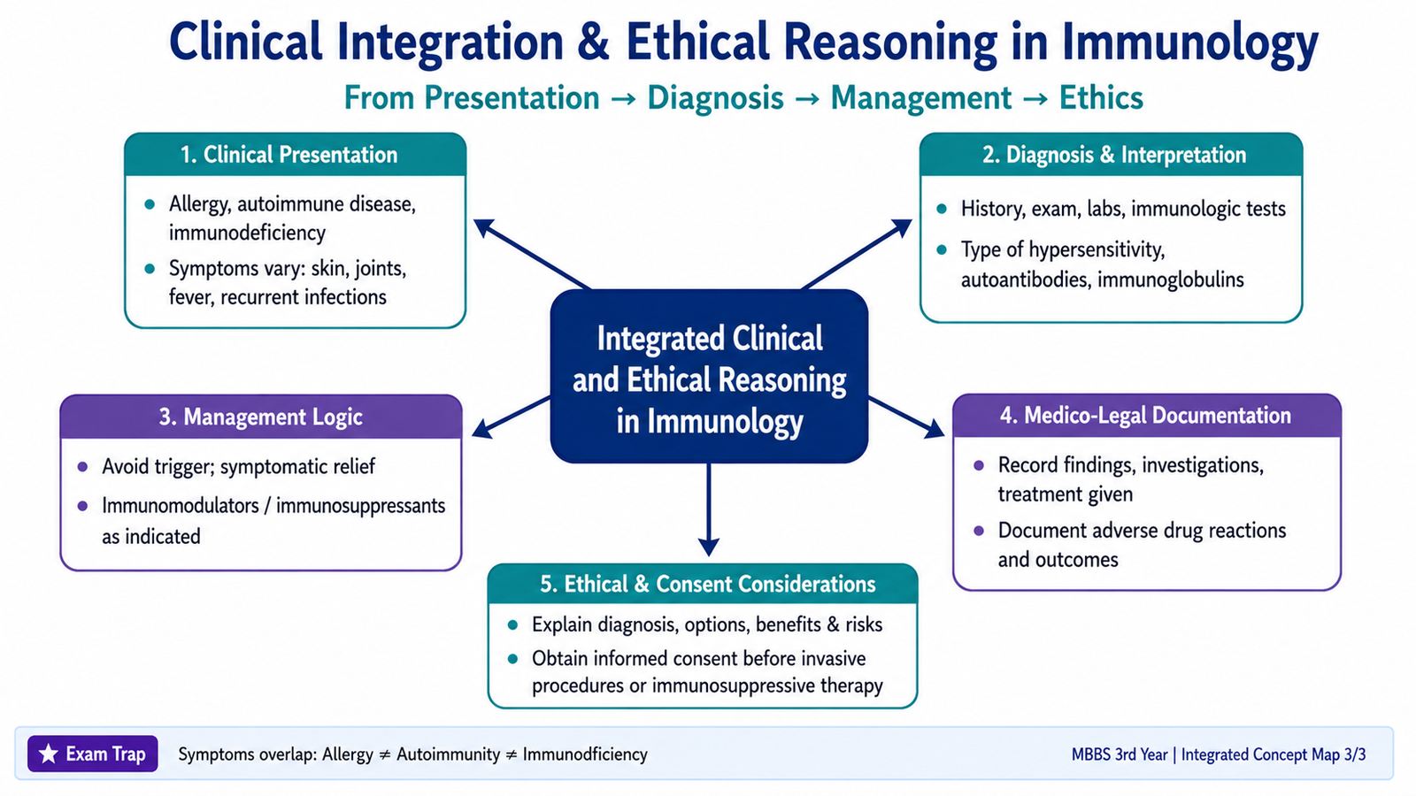
E. Immunodeficiency: Congenital, Acquired and HIV Infection
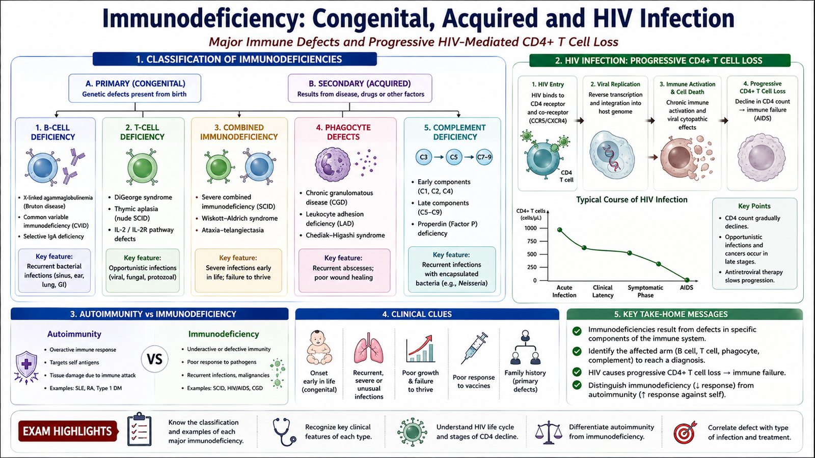
Immunodeficiency is a state in which one or more components of the immune system are absent or function inadequately. The major consequence is increased susceptibility to infection. Depending on the component affected, patients may also develop persistent infections, unusual infections, recurrent infections or an increased risk of certain malignancies.
Congenital or Primary Immunodeficiencies
Primary immunodeficiency disorders result from inherited or developmental defects of the immune system. They can be classified according to the major component affected.
- B-cell or antibody deficiencies: defective antibody production produces particular susceptibility to extracellular bacterial infections. Examples include X-linked agammaglobulinemia and selective IgA deficiency.
- T-cell deficiencies: impaired cell-mediated immunity particularly affects defense against intracellular pathogens. DiGeorge syndrome is an important example.
- Combined immunodeficiencies: both cellular and humoral immunity are severely impaired. Severe combined immunodeficiency is the classic example.
- Phagocyte defects: abnormalities of phagocyte number or function interfere with killing of microbes. Chronic granulomatous disease is an important example.
- Complement deficiencies: loss of complement components may impair opsonization, inflammation or membrane attack complex activity.
Acquired or Secondary Immunodeficiency
Secondary immunodeficiency develops because of another disease, infection, nutritional problem or treatment. Important causes include HIV infection, severe malnutrition, malignancy and immunosuppressive therapy. Secondary immunodeficiencies are more common overall than inherited disorders.
Pathogenesis of HIV Infection
Human immunodeficiency virus mainly causes disease by progressively impairing CD4+ T-cell-mediated immunity. The virus also infects macrophages and related cells, which can contribute to persistence of infection.
The viral envelope protein gp120 attaches to CD4 and commonly uses chemokine co-receptors such as CCR5 or CXCR4. Viral fusion permits entry into the cell. Reverse transcriptase forms viral DNA, which becomes integrated into the host genome. This integrated viral DNA allows persistent infection.
CD4+ T cells are lost through several mechanisms, including direct viral effects, immune-mediated destruction of infected cells and chronic immune activation. As helper T-cell function declines, macrophage activation, cytotoxic T-cell responses and effective B-cell responses also become less coordinated.
The final result is increasing susceptibility to opportunistic infections and certain malignancies.
| Feature | Autoimmune Disease | Immunodeficiency |
|---|---|---|
| Basic problem | Immune response against self | Inadequate immune defense |
| Underlying mechanism | Loss of self-tolerance | Defect or loss of an immune component |
| Main consequence | Inflammation, cell injury or abnormal organ function | Recurrent or unusual infections |
| Example | SLE, Graves disease | SCID, HIV infection |
F. Immunosuppressive Drugs: Corticosteroids, Immunophilin Ligands, Enzyme Inhibitors and Cytotoxic Agents
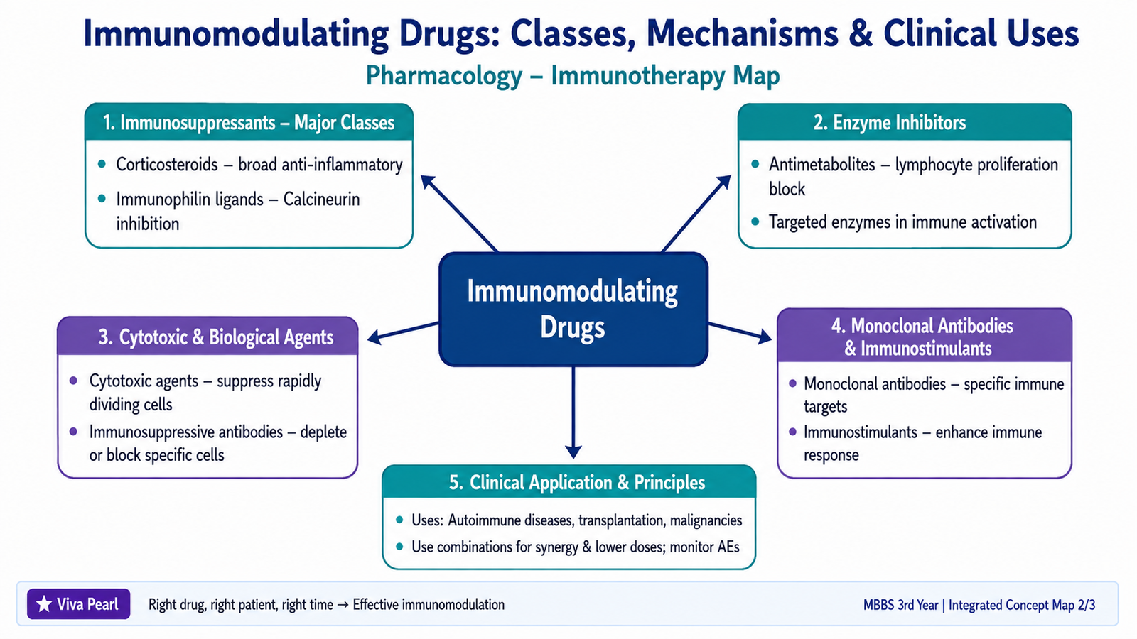
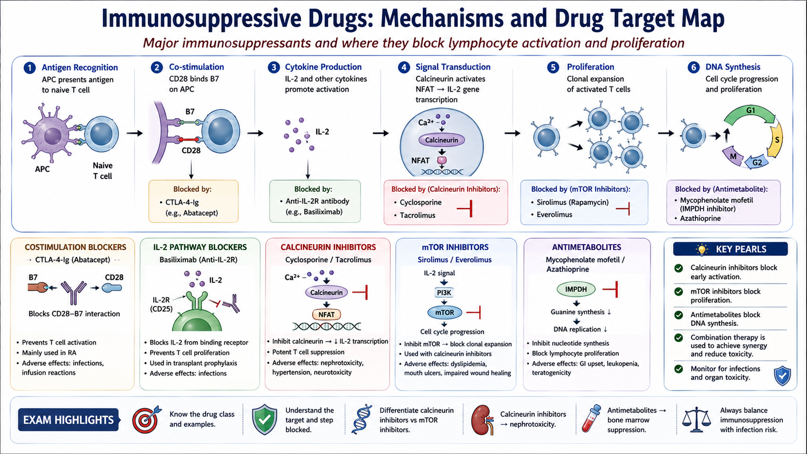
Immunosuppressive drugs reduce immune-cell activation, proliferation or effector function. They are important when immune activity itself is causing harm, particularly in transplantation and autoimmune disease. Different drug classes act at different stages of lymphocyte activation, so understanding their targets explains both their therapeutic effects and their toxicities.
Major Classification
- Corticosteroids
- Immunophilin-binding drugs
- Enzyme and nucleotide-synthesis inhibitors
- Cytotoxic or antimetabolite agents
- Immunosuppressive antibodies and other biological agents
Corticosteroids
Corticosteroids such as prednisolone and methylprednisolone produce broad anti-inflammatory and immunosuppressive effects. Their intracellular receptors alter gene transcription and reduce production of many inflammatory cytokines. They also reduce lymphocyte activation, macrophage function and movement of inflammatory cells into tissues.
Clinical uses include autoimmune and inflammatory diseases and prevention or treatment of transplant-related immune reactions.
Important adverse effects with substantial or prolonged exposure include:
- increased susceptibility to infection;
- hyperglycemia;
- hypertension and fluid-related effects;
- osteoporosis;
- muscle wasting and skin changes; and
- suppression of the hypothalamic-pituitary-adrenal axis.
Cyclosporine and Tacrolimus
Cyclosporine binds the immunophilin cyclophilin, whereas tacrolimus binds FK-binding protein. The drug-protein complexes inhibit calcineurin. Calcineurin is required for activation of transcription factors involved in IL-2 production. Reduced IL-2 formation therefore suppresses T-cell activation and proliferation.
Both drugs are important in transplantation and selected immune-mediated diseases. A major toxicity of both is nephrotoxicity. Hypertension and neurotoxicity may also occur. Cyclosporine can characteristically cause gingival overgrowth and increased hair growth, while tacrolimus is more associated with disturbances of glucose metabolism.
Sirolimus
Sirolimus also binds FK-binding protein, but unlike tacrolimus it does not inhibit calcineurin. Its complex inhibits mTOR, thereby interfering with the response of T cells to IL-2 and reducing lymphocyte proliferation.
Important adverse effects include bone-marrow suppression, hyperlipidemia and impaired wound healing. Its mechanism therefore differs clearly from that of calcineurin inhibitors.
Mycophenolate Mofetil
Mycophenolate is converted to mycophenolic acid, which inhibits inosine monophosphate dehydrogenase. This decreases de novo guanine nucleotide synthesis. Activated T and B lymphocytes depend strongly on this pathway, so their proliferation is selectively reduced.
It is used particularly as part of combination immunosuppression after transplantation and in selected autoimmune diseases. Major adverse effects include gastrointestinal disturbance, bone-marrow suppression and increased susceptibility to infection.
Azathioprine and Other Cytotoxic or Antimetabolite Drugs
Azathioprine is converted to metabolites related to 6-mercaptopurine. These interfere with purine synthesis and therefore inhibit proliferation of rapidly dividing cells, including activated lymphocytes. Important toxicities include bone-marrow suppression, infection and hepatotoxicity.
An important interaction occurs because drugs that inhibit xanthine oxidase can reduce the metabolism of 6-mercaptopurine-related metabolites and markedly increase toxicity.
Cyclophosphamide is an alkylating drug that suppresses rapidly proliferating immune cells. It may be used when strong immunosuppression is needed in severe autoimmune disease. Major toxicities include bone-marrow suppression, infection, infertility and hemorrhagic cystitis.
Methotrexate is an antimetabolite with important immunomodulatory effects and is widely used in selected autoimmune and inflammatory disorders. Major toxicities include bone-marrow suppression, mucosal injury, hepatotoxicity and fetal toxicity.
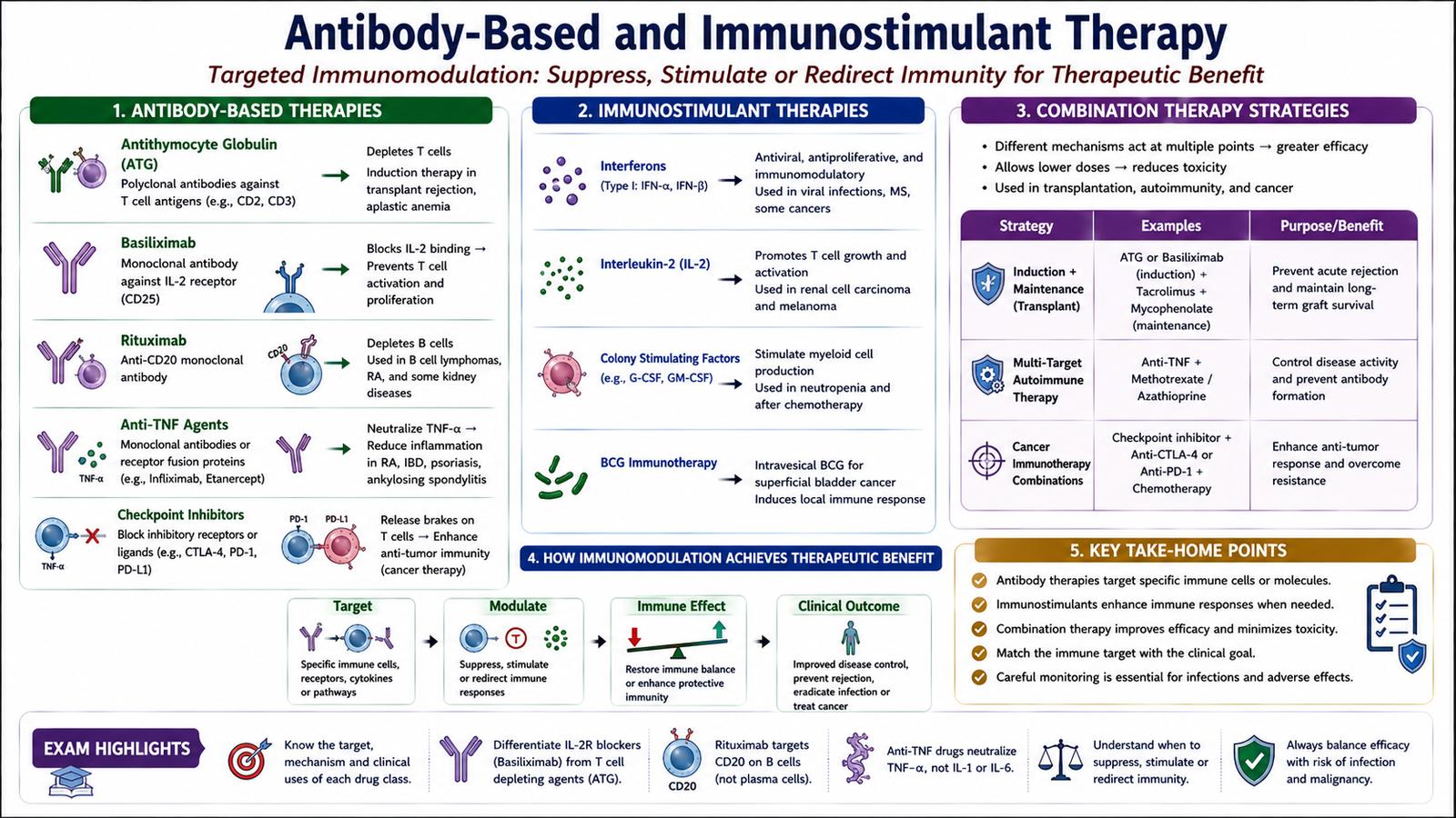
G. Antibody-Based and Immunostimulant Therapy, Combination Strategies and Therapeutic Rationale
Modern immunomodulation can target specific immune cells, cytokines or regulatory pathways. Antibody-based drugs may suppress a selected immune response more precisely than conventional cytotoxic drugs. Other agents stimulate immune function and are useful when increasing a particular immune response provides therapeutic benefit.
Immunosuppressive Antibodies
Antithymocyte globulin contains antibodies directed against multiple antigens on human T lymphocytes. Binding promotes depletion and functional suppression of T cells. It is used particularly for strong immunosuppression in transplantation.
Important adverse effects include:
- infusion reactions;
- cytokine-related systemic reactions;
- serum-sickness-type reactions;
- leukopenia or thrombocytopenia; and
- increased susceptibility to infection.
Basiliximab is a monoclonal antibody directed against the IL-2 receptor alpha chain, also called CD25, on activated T lymphocytes. Blocking this receptor interferes with IL-2-driven T-cell proliferation and can therefore reduce activation of the immune response after transplantation.
Selected Monoclonal Antibodies
Rituximab binds CD20 on B lymphocytes and causes B-cell depletion. It is used in selected B-cell malignancies and autoimmune diseases. Important adverse effects include infusion reactions and increased risk of infection.
Anti-TNF monoclonal antibodies such as infliximab and adalimumab neutralize tumor necrosis factor and reduce inflammatory signaling. They are useful in several chronic immune-mediated inflammatory diseases. Because TNF is important in host defense, treatment can increase susceptibility to serious infection, including reactivation of latent infection.
Monoclonal antibodies can also be used to increase antitumor immunity. Immune checkpoint inhibitors block inhibitory signals that normally restrain T cells. For example, antibodies directed against PD-1 or CTLA-4 can enhance T-cell responses against malignant cells.
Immunostimulant Drugs
Immunostimulants increase selected immune functions rather than suppress them. Their actions differ according to the pathway targeted.
- Interferons: cytokines that can produce antiviral, antiproliferative and immunomodulatory effects. Different interferons have different therapeutic applications.
- Interleukin-2: promotes activation and proliferation of T lymphocytes and can enhance immune responses against selected malignancies.
- Colony-stimulating factors: stimulate production of selected leukocyte populations and can help restore neutrophil numbers when bone-marrow function is suppressed.
- BCG: can stimulate a local antitumor immune response when used in selected bladder malignancies.
Why Immunomodulation Is Used in Autoimmune Disease
In autoimmune disease, the therapeutic goal is to reduce the damaging immune response sufficiently to protect tissues while preserving as much protective immunity as possible.
Broad agents such as corticosteroids suppress multiple pathways, whereas targeted drugs such as monoclonal antibodies interfere with specific cells or mediators. Targeted therapy may therefore be chosen when a particular immune pathway has an important role in the disease.
Why Immunomodulation Is Used in Malignancy
Malignant cells may evade immune destruction by reducing immune recognition or by activating inhibitory pathways. Immunotherapy can restore or strengthen antitumor immune responses. Alternatively, monoclonal antibodies can directly identify molecules expressed by malignant cells and help eliminate those cells.
Advantages and Disadvantages of Combination Therapy
Immunomodulating drugs are frequently combined because different agents act at different points in the immune response.
Advantages include:
- suppression of several immune pathways at the same time;
- greater therapeutic effectiveness;
- ability to reduce reliance on a very high dose of a single drug; and
- better control of complex immune reactions such as graft rejection.
Disadvantages include:
- greater susceptibility to infection;
- possible increase in malignancy risk with prolonged profound immunosuppression;
- overlapping organ toxicity or bone-marrow suppression;
- drug interactions; and
- greater monitoring and treatment complexity.
⭐ AIM High-Yield Review
- Type I hypersensitivity is an IgE- and mast-cell-mediated immediate reaction.
- Type II hypersensitivity involves IgG or IgM against cellular or tissue antigens and may cause cell destruction, inflammation or receptor dysfunction.
- Type III hypersensitivity results from deposition of antigen-antibody immune complexes followed by complement activation and inflammation.
- Type IV hypersensitivity is T-cell mediated and is the classic delayed hypersensitivity mechanism.
- Central tolerance removes or modifies self-reactive lymphocytes during development; peripheral tolerance uses anergy, regulatory T cells and deletion.
- ⭐ Hyperacute graft rejection is caused by pre-existing antibodies and produces vascular thrombosis and rapid graft failure.
- Chronic rejection is characterized particularly by progressive graft vascular narrowing and fibrosis.
- ⭐ In GVHD, donor immune cells attack recipient tissues; in ordinary graft rejection, recipient immunity attacks the graft.
- Autoimmunity develops when self-tolerance fails in a genetically susceptible person, often with additional environmental triggers.
- Immunodeficiency may involve B cells, T cells, combined immunity, phagocytes or complement and may be congenital or acquired.
- ⭐ HIV progressively impairs immunity mainly through infection, dysfunction and loss of CD4+ T cells.
- Cyclosporine and tacrolimus inhibit calcineurin and reduce IL-2 production; nephrotoxicity is a major adverse effect.
- Sirolimus inhibits mTOR and blocks IL-2-driven lymphocyte proliferation rather than inhibiting calcineurin.
- Mycophenolate inhibits inosine monophosphate dehydrogenase and restricts guanine nucleotide synthesis in activated lymphocytes.
- ⭐ Combination immunomodulatory therapy can improve efficacy but increases the risks of infection, toxicity and drug interactions.
Hypersensitivity, Immune Tolerance, Autoimmunity, Immunodeficiency & Immunomodulatory Drugs
Use these focused videos after reading the AIM Learning Material to reinforce the major immunology mechanisms and pharmacological targets.
Immune Tolerance, Allergy and Hypersensitivity
Focus on immune tolerance, failure of self-tolerance, allergy, immediate and delayed hypersensitivity, anaphylaxis, transplant rejection and immunodeficiency.
Immunosuppressants — Mechanisms Made Easy
Focus on T-cell activation, calcineurin inhibitors, mTOR inhibitors, antimetabolites, corticosteroids and antibody-based immunosuppressive therapy.
HIV — Life Cycle, Pathophysiology and AIDS
Focus on viral entry, replication, integration, progressive immune-system damage and development of HIV-associated immunodeficiency.