
Forensic Hematology: Blood Groups, Bloodstains and Biological Trace Evidence
Topic Introduction
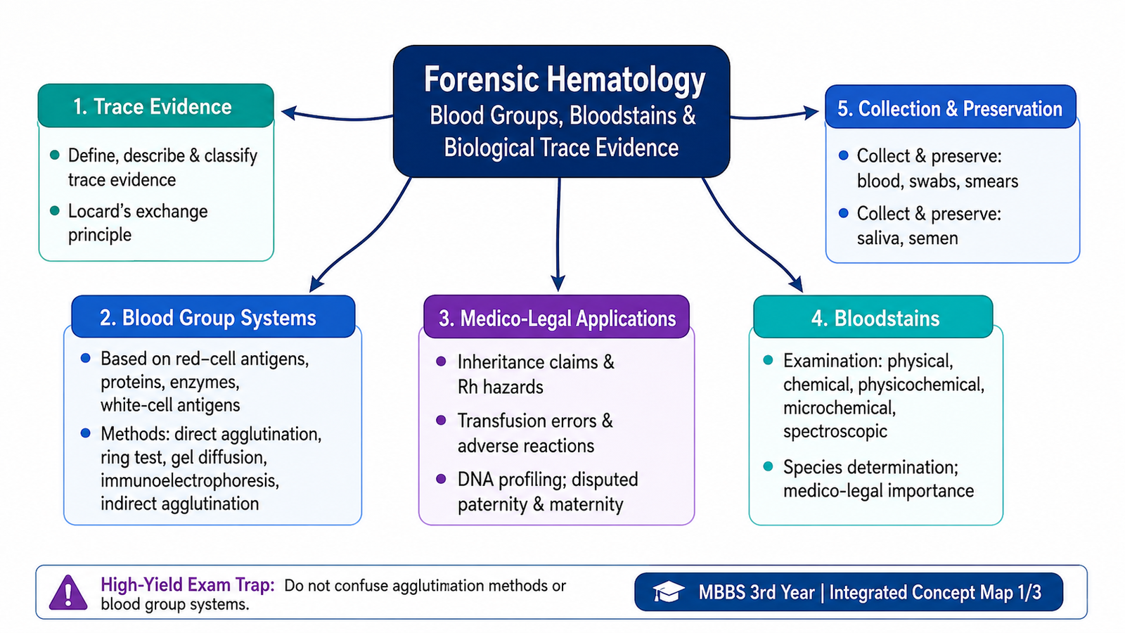
Forensic hematology applies knowledge of blood and other biological materials to medico-legal investigation. A small bloodstain, saliva deposit or semen stain may become important trace evidence when it helps associate a person, object or place with an event. Blood can be examined at several levels: its physical appearance, chemical properties, cellular composition, inherited blood-group markers and, where appropriate, DNA. Blood-group evidence has important historical and practical applications, but its value must always be interpreted carefully because many individuals may share the same blood group. In this chapter, you will learn how trace evidence is classified, how blood groups are inherited and detected, how suspected bloodstains are examined, how species origin may be investigated, and how biological specimens should be collected and preserved without contamination.
A. Trace Evidence and Locard’s Exchange Principle
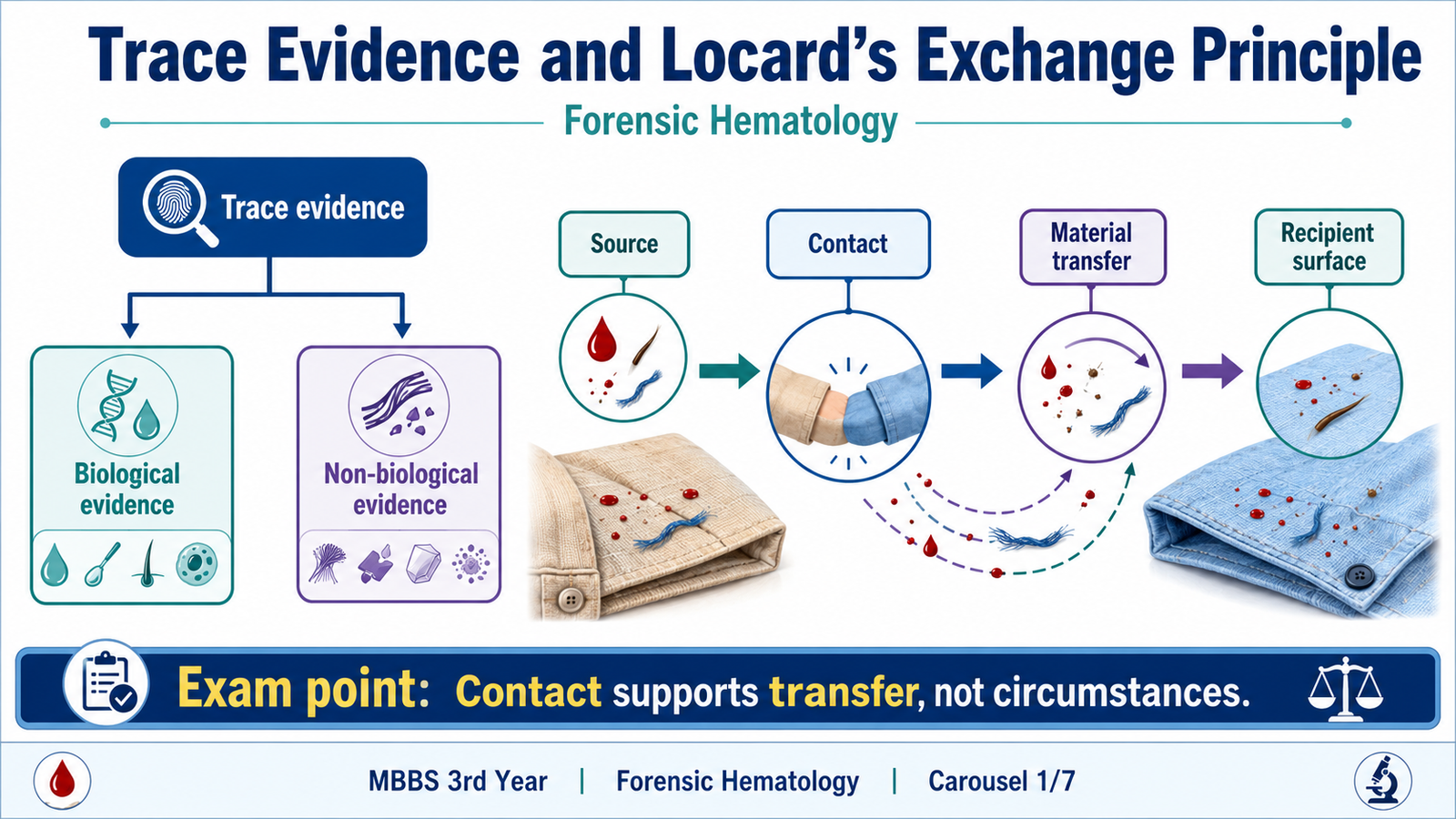
Trace evidence is a small quantity of material transferred during contact between people, objects or environments. Its importance does not depend simply on its size. Even a minute biological or non-biological material may provide useful information when it is collected correctly and interpreted in the context of an investigation.
Definition and classification
Trace evidence may broadly be divided into biological and non-biological material. Biological trace evidence originates from living organisms and may contain cells, proteins, enzymes, antigens or genetic material. Non-biological trace evidence comes from materials or objects in the environment.
- Biological trace evidence: blood, saliva, semen, hair and other biological material.
- Non-biological trace evidence: fibres, glass fragments, paint, soil and similar transferred material.
In forensic hematology, blood is especially important because it may provide information through its appearance, cellular components, blood-group markers and DNA-containing cells.
Locard’s exchange principle
Locard’s exchange principle states that contact between two persons, objects or environments can result in an exchange of material. In simple terms, contact may leave something behind and may also carry something away.
For example, blood transferred to clothing may support contact between the clothing and a bleeding source. However, the presence of a trace should not automatically be treated as proof of how, when or why the contact occurred. The evidentiary meaning depends on the circumstances, location of the material, laboratory findings and proper handling of the specimen.
B. Blood as Biological Trace Evidence
Blood is a specialized fluid connective tissue composed of a liquid portion, plasma, and formed cellular elements. Understanding its basic composition helps explain why different parts of blood can provide different kinds of forensic information.
Plasma
Plasma is the fluid component of blood. It contains water, electrolytes and several proteins. Some inherited variations in blood proteins have historically been used as forensic markers because different individuals may possess different genetically determined forms of these proteins.
Red blood cells
Red blood cells are the most numerous formed elements of blood. Mature red cells do not contain nuclei. Their membranes carry important inherited antigens, including those responsible for the ABO and Rh blood-group systems. This explains why red-cell antigens can be tested for blood grouping. A reaction occurs when a red-cell antigen meets its corresponding antibody under appropriate testing conditions.
White blood cells
White blood cells are nucleated cells. Their nuclei contain DNA, making leukocytes an important source of nuclear genetic material in blood. White blood cells also possess inherited surface antigens, including human leukocyte antigens, which historically had value in biological relationship testing before modern DNA profiling became dominant.
Platelets
Platelets are small cytoplasmic fragments derived from megakaryocytes. Their principal biological role is in hemostasis. Unlike leukocytes, platelets do not contain a nucleus and therefore are not the main source of nuclear DNA in a blood sample.

C. Blood-Group Systems and Their Genetic Basis
Blood-group characteristics are inherited. This means that the antigens, proteins or enzyme variants detected in an individual are influenced by genes received from the parents. Forensic interpretation therefore depends on understanding basic inheritance and recognizing that a blood group is a class characteristic: many unrelated people may share the same group.
Basic genetic principles
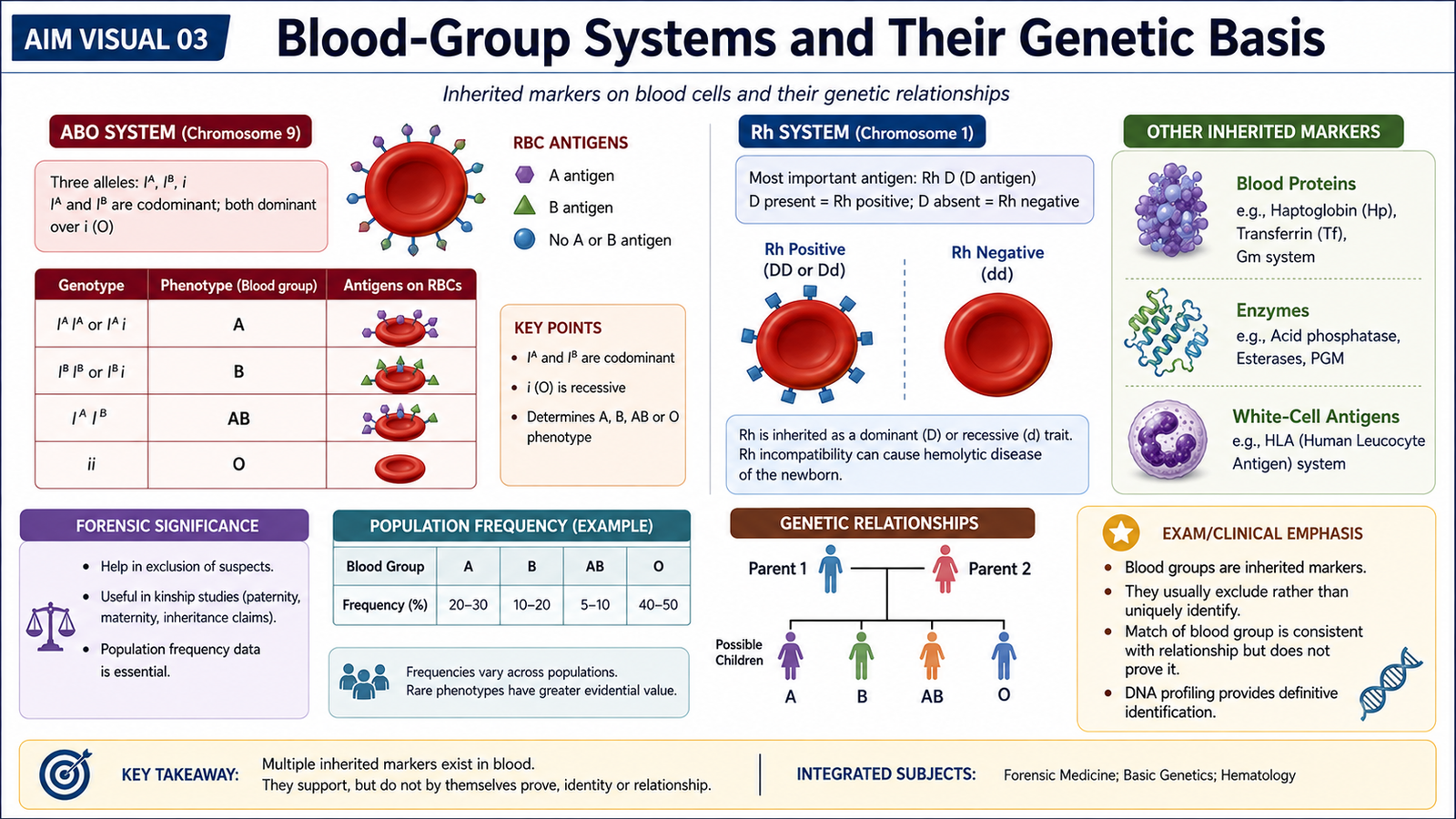
A person usually receives one allele at a particular genetic locus from each parent. Alleles may be dominant, recessive or codominant depending on the system being considered. The ABO system is the classic example. The A and B alleles are codominant, meaning that if both are inherited, both are expressed and the phenotype is AB. The O allele does not produce A or B antigen and is recessive to A and B.
- Group A: A antigen is expressed on red cells.
- Group B: B antigen is expressed on red cells.
- Group AB: both A and B antigens are expressed.
- Group O: neither A nor B antigen is expressed.
The Rh system contains several antigens, of which the D antigen is the most important in routine classification. A person whose red cells express D antigen is described as Rh positive, while absence of detectable D antigen is described as Rh negative.
Different inherited blood markers
Forensic blood grouping is broader than ABO and Rh alone. Inherited biological variation can be demonstrated in red-cell antigens, plasma or serum proteins, enzymes and white-cell antigens. Before DNA profiling, combinations of these polymorphic markers increased the ability to distinguish between individuals or to exclude an alleged biological relationship.
| Marker category | Main location | Examples / concept | Forensic relevance |
|---|---|---|---|
| Red-cell antigens | Red-cell membrane | ABO, Rh and other inherited antigen systems | Blood grouping and exclusionary comparisons |
| Blood proteins | Plasma or serum | Inherited protein polymorphisms | Historical individualization and relationship studies |
| Enzymes | Blood cells or plasma | Genetically determined enzyme variants | Additional inherited markers |
| White-cell antigens | Leukocyte surface | Human leukocyte antigen systems | Historically useful in relationship testing |
These markers do not usually identify one unique person by themselves. Their value increases when several independent inherited systems are considered together. Modern DNA profiling provides much greater discriminatory power.
D. Methods of Blood-Group Determination
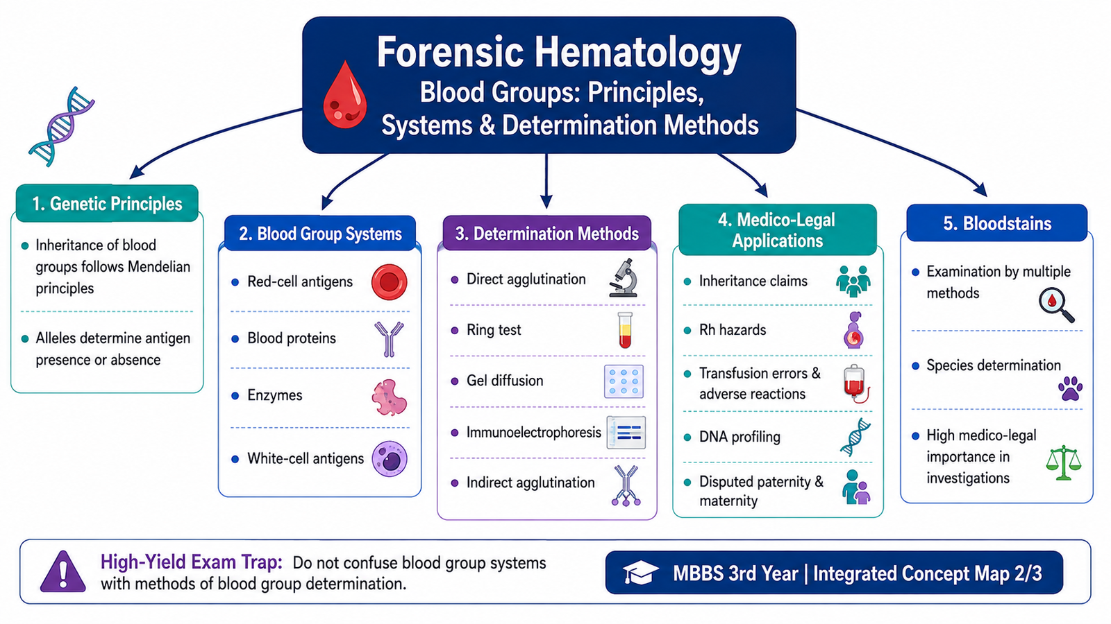
Blood-group determination depends on detecting a specific antigen, antibody, protein or other inherited marker. Different laboratory methods are used because not all markers behave in the same way. Some are present directly on intact cells, while others are soluble proteins or require an indirect system to make the reaction visible.
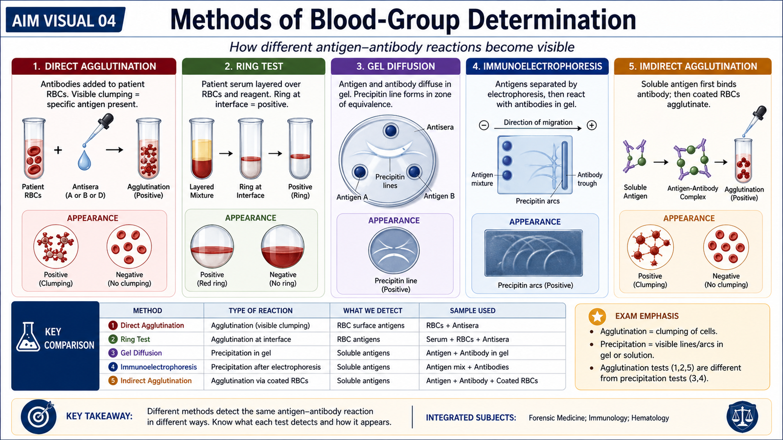
Direct agglutination
Agglutination means visible clumping of cells or particles caused by an antigen-antibody reaction. In direct blood grouping, red cells carrying a particular surface antigen are mixed with a known antibody directed against that antigen.
For example, if red cells agglutinate with anti-A reagent, this indicates the presence of A antigen on those cells. The same principle underlies conventional antigen-based blood-group testing.
Ring test
The ring test demonstrates an antigen-antibody reaction at the interface between two liquid layers. When a soluble antigen and its corresponding antibody react under suitable conditions, a visible ring of precipitate may appear where the two meet. This is a precipitation reaction rather than clumping of intact red cells. It is therefore useful for understanding tests involving soluble antigen-antibody systems.
Gel diffusion
In gel diffusion, antigen and antibody move through a gel medium. When they meet in appropriate proportions, they form a visible precipitation line. The position and pattern of the line demonstrate that a specific antigen-antibody reaction has occurred.
Immunoelectrophoresis
Immunoelectrophoresis combines two principles. First, proteins are separated in an electric field according to their physical and electrical properties. They are then allowed to react with specific antibodies. The resulting precipitation pattern helps demonstrate particular protein components.
Indirect agglutination
Some antigens are soluble and do not naturally form visible clumps. In indirect or passive agglutination, a soluble antigen or antibody is attached to carrier particles so that an otherwise invisible antigen-antibody reaction can be converted into visible agglutination. The important principle is therefore the same: a specific immunological reaction is made visible, but the method used depends on the physical form of the marker being detected.
E. Medico-Legal Applications of Blood Groups
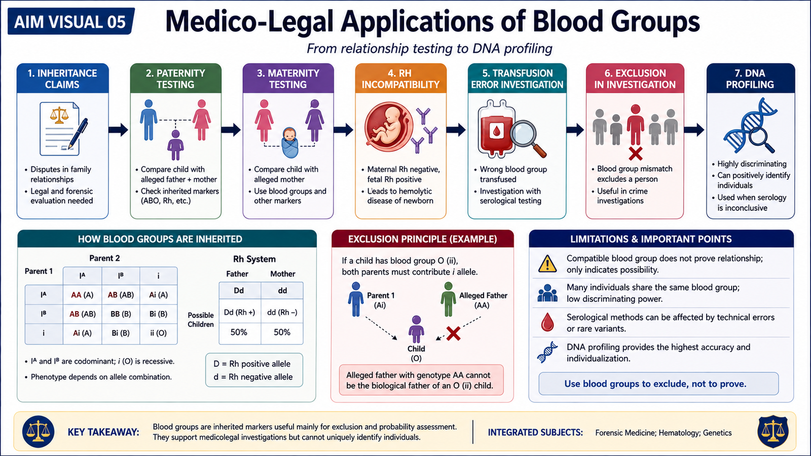
Blood-group evidence has several medico-legal applications because blood-group characteristics are inherited and can be compared between individuals. Its greatest value is often exclusion. If an inherited marker is incompatible with a claimed biological relationship or with a questioned blood source, that incompatibility may be important. A compatible blood group, however, does not by itself prove identity because the same group may occur in many people.
Inheritance claims
In disputes involving biological inheritance, inherited blood-group characteristics can be compared among family members. The interpretation is based on whether the observed phenotype could reasonably have resulted from the parental combinations being considered. If a child’s inherited marker is incompatible with the alleged parental combination, the relationship may be excluded by that system. If it is compatible, the finding only shows that the relationship is possible; it does not establish it conclusively.
Disputed paternity and maternity
The same genetic principle applies to disputed paternity and maternity. Historically, ABO and other inherited blood markers were used to assess whether an alleged parent could have contributed the child’s inherited characteristics.
Rh hazards
Rh status becomes important when an Rh-negative individual is exposed to Rh-positive red cells. Such exposure may stimulate formation of antibodies against the D antigen. Once sensitization has occurred, later exposure to Rh-positive cells can produce clinically important immune reactions. Therefore, inherited Rh status has significance in transfusion medicine and in understanding maternal-fetal blood-group incompatibility.
Transfusion errors and adverse reactions
Correct blood grouping is essential before transfusion because incompatible red-cell antigens and antibodies can react with one another. If incompatible blood is transfused, antibodies may bind to donor red cells and cause agglutination and destruction of those cells.
In a medico-legal investigation of a suspected transfusion error, documentation of patient identity, donor-unit identity, blood grouping and compatibility testing may therefore be important.
DNA profiling
Conventional blood groups classify a person into relatively broad inherited categories. DNA profiling examines highly variable genetic regions and provides much greater discriminatory power. In blood, nucleated white blood cells provide the major source of nuclear DNA. DNA profiling can therefore be used to compare biological material with individuals and to investigate biological relationships with much greater precision than conventional blood grouping alone.
F. Examination and Medico-Legal Importance of Bloodstains
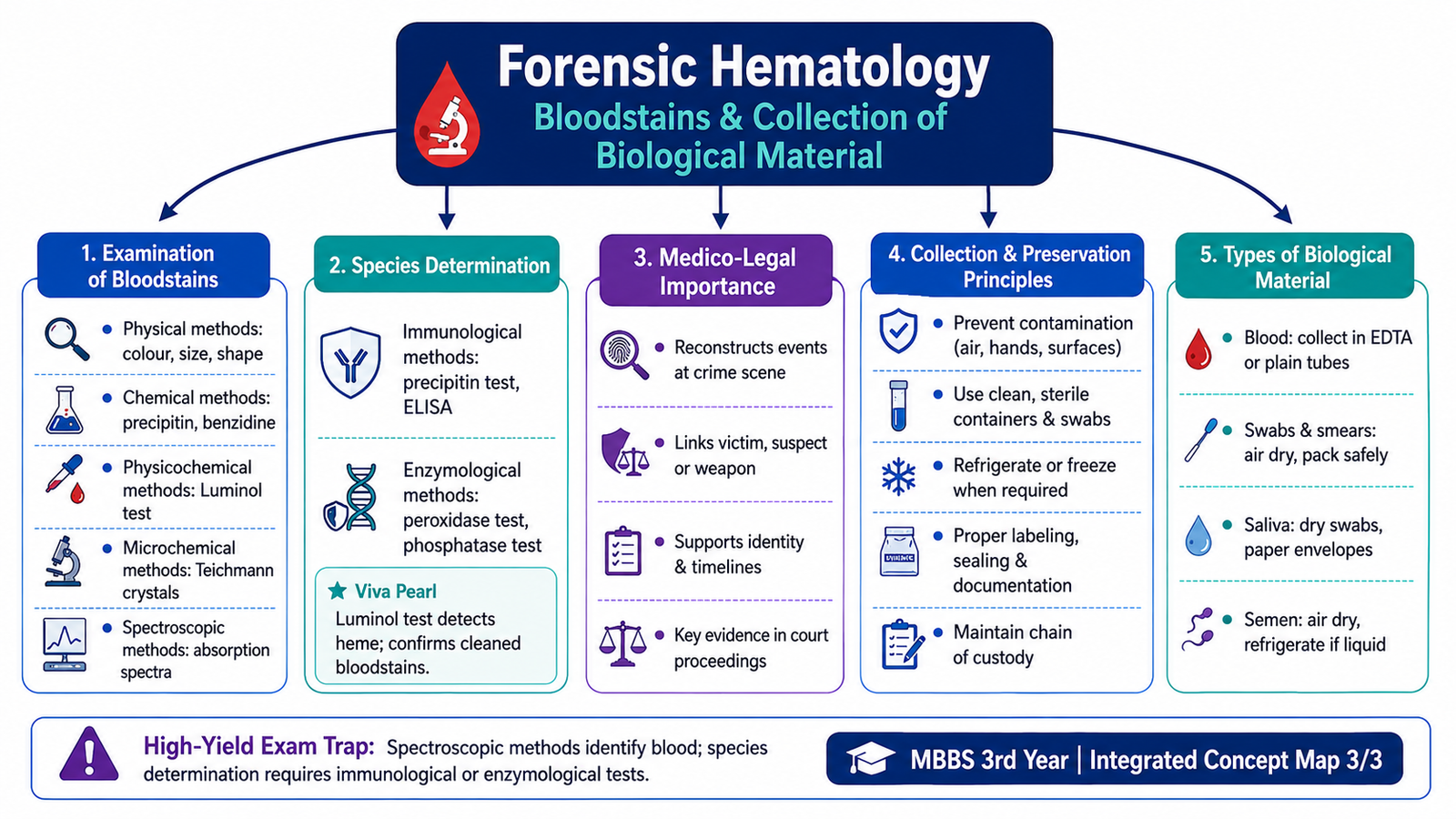
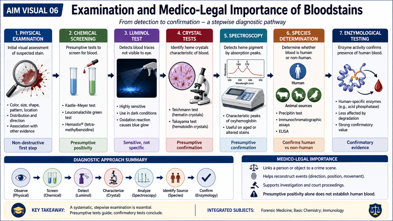
A suspected bloodstain cannot be interpreted from appearance alone. Forensic examination proceeds from observation to laboratory testing. The main questions are whether the material could be blood, whether blood can be confirmed, whether its biological origin can be assessed, and what additional information may reasonably be obtained from it.
Physical examination
Physical examination records the visible characteristics of a suspected bloodstain before laboratory testing changes or consumes the material. Important observations include its location, colour, size, shape, distribution and relationship to the surface on which it is found. Fresh blood is typically red. As a stain ages and hemoglobin undergoes chemical change, it commonly becomes darker and may appear reddish-brown or brown. Colour alone cannot reliably determine the exact age of a stain because environmental conditions influence its appearance. The shape and distribution of stains may provide contextual information about deposition or movement, but such patterns should be interpreted cautiously and together with other evidence.
Chemical methods
Chemical screening methods commonly depend on the activity of the heme component of hemoglobin, which can promote oxidation reactions that produce a visible colour change. These tests are useful for detecting possible blood, including stains that are not obvious by simple inspection. Examples of presumptive chemical approaches include colour reactions such as the phenolphthalein-based test and other oxidation-reduction tests historically used for blood detection. A positive presumptive test indicates that blood may be present, but it is not automatically proof of human blood because some other substances may produce similar reactions.
Physicochemical methods
Physicochemical methods combine a chemical reaction with a physical signal that makes a suspected stain easier to locate or examine. A well-known example is a luminescence-based test such as luminol, in which a reaction associated with heme can produce visible light in appropriate conditions. Such methods are especially useful for locating small or poorly visible suspected blood traces. The result remains a screening finding and should be followed by appropriate confirmatory and interpretive testing.
Microchemical methods
Microchemical or crystal tests demonstrate characteristic crystalline derivatives of hemoglobin under microscopic examination. Classical examples include Teichmann hemin crystals and Takayama hemochromogen crystals. Their importance is that hemoglobin is converted into recognizable crystalline products. Demonstration of characteristic crystals provides stronger support for the presence of blood than simple colour-based screening alone.
Spectroscopic methods
Hemoglobin and its derivatives absorb light at characteristic wavelengths. Spectroscopic examination evaluates these absorption patterns. Because different derivatives of hemoglobin produce characteristic spectra, spectroscopy can support the identification of blood pigments in a suspected stain.
Immunological methods for species determination
After confirming that a stain is blood, an important forensic question may be whether it is of human or non-human origin. Immunological methods use the specificity of antigen-antibody reactions to investigate the species origin of blood. The classical principle is demonstrated by the precipitin reaction. Proteins from the questioned blood are reacted with antibodies directed against proteins of a particular species. Formation of a specific precipitate supports the presence of proteins corresponding to that species.
Enzymological methods
Enzymological examination investigates enzyme activity or genetically determined enzyme variants present in biological material. Such systems may provide additional information about a blood sample, although their discriminatory value is considerably lower than modern DNA profiling.
| Method | Main principle | Main information |
|---|---|---|
| Physical | Visual examination of stain characteristics | Location, colour, shape and distribution |
| Chemical | Hemoglobin-associated chemical reactions | Presumptive evidence of blood |
| Physicochemical | Chemical reaction producing a physical signal | Localization of suspected blood traces |
| Microchemical | Formation of characteristic hemoglobin-derived crystals | Support for confirmation of blood |
| Spectroscopic | Characteristic light absorption of hemoglobin derivatives | Identification of blood pigments |
| Immunological | Specific antigen-antibody reaction | Species determination |
| Enzymological | Study of enzyme activity or inherited variants | Additional biological characterization |
Medico-legal importance of bloodstains
Bloodstains may provide several types of information when properly examined. They may establish that blood is present, support assessment of its species origin, permit blood-group or genetic examination when sufficient material remains, and help associate biological material with a person, object or location.
- May demonstrate the presence of blood on an object or surface.
- May help distinguish human from non-human blood.
- May permit conventional blood-group examination in suitable material.
- May provide leukocyte-derived DNA for profiling when usable genetic material is present.
- May support reconstruction of contact or events when interpreted together with other evidence.
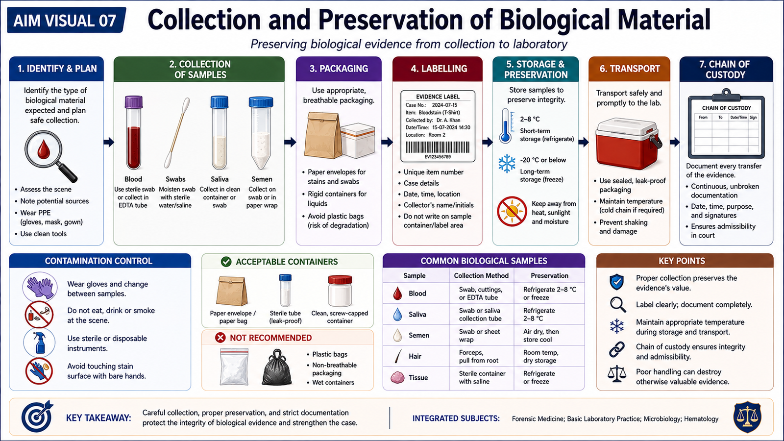
G. Collection and Preservation of Biological Material
The evidentiary value of biological material depends not only on laboratory technology but also on how the specimen is collected and preserved. Poor handling can introduce contamination, mix samples from different sources, promote degradation or make it difficult to prove that the specimen tested in the laboratory is the same specimen that was originally collected.
General principles
Collection should preserve the biological material while protecting its identity and integrity. Each specimen should be handled with clean appropriate equipment, packaged separately where cross-contamination is possible, clearly identified and sealed in a manner that allows subsequent handling to be documented.
- Avoid contamination from the collector, surrounding surfaces or other specimens.
- Use a separate clean collection method for each independent sample.
- Keep specimens from different sources separate.
- Label each specimen clearly with relevant identifying information.
- Preserve the sample in a condition suitable for the intended examination.
- Seal and document transfer of the specimen to maintain chain of custody.
Blood
Liquid blood should be collected into an appropriate clean specimen container selected according to the intended laboratory examination. The sample must be properly labelled and protected from conditions that may promote degradation or contamination. When blood is present as a stain on clothing or another movable object, the stained item itself may be submitted where appropriate. If a stain must be sampled from a surface, the method should minimize loss of material and prevent transfer from neighbouring areas.
Swabs and smears
Swabs are useful for collecting biological material from body surfaces or objects. The collection method should obtain the questioned material without unnecessarily mixing it with material from another site. Moist biological material is more vulnerable to microbial growth and degradation. Therefore, swabs containing wet biological material are generally allowed to dry appropriately before final dry storage or packaging when the testing method permits. Each swab should remain individually identified. Smears should be prepared and handled in a way that protects the deposited cellular material from contamination and mechanical damage.
Saliva
Saliva may be present on surfaces or objects that have contacted the mouth. It can contain epithelial cells and therefore may provide genetic material. Collection should target the relevant area using a clean technique and should avoid contamination from handling or nearby surfaces. After collection, the specimen should be preserved in a condition that limits degradation and permits the intended laboratory examination.
Semen
Semen may be recovered as liquid material, a swab specimen or a dried stain. Collection should preserve both cellular and non-cellular components that may be relevant to laboratory examination. Wet material should not be sealed in a way that encourages moisture-related deterioration. Dried material should be protected from contamination, loss and unnecessary handling. Separate items should remain separately packaged and labelled.
Chain of custody
Chain of custody is the documented history of possession, transfer and handling of an evidentiary specimen. Its purpose is to demonstrate continuity from collection to examination and, where required, presentation in medico-legal proceedings.
Even an excellent laboratory result may lose evidentiary value if the sample cannot be reliably linked to its source because of poor identification, contamination or undocumented handling.
⭐ AIM High-Yield Review
- Trace evidence is small transferred material that may associate a person, object or place with an event.
- Locard’s exchange principle: contact can result in transfer of material between interacting surfaces or persons.
- Blood contains plasma, red cells, white cells and platelets; each has different forensic relevance.
- ⭐ Red-cell membranes carry important blood-group antigens, whereas nucleated leukocytes are a major source of nuclear DNA.
- In the ABO system, A and B are codominant; O is recessive to A and B.
- Inherited forensic blood markers include red-cell antigens, blood proteins, enzymes and white-cell antigens.
- Direct agglutination demonstrates antigen-antibody reactions through visible clumping of cells.
- Ring tests and gel diffusion are based on precipitation between soluble antigen and antibody.
- ⭐ Conventional blood-group evidence is particularly useful for exclusion; compatibility alone does not prove identity or parentage.
- An incompatible transfusion may cause an antigen-antibody reaction leading to red-cell agglutination and hemolysis.
- Bloodstain examination may use physical, chemical, physicochemical, microchemical, spectroscopic, immunological and enzymological methods.
- Teichmann and Takayama tests demonstrate characteristic hemoglobin-derived crystals.
- The classical precipitin reaction uses antigen-antibody specificity to help determine species origin.
- ⭐ A positive presumptive blood test does not by itself prove that the material is human blood or identify its source.
- Correct collection, separate packaging, labelling, preservation and chain of custody protect the evidentiary value of blood, swabs, saliva and semen.
Forensic Hematology: Blood Groups, Bloodstains and Biological Trace Evidence
Watch these videos in sequence after reading the AIM Learning Material. Together they reinforce trace evidence and Locard’s principle, blood-group determination, examination of bloodstains, and biological-evidence handling.
Trace Evidence & Locard’s Exchange Principle
Covers definition and types of trace evidence, biological versus non-biological traces, significance of trace evidence, and Locard’s exchange principle.
Techniques for Determination of Blood Group
Reinforces forensic serology and the principles used to determine blood groups, complementing the chapter’s discussion of inherited blood markers and agglutination-based methods.
Examination of Blood Stains — Forensic Serology
Focuses on forensic examination of bloodstains and is the closest video match for the physical, chemical and laboratory examination component of this KMU topic.
Biological Evidence Packaging — NIJ Demonstration
Official National Institute of Justice training demonstration reinforcing contamination prevention and appropriate packaging of biological evidence.