
A. Normal Hemostasis and Coagulation Pathways
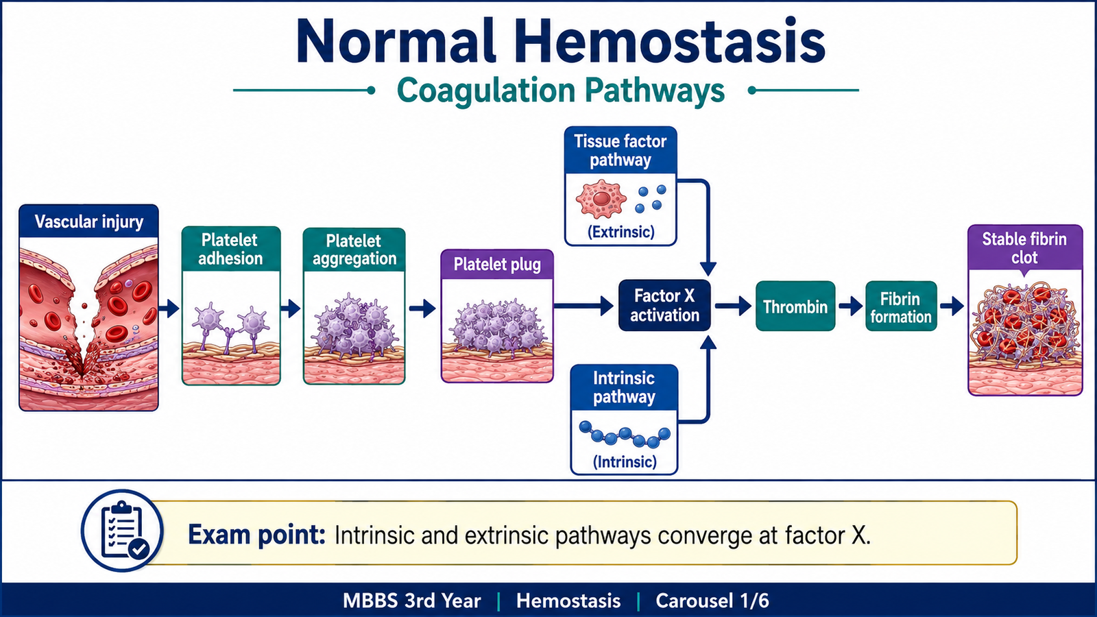
Hemostasis is the normal process that stops bleeding after a blood vessel is injured while keeping blood fluid inside uninjured vessels. It depends on a coordinated interaction between the vessel wall, platelets and plasma coagulation factors. For understanding platelet disorders, von Willebrand disease, hemophilia and disseminated intravascular coagulation, it is useful to first separate hemostasis into primary hemostasis and secondary hemostasis.
Primary hemostasis
Primary hemostasis produces the initial platelet plug. When vascular endothelium is damaged, the underlying extracellular matrix becomes exposed. von Willebrand factor (vWF) binds to exposed subendothelial collagen and provides a bridge for platelet adhesion through the platelet GPIb receptor.
Adherent platelets become activated, change shape and release mediators such as ADP. Platelet activation also promotes formation of thromboxane A2. These signals recruit and activate additional platelets. Activated platelets express functional GPIIb/IIIa receptors, which bind fibrinogen and connect adjacent platelets. This produces platelet aggregation.
Secondary hemostasis
The platelet plug is initially fragile. Secondary hemostasis strengthens it by generating fibrin through the coagulation cascade. Coagulation is traditionally divided into intrinsic, extrinsic and common pathways. Although the pathways interact extensively in the body, this classification remains important for understanding coagulation-factor deficiencies and laboratory tests.
| Pathway | Important Factors | Main Laboratory Test |
|---|---|---|
| Extrinsic | Tissue factor and factor VII | PT |
| Intrinsic | Factors XII, XI, IX and VIII | aPTT |
| Common | Factors X, V, II and fibrinogen | Can affect both PT and aPTT |
Both pathways lead to activation of factor X. Activated factor X contributes to conversion of prothrombin into thrombin. Thrombin then converts fibrinogen into fibrin. Factor XIII stabilizes the fibrin network by cross-linking fibrin strands. Therefore, disorders of platelets and vWF mainly disturb the initial platelet plug, while hemophilia mainly affects fibrin formation.
B. Thrombocytopenia, Immune Thrombocytopenia and Thrombotic Microangiopathies
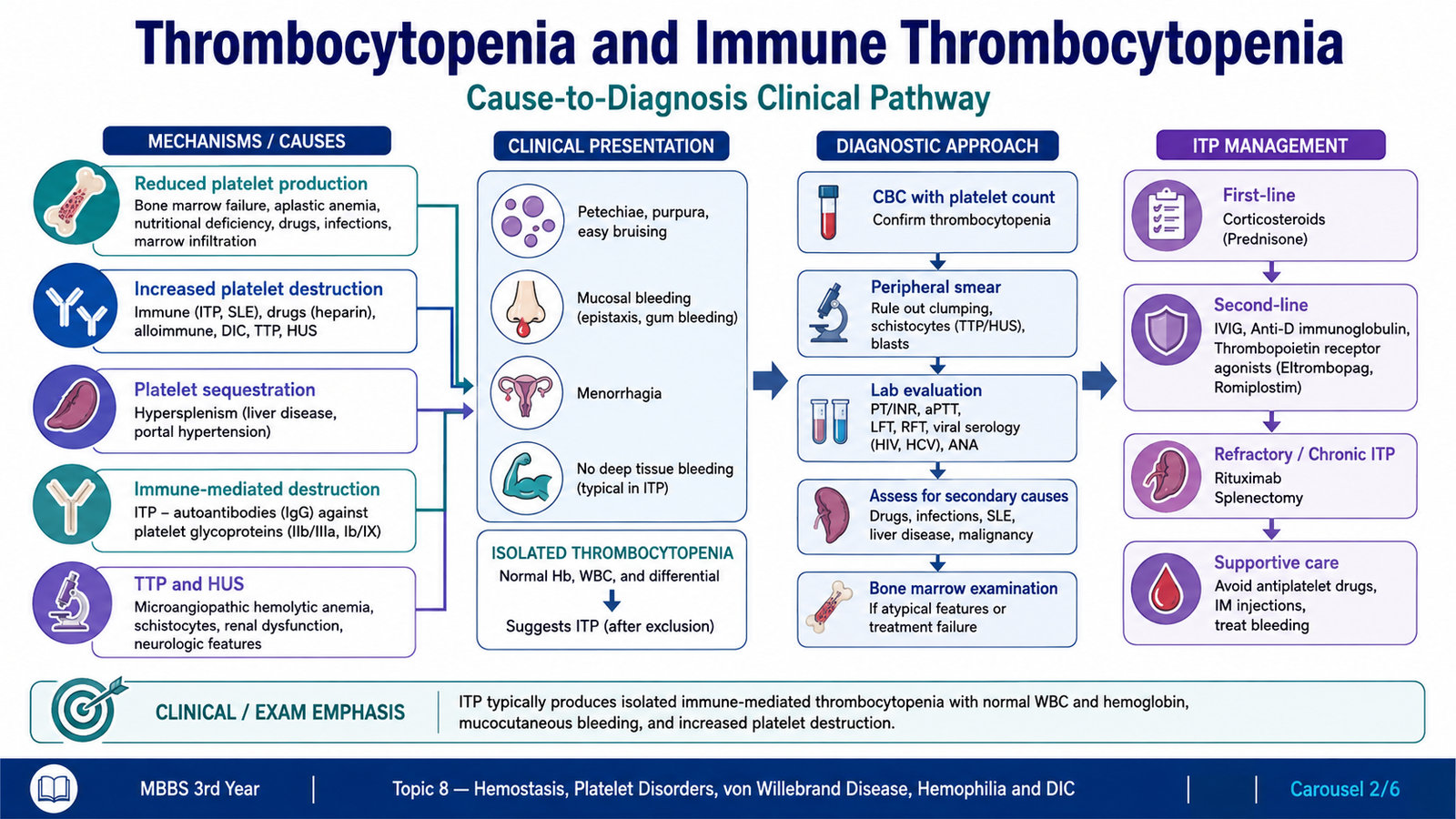
Thrombocytopenia means a reduction in the circulating platelet count. Because platelets are essential for primary hemostasis, marked platelet deficiency typically produces mucocutaneous bleeding rather than the deep muscle and joint bleeding characteristic of coagulation-factor deficiencies.
Major causes of thrombocytopenia
The causes become easier to understand when classified according to what has happened to the platelet population.
- Reduced platelet production: bone-marrow failure, marrow infiltration or suppression of megakaryocyte production.
- Increased destruction: immune destruction, including immune thrombocytopenia.
- Increased consumption: platelets are consumed in processes such as DIC and thrombotic microangiopathies.
- Splenic sequestration: an enlarged spleen can retain an excessive proportion of circulating platelets.
- Dilution: major replacement of blood volume can dilute circulating platelets.
Immune thrombocytopenia
Immune thrombocytopenia (ITP) is an acquired immune disorder in which platelets are destroyed prematurely. Autoantibodies, usually IgG, are directed against platelet membrane glycoproteins. Important targets include the GPIIb/IIIa and GPIb-containing complexes.
Antibody-coated platelets are removed mainly by macrophages in the spleen. Platelet survival therefore becomes shortened. The bone marrow attempts to compensate by increasing platelet production, so megakaryocytes may be normal or increased if marrow examination is performed.
Clinical features of ITP
Because the defect affects primary hemostasis, bleeding usually occurs from the skin and mucosal surfaces.
- Petechiae and purpura
- Easy bruising
- Epistaxis or gingival bleeding
- Heavy menstrual bleeding in affected women
- Prolonged bleeding following minor trauma
Deep muscle hematomas and recurrent hemarthroses are much more suggestive of a coagulation-factor deficiency such as hemophilia than isolated ITP.
Diagnostic approach to ITP
ITP is mainly a diagnosis of exclusion. The typical pattern is isolated thrombocytopenia without another clear explanation for the low platelet count.
- CBC: decreased platelet count with otherwise relatively preserved blood-cell lines in uncomplicated ITP.
- Peripheral smear: confirms thrombocytopenia and may show relatively large young platelets.
- PT and aPTT: usually normal because plasma coagulation factors are not primarily defective.
- Clinical assessment: looks for drugs, infections, systemic disease, splenomegaly or other causes of thrombocytopenia.
- Bone marrow: is not required in every straightforward case; when examined, megakaryocytes may be increased.
Basic management of ITP
Treatment depends on the severity of thrombocytopenia, the presence of clinically important bleeding and the individual clinical setting. Therapy is aimed at reducing immune-mediated platelet destruction and, when necessary, increasing platelet production.
- Corticosteroids reduce immune-mediated platelet destruction.
- Intravenous immunoglobulin (IVIG) can produce a relatively rapid increase in platelet count when rapid improvement is required.
- Persistent or refractory disease may require therapies directed at splenic platelet destruction or stimulation of platelet production.
- Platelet transfusion is generally reserved for major or life-threatening bleeding because transfused platelets may also undergo immune destruction.
Major thrombotic microangiopathies
Thrombotic microangiopathies are disorders in which platelet-rich thrombi develop within small vessels. Platelets are consumed in these microthrombi, producing thrombocytopenia, while red cells may become mechanically fragmented as they pass through affected vessels.
- Thrombotic thrombocytopenic purpura (TTP)
- Hemolytic uremic syndrome (HUS)
TTP is closely associated with severe deficiency of the vWF-cleaving protease ADAMTS13, allowing unusually large vWF multimers to promote platelet aggregation in small vessels. HUS is another major thrombotic microangiopathy in which renal involvement is particularly important.
Oprelvekin and platelet support
Oprelvekin is a recombinant form of interleukin-11. It stimulates the development and maturation of megakaryocytes and thereby increases platelet production. Its pharmacological role is therefore different from treatment of ITP: it supports platelet production rather than directly suppressing antiplatelet autoantibodies. Important adverse effects are related particularly to fluid retention and cardiovascular effects.
C. von Willebrand Disease
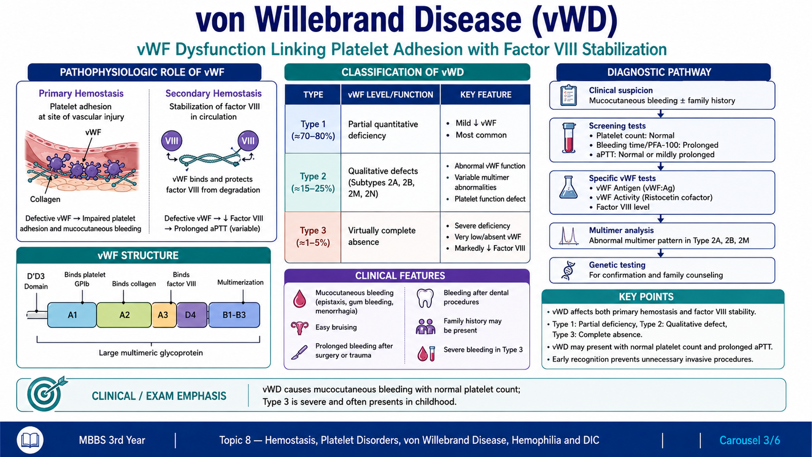
von Willebrand disease (vWD) is an inherited bleeding disorder caused by a quantitative deficiency or functional abnormality of von Willebrand factor. vWF has two major hemostatic functions. It promotes platelet adhesion to damaged vascular surfaces and also carries and stabilizes factor VIII in the circulation. A defect in vWF can therefore disturb primary hemostasis and, in more severe disease, reduce factor VIII activity.
Classification
| Type | Basic Defect | Key Concept |
|---|---|---|
| Type 1 | Partial quantitative deficiency | Reduced amount of otherwise functioning vWF |
| Type 2 | Qualitative abnormality | vWF is present but functions abnormally |
| Type 3 | Severe quantitative deficiency | Very little functional vWF is present |
Type 2 disease contains several subtypes based on the particular functional defect. For undergraduate understanding, the important principle is that Type 1 is mainly quantitative, Type 2 is qualitative and Type 3 is a severe quantitative deficiency.
Clinical pattern
Because vWF is essential for platelet adhesion, patients commonly develop a primary-hemostasis pattern of bleeding. This includes easy bruising, epistaxis, gingival bleeding, heavy menstrual bleeding and excessive bleeding following dental extraction or surgery.
Investigations
Diagnosis requires evidence that vWF quantity or function is abnormal. No single screening test is sufficient to characterize every form of vWD.
- Platelet count: usually normal in the common forms because the principal defect is vWF rather than platelet number.
- PT: usually normal.
- aPTT: may be prolonged when factor VIII activity is sufficiently reduced.
- vWF antigen: estimates the amount of circulating vWF.
- vWF activity testing: evaluates the functional ability of vWF to support platelet interaction.
- Factor VIII activity: may be reduced because vWF normally stabilizes circulating factor VIII.
- Specialized vWF studies: can help distinguish qualitative subtypes when required.
D. Hemophilia A and Hemophilia B
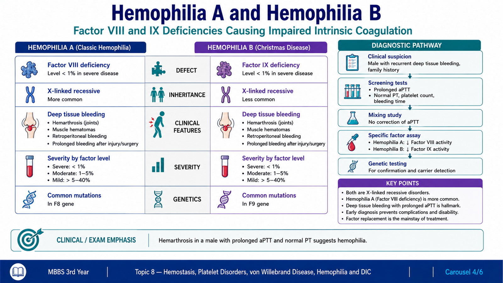
Hemophilia is an inherited disorder of secondary hemostasis caused by deficiency of a coagulation factor in the intrinsic pathway. Hemophilia A results from factor VIII deficiency, while hemophilia B results from factor IX deficiency. Both usually show X-linked recessive inheritance and therefore predominantly affect males, while females are commonly carriers.
Clinical course
Platelet plug formation is initially possible in hemophilia, but it is not adequately reinforced by fibrin. Bleeding can therefore continue or recur after an apparently minor injury.
- Hemarthrosis: bleeding into joints is a characteristic manifestation.
- Deep muscle hematomas: occur because secondary hemostasis is defective.
- Prolonged or delayed bleeding following trauma, surgery or dental procedures.
- Repeated joint bleeding may eventually damage the affected joints.
- Severity generally reflects how much functional coagulation factor remains.
Petechiae are not a typical feature because platelet number and initial platelet-plug formation are usually preserved.
Laboratory diagnosis
- Platelet count: usually normal.
- PT: usually normal because the extrinsic pathway is intact.
- aPTT: prolonged because factors VIII and IX belong to the intrinsic pathway.
- Mixing study: correction of a prolonged aPTT after mixing patient plasma with normal plasma supports a coagulation-factor deficiency rather than an inhibitor.
- Specific factor assay: demonstrates reduced factor VIII activity in hemophilia A or factor IX activity in hemophilia B.
| Feature | Hemophilia A | Hemophilia B |
|---|---|---|
| Deficient factor | Factor VIII | Factor IX |
| Main pathway affected | Intrinsic | Intrinsic |
| aPTT | Prolonged | Prolonged |
| Confirmation | Factor VIII assay | Factor IX assay |
Pharmacological treatment of hemophilia
Treatment is directed at replacing or increasing the missing hemostatic activity and controlling bleeding. The choice depends on the deficient factor and the clinical situation.
- Hemophilia A: factor VIII replacement directly corrects the deficient coagulation factor.
- Hemophilia B: factor IX replacement directly corrects the deficient factor.
- Desmopressin: can be useful in selected patients with mild hemophilia A because it promotes release of endogenous vWF and factor VIII from endothelial stores.
- Desmopressin does not correct hemophilia B because it does not increase factor IX.
Antifibrinolytic drugs
Once a fibrin clot has formed, the fibrinolytic system gradually breaks it down. Antifibrinolytic drugs such as tranexamic acid and aminocaproic acid reduce fibrin breakdown by interfering with plasminogen activation and the action of plasmin on fibrin.
These drugs are particularly useful as adjuncts when bleeding occurs from areas with high fibrinolytic activity, such as the oral cavity, including bleeding associated with dental procedures. They do not replace the missing coagulation factor in severe hemophilia.
Adverse effects may include gastrointestinal symptoms. Because these drugs inhibit fibrinolysis, excessive clot stabilization and thrombosis are important considerations in susceptible patients.
E. Disseminated Intravascular Coagulation
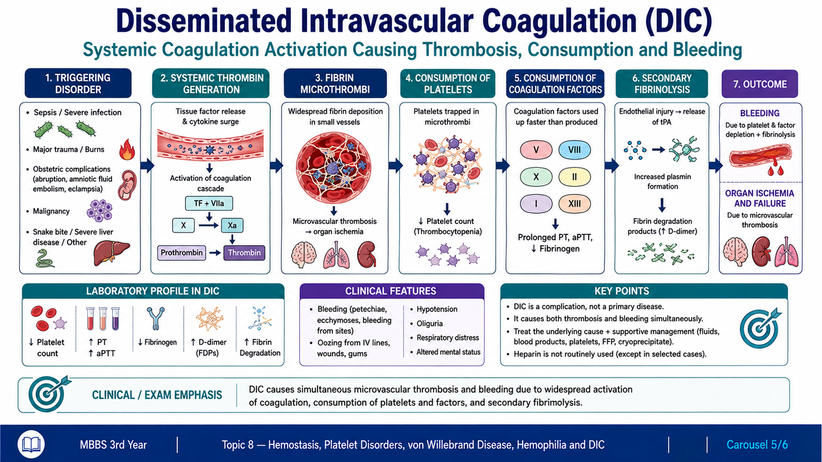
Disseminated intravascular coagulation (DIC) is an acquired systemic disorder in which coagulation becomes activated throughout the circulation. Large amounts of thrombin are generated, fibrin is deposited in small vessels and platelets and coagulation factors are consumed. The same patient can therefore develop both microvascular thrombosis and bleeding.
Major disorders associated with DIC
DIC is not usually a primary disease. It develops as a complication of disorders that strongly activate coagulation or cause extensive tissue injury.
- Severe infections, particularly systemic infections associated with marked activation of coagulation
- Major obstetric complications
- Certain malignancies, including acute promyelocytic leukemia and some advanced carcinomas
- Major trauma and extensive tissue injury
Pathophysiology
The central abnormality is excessive generation of thrombin. This produces widespread fibrin deposition in the microcirculation. As fibrin thrombi form, platelets and coagulation factors are consumed. At the same time, fibrinolytic mechanisms are activated in an attempt to remove the fibrin.
Microthrombi can obstruct small vessels and reduce tissue perfusion. Meanwhile, consumption of platelets and coagulation proteins impairs normal hemostasis. Breakdown of fibrin also generates fibrin degradation products, which provide an important laboratory clue to the presence of widespread coagulation and fibrinolysis.
Morphological changes
The characteristic pathological feature is the presence of fibrin-rich microthrombi in small vessels and capillaries. These deposits may be found in several organs.
- Kidneys
- Lungs
- Brain
- Heart
- Adrenal glands and other organs
Vascular occlusion may produce tissue ischemia and organ dysfunction. At the same time, depletion of platelets and coagulation factors can produce widespread hemorrhage. Red cells passing through fibrin-containing narrowed vessels may become mechanically fragmented, producing schistocytes on the peripheral blood smear.
Diagnostic approach
Diagnosis depends on recognizing an appropriate underlying clinical condition together with a pattern of coagulation abnormalities. There is no single laboratory finding that should be interpreted completely in isolation.
- Platelet count: commonly reduced because platelets are consumed.
- PT: commonly prolonged because coagulation factors are being consumed.
- aPTT: may also be prolonged.
- Fibrinogen: may be decreased because it is converted into fibrin and consumed.
- D-dimer and fibrin degradation products: increased because fibrin formation is followed by increased fibrinolysis.
- Peripheral smear: may show schistocytes due to mechanical fragmentation of red cells within abnormal small vessels.
F. Pharmacological Management of Bleeding Disorders and Thrombocytopenia
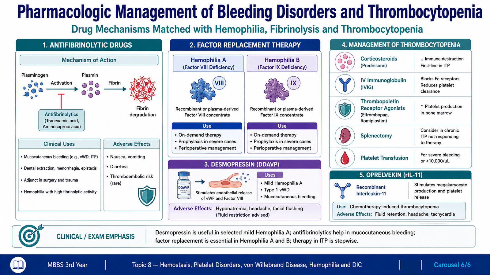
Pharmacological management of bleeding disorders depends on identifying the part of hemostasis that is defective. In hemophilia, treatment aims to replace or increase the deficient coagulation factor. Antifibrinolytic drugs do not replace a missing factor; instead, they help preserve fibrin that has already formed. In selected patients with mild hemophilia A, desmopressin can increase endogenous factor VIII activity. In thrombocytopenia caused by inadequate platelet production, hematopoietic support such as oprelvekin can increase platelet formation by stimulating megakaryocytes.
Antifibrinolytic Drugs
Fibrinolysis is the normal process by which fibrin clots are broken down. Plasmin is the major enzyme responsible for fibrin degradation and is formed from its inactive precursor, plasminogen. Tranexamic acid and aminocaproic acid are antifibrinolytic drugs that interfere with this process and therefore help stabilize an already formed clot.
→ interferes with plasminogen activation and plasmin action
→ reduced fibrin degradation
→ greater clot stability
→ reduced bleeding
Clinical Uses
Antifibrinolytic drugs are particularly useful when excessive fibrinolysis contributes to bleeding. They may also be used as adjuncts in patients with coagulation-factor deficiencies, especially when bleeding occurs from mucosal surfaces where fibrinolytic activity is relatively high.
- Control of mucosal bleeding.
- Reduction of bleeding associated with dental procedures.
- Adjunctive treatment in selected patients with hemophilia.
These drugs do not replace factor VIII or factor IX. Their role is to protect the fibrin clot from premature breakdown.
Adverse Effects
Gastrointestinal symptoms such as nausea and abdominal discomfort may occur. Because these drugs reduce fibrinolysis, excessive persistence of fibrin may also increase concern about thrombotic complications in susceptible patients.
Pharmacological Treatment of Hemophilia
Hemophilia A and hemophilia B are deficiencies of different coagulation factors, so treatment must be matched to the missing factor. The aim is to restore sufficient coagulation activity for stable fibrin formation and control of bleeding.
| Disorder | Defect | Treatment Principle |
|---|---|---|
| Hemophilia A | Factor VIII deficiency | Factor VIII replacement |
| Hemophilia B | Factor IX deficiency | Factor IX replacement |
Factor replacement directly corrects the deficient component of the intrinsic coagulation pathway. Antifibrinolytic therapy may be added when preservation of an already formed fibrin clot is particularly useful, but it does not substitute for appropriate factor replacement when the coagulation-factor deficiency is clinically important.
Role of Desmopressin in Hemophilia
Desmopressin is useful in selected patients with mild hemophilia A. It promotes the release of stored von Willebrand factor and factor VIII from endothelial cells. The resulting increase in circulating factor VIII activity can improve coagulation when the patient already has some endogenous factor VIII available.
→ increased release of vWF and factor VIII
→ increased circulating factor VIII activity
→ improved secondary hemostasis in selected mild hemophilia A
Desmopressin is not an effective treatment for hemophilia B because hemophilia B results from factor IX deficiency, and desmopressin does not replace or significantly increase factor IX.
Oprelvekin in Thrombocytopenia
Oprelvekin is a recombinant form of interleukin-11 (IL-11). Unlike treatments that act on coagulation factors or fibrinolysis, oprelvekin acts on platelet production. It stimulates the development and maturation of megakaryocytes, the bone-marrow cells that produce platelets.
→ IL-11 activity
→ stimulation of megakaryocyte development and maturation
→ increased platelet production
→ support of platelet recovery
Its role is therefore most logically understood as hematopoietic support. It increases platelet production rather than directly preventing immune destruction of platelets. Important adverse effects include fluid retention and edema, with cardiovascular effects being particularly relevant in susceptible patients.
Factor VIII deficiency → factor VIII replacement
Factor IX deficiency → factor IX replacement
Selected mild hemophilia A → desmopressin
Need to preserve fibrin clot → antifibrinolytic drug
Need to stimulate platelet production → oprelvekin
Drug-action and treatment-selection pathway showing antifibrinolytics reducing fibrin breakdown, factor VIII replacement for hemophilia A, factor IX replacement for hemophilia B, desmopressin increasing vWF and factor VIII in selected mild hemophilia A, and oprelvekin stimulating megakaryocytes to increase platelet production.
Hemostasis, Platelet Disorders, von Willebrand Disease, Hemophilia and DIC
Use these focused videos after studying the AIM learning material to reinforce the major mechanisms, clinical patterns and diagnostic principles.
Thrombocytopenia
Reinforces thrombocytopenia, DIC and thrombotic microangiopathic disorders.
von Willebrand Disease & Qualitative Platelet Disorders
Focuses on vWF function, pathogenesis, clinical presentation and diagnostic principles of von Willebrand disease.
Hemophilia & Other Coagulation Deficiencies
Reinforces hemophilia A and B, coagulation-factor deficiencies and the laboratory approach to secondary hemostatic disorders.