
This chapter follows the supplied KMU learning outcomes and builds the different hemolytic anemias in a logical sequence. First understand why red cells are destroyed in each disorder, then use the morphology and laboratory findings to distinguish them. Finish with the AIM High-Yield Review for rapid revision.
Topic 2 — Hemolytic Anemias and Hemoglobinopathies
A mechanism-based approach to hereditary spherocytosis, sickle cell anemia, thalassemia, G6PD deficiency, paroxysmal nocturnal hemoglobinuria and immune hemolytic anemia, with emphasis on morphology, diagnostic reasoning and the required clinical management principles.
Topic Introduction
Hemolytic anemias are disorders in which red blood cells are destroyed faster than the bone marrow can replace them. The defect may lie inside the red cell itself, as in membrane disorders, hemoglobin abnormalities and enzyme deficiencies, or may arise from external factors such as antibodies or complement. Although these conditions share evidence of increased red-cell destruction, they differ greatly in their mechanisms, morphology and diagnostic tests. This chapter explains these differences through six major disorders. The central learning approach is simple: identify the abnormality, understand how it damages the red cell, recognize the resulting blood-film appearance and then select the investigation that confirms the diagnosis.
A. Understanding Hemolytic Anemias: The Core Framework
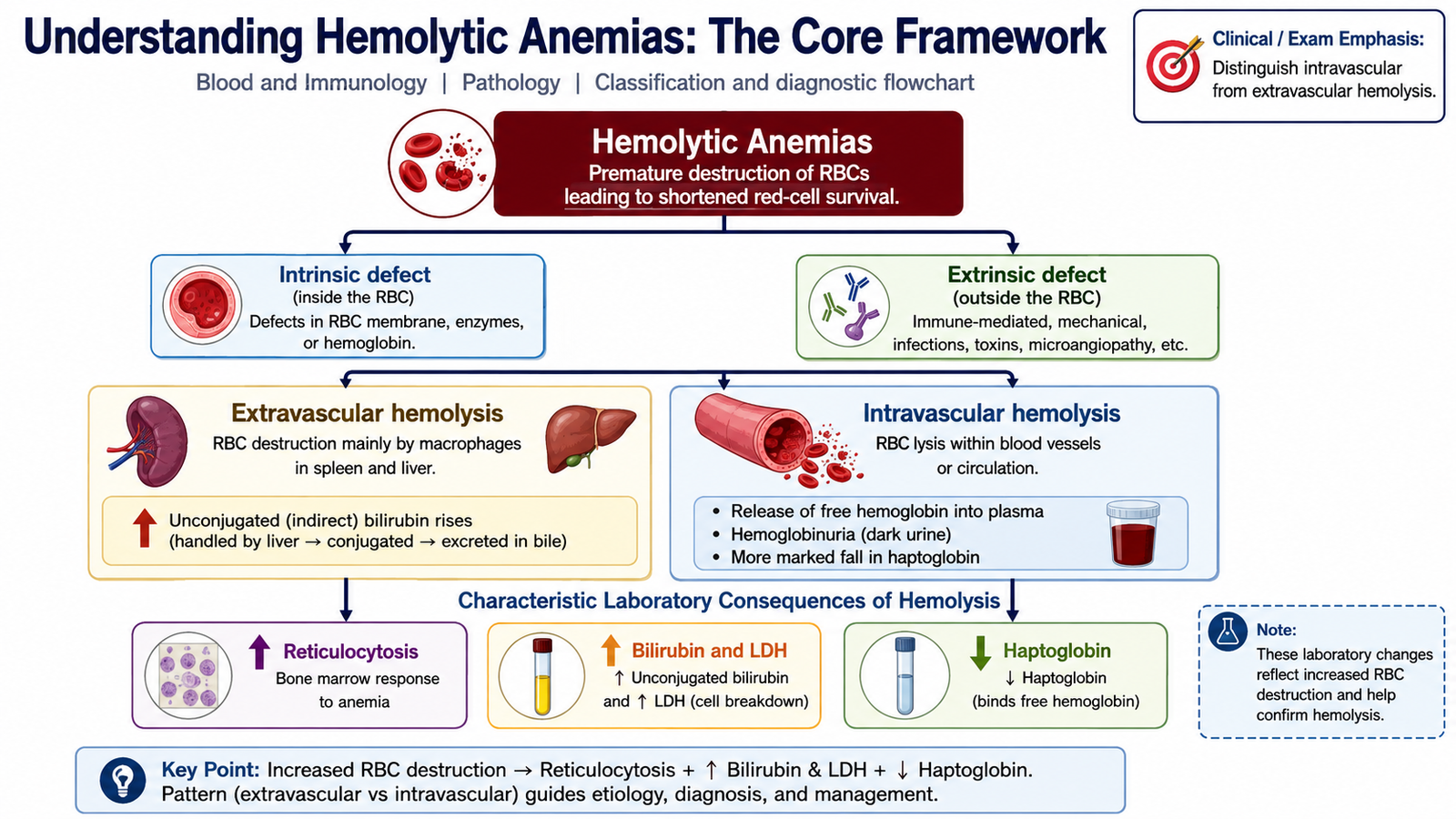
Normal red blood cells circulate for approximately 120 days before they are removed mainly by macrophages of the spleen and other parts of the reticuloendothelial system. In hemolytic anemia, red cells have a shortened survival. The marrow responds by increasing erythropoiesis, so the reticulocyte count usually rises if marrow function is adequate.
Hemolysis can be understood in two complementary ways: according to the origin of the red-cell abnormality and according to the site where destruction occurs. This framework makes the individual diseases in this chapter easier to understand.
Intrinsic and extrinsic causes
- Intrinsic red-cell defects arise from abnormalities within the RBC itself. Examples in this topic include hereditary spherocytosis, sickle cell disease, thalassemia and G6PD deficiency.
- Acquired intrinsic defects can also occur. Paroxysmal nocturnal hemoglobinuria is an acquired clonal stem-cell disorder in which circulating blood cells become unusually sensitive to complement.
- Extrinsic hemolysis occurs when structurally normal or near-normal red cells are damaged by an outside process. Immune hemolytic anemia is an important example.
Extravascular hemolysis
Extravascular hemolysis occurs mainly when abnormal or antibody-coated RBCs are removed by macrophages in the spleen and liver. The spleen is especially important because red cells must deform considerably to pass through its narrow vascular spaces. Cells that are rigid, spherical or coated with IgG are therefore preferentially trapped and destroyed.
Important consequences include anemia, reticulocytosis, unconjugated hyperbilirubinemia and, when persistent, splenomegaly. Increased bilirubin production may also promote pigment gallstone formation.
Intravascular hemolysis
In intravascular hemolysis, red cells rupture within the circulation. Free hemoglobin is released into plasma. Haptoglobin binds this free hemoglobin and is therefore reduced when significant intravascular hemolysis occurs. If the amount of free hemoglobin exceeds the binding capacity of haptoglobin, hemoglobinemia and hemoglobinuria may occur.
B. Hereditary Spherocytosis
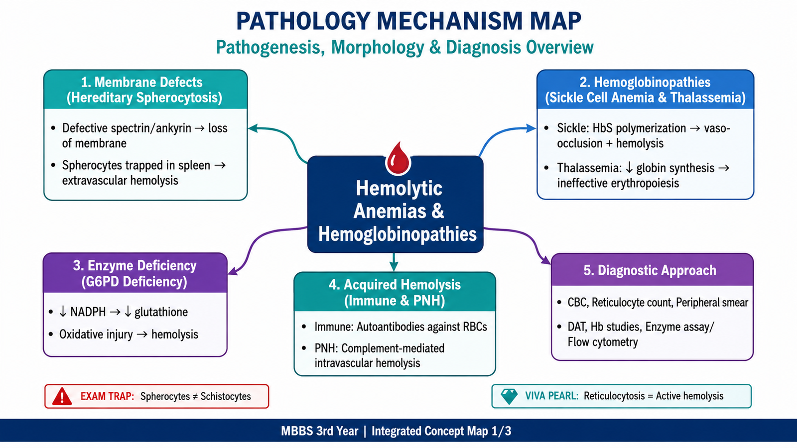
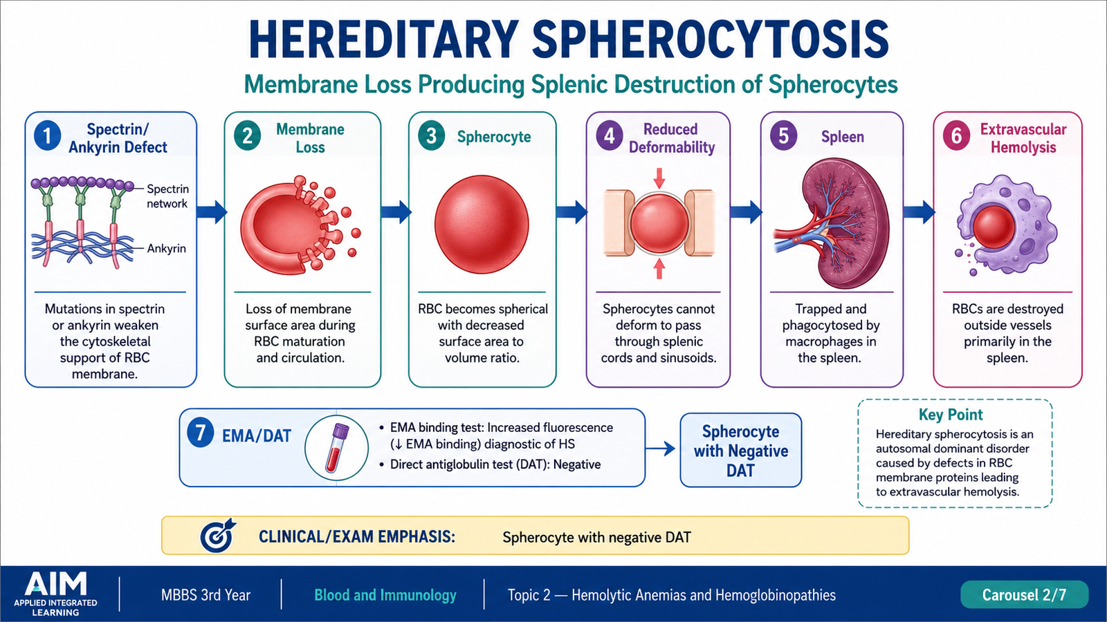
Hereditary spherocytosis is an inherited disorder of the red-cell membrane in which loss of membrane surface area converts the normal flexible biconcave RBC into a spherical cell. The spherical cell can transport oxygen, but it cannot deform normally while passing through the spleen. The spleen therefore removes these cells prematurely, producing predominantly extravascular hemolysis.
Pathogenesis
The RBC membrane skeleton normally maintains cell shape and mechanical stability. Hereditary abnormalities involving membrane-associated proteins such as spectrin, ankyrin, band 3 or protein 4.2 weaken the attachment between the membrane skeleton and lipid bilayer. Small portions of membrane are progressively lost.
Because membrane loss occurs without an equivalent loss of intracellular volume, the cell becomes increasingly spherical. The spleen further aggravates membrane loss as affected RBCs repeatedly attempt to pass through the splenic circulation. This produces chronic extravascular hemolysis.
Characteristic Morphology
The most characteristic peripheral blood finding is the spherocyte. A spherocyte is smaller and more densely stained than a normal RBC and lacks the normal central pallor because its biconcave shape has been lost.
- Spherocytes with absent or markedly reduced central pallor
- Reticulocytosis due to compensatory marrow response
- Polychromasia may accompany reticulocytosis
- Erythroid hyperplasia may be present in bone marrow when hemolysis is significant
Diagnostic Approach
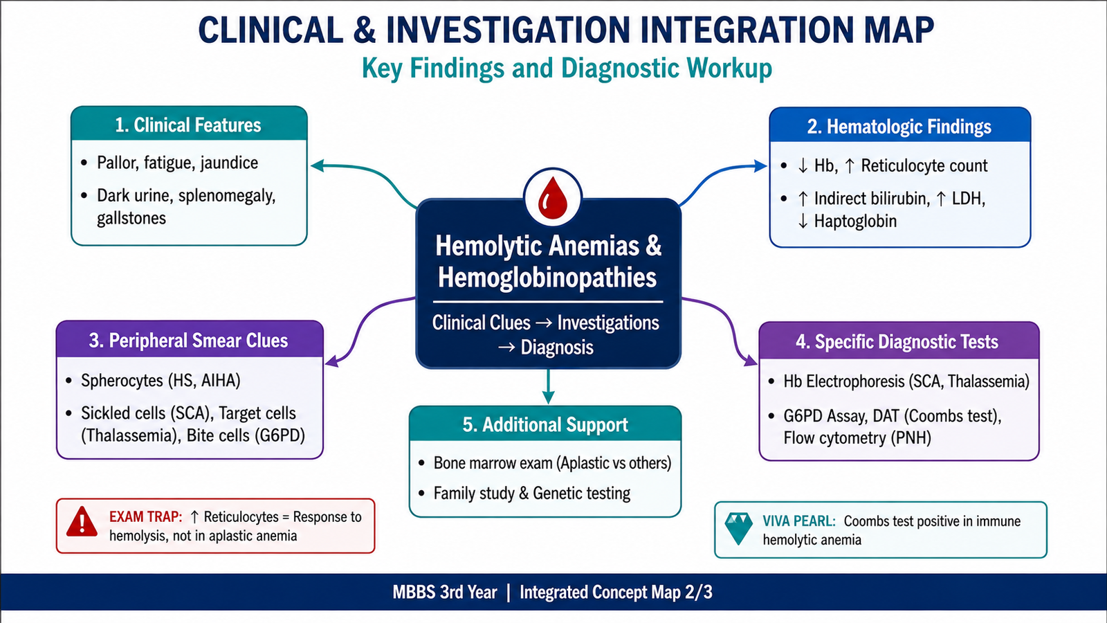
The diagnosis combines evidence of hemolysis with evidence of a membrane defect. A CBC may show anemia with reticulocytosis. The mean corpuscular hemoglobin concentration may be increased because membrane surface area has been lost while cellular hemoglobin is relatively preserved.
- Peripheral smear: spherocytes lacking central pallor.
- Hemolysis profile: reticulocytosis, increased unconjugated bilirubin and LDH, with reduced haptoglobin when hemolysis is significant.
- Eosin-5-maleimide binding test: flow-cytometric assessment of RBC membrane-protein abnormalities and an important confirmatory investigation.
- Osmotic fragility: increased because spherocytes have less membrane reserve and tolerate hypotonic swelling poorly.
- Direct antiglobulin test: usually negative. This helps distinguish hereditary spherocytosis from immune hemolytic anemia, in which spherocytes may also occur.
C. Sickle Cell Anemia
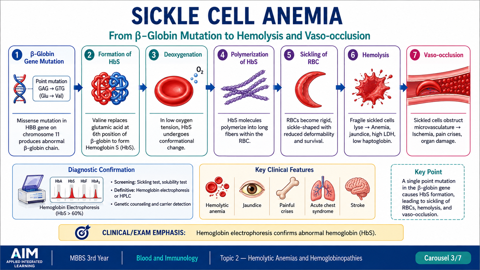
Sickle cell anemia is an inherited hemoglobin disorder caused by production of hemoglobin S. When HbS is deoxygenated, its molecules polymerize and distort the RBC into the characteristic sickle shape. Repeated episodes of sickling damage the cell membrane, producing chronic hemolytic anemia, while rigid sickled cells can also obstruct small vessels and cause tissue ischemia.
Etiology and Molecular Basis
Sickle cell disease results from a mutation in the β-globin gene. The abnormal β-globin chain contains valine instead of glutamic acid at position 6. This apparently small amino-acid substitution creates a hydrophobic region that allows deoxygenated HbS molecules to interact and form long polymers.
Sickle cell anemia refers to the severe homozygous form in which the patient inherits an HbS-producing β-globin gene from both parents.
Pathogenesis
Polymer formation occurs most readily when HbS is deoxygenated. The polymers push against the RBC membrane and convert the normally flexible cell into an elongated or sickled form. Initially, sickling can reverse when oxygenation improves. Repeated cycles, however, injure the membrane and eventually create permanently damaged cells.
Two major pathological processes follow:
- Hemolysis: damaged and poorly deformable cells are removed, producing chronic anemia.
- Vaso-occlusion: rigid cells obstruct the microcirculation, producing ischemic pain and organ injury.
Conditions that promote deoxygenation or increase intracellular HbS concentration can favor sickling. Dehydration, hypoxia and acidosis therefore have physiological importance because they can increase polymer formation.
RBC Morphology
- Sickle cells: elongated, crescent-shaped RBCs caused by HbS polymerization.
- Target cells: may be present because of altered RBC membrane-to-volume relationships.
- Polychromasia: reflects increased reticulocyte release from the marrow.
- Nucleated RBCs: may appear when erythropoietic stress is marked.
- Howell–Jolly bodies: may appear when repeated splenic infarction leads to functional asplenia.
Diagnostic Approach and Investigations
The investigation begins by confirming hemolytic anemia and then demonstrating HbS.
- CBC: anemia, usually with increased reticulocytes.
- Peripheral blood film: sickled cells, target cells and evidence of increased erythropoiesis.
- Hemolysis markers: increased bilirubin and LDH with reduced haptoglobin.
- Hemoglobin analysis: hemoglobin electrophoresis or high-performance liquid chromatography identifies and quantifies abnormal hemoglobin fractions.
- Additional investigations: are selected according to suspected complications and organ involvement.
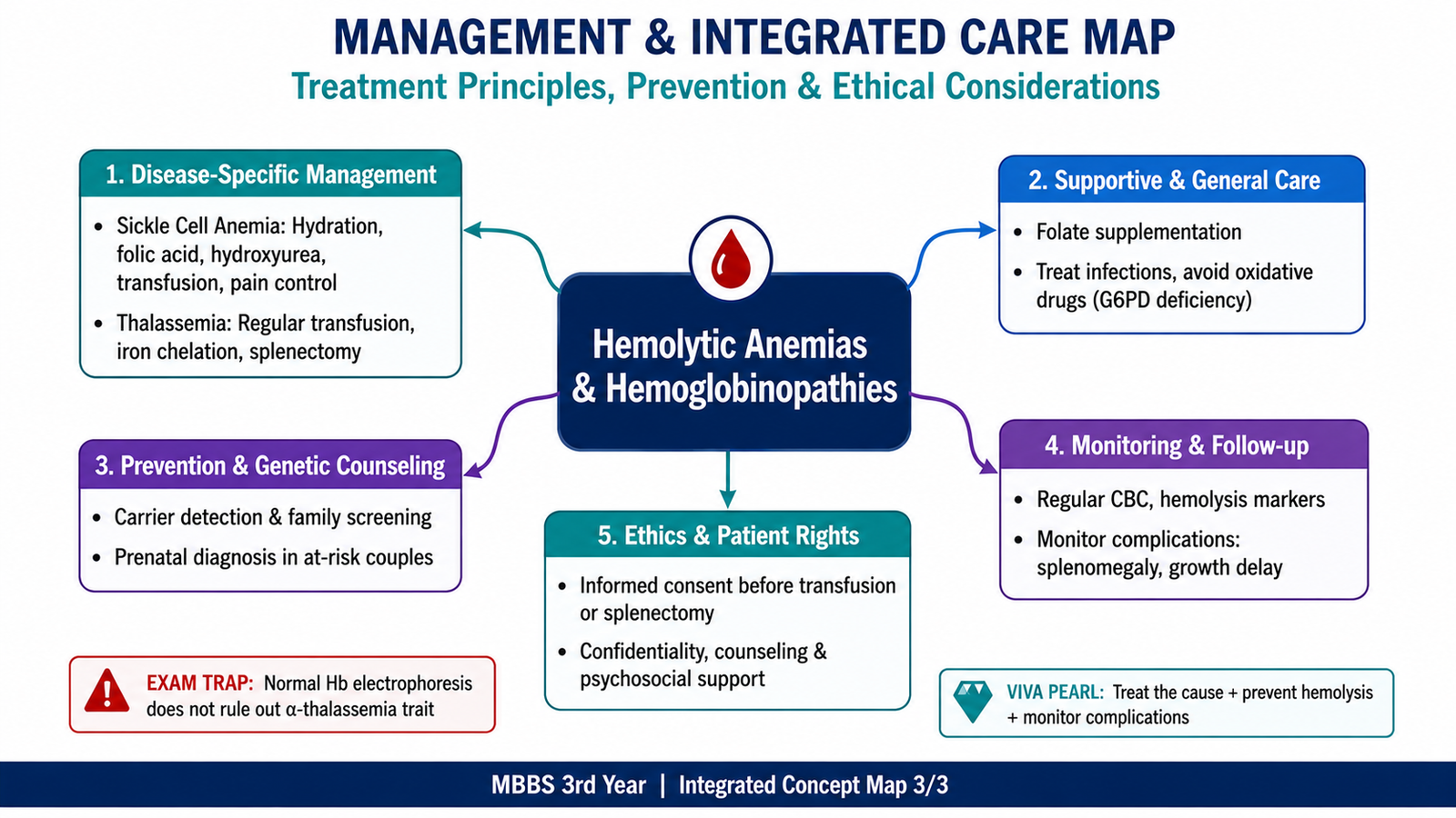
Management Principles
Management aims to reduce sickling, treat acute complications and prevent progressive organ injury. The exact treatment depends on the patient’s presentation and severity.
- Maintain adequate hydration and correct factors that worsen tissue hypoxia.
- Provide effective analgesia during painful vaso-occlusive episodes.
- Recognize and treat infection promptly, particularly because functional asplenia increases susceptibility to severe infection.
- Hydroxyurea is used in appropriate patients because it increases fetal hemoglobin, which interferes with HbS polymerization and reduces sickling-related complications.
- Red-cell transfusion is used when clinically indicated, especially for selected severe complications.
- Long-term care includes prevention and surveillance for organ complications.
- Hematopoietic stem-cell transplantation can provide definitive treatment in appropriately selected patients.
D. Thalassemia
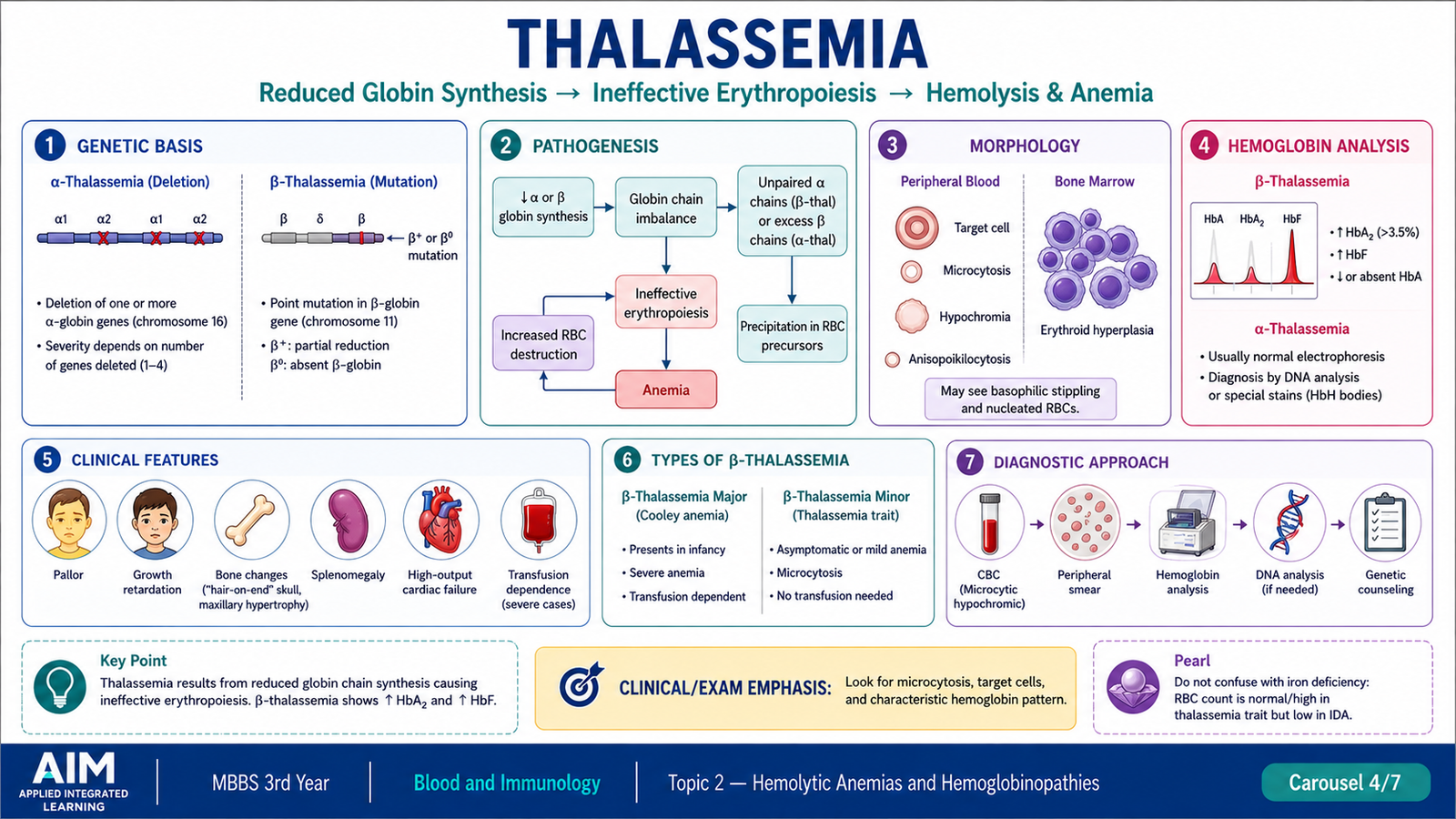
Thalassemias are inherited disorders in which synthesis of one of the globin chains of hemoglobin is reduced or absent. The central problem is therefore quantitative: the globin chain has a normal basic structure, but too little of it is produced. Reduced α-chain production causes α-thalassemia, whereas reduced β-chain production causes β-thalassemia.
Classification
| Group | Underlying defect | Important forms |
|---|---|---|
| α-Thalassemia | Reduced α-globin synthesis | Silent carrier, α-thalassemia trait, HbH disease, severe fetal disease with Hb Bart’s |
| β-Thalassemia | Reduced or absent β-globin synthesis | β-thalassemia trait/minor, intermedia and major |
Genetics of α-Thalassemia
There are normally four α-globin genes, two on each chromosome 16. α-Thalassemia most commonly results from deletion of one or more of these genes. Clinical severity therefore generally increases as more α-globin genes are lost.
- One gene affected: silent carrier state with little or no anemia.
- Two genes affected: α-thalassemia trait, usually causing mild microcytic anemia.
- Three genes affected: HbH disease, in which excess β chains form β4 tetramers.
- All four genes affected: profound α-chain deficiency with formation of γ4 tetramers, called Hb Bart’s, during fetal life.
Genetics of β-Thalassemia
β-globin is encoded by genes on chromosome 11. β-thalassemia usually results from mutations that reduce or prevent production of β-globin rather than from large gene deletions.
- β+ mutation: some β-globin synthesis remains.
- β0 mutation: β-globin production from the affected gene is absent.
The phenotype depends on the combination of inherited mutations and the amount of β-globin that can still be produced.
Pathogenesis of β-Thalassemia
The disease is not caused simply by having less hemoglobin. Reduced β-chain production leaves excess unpaired α chains. These free α chains are unstable and precipitate inside developing erythroid cells. The precipitated material damages cell membranes.
Many abnormal erythroid precursors die inside the bone marrow before reaching the circulation. This is called ineffective erythropoiesis. Red cells that do enter the circulation remain abnormal and may be removed prematurely by the spleen.
Severe anemia stimulates erythropoietin production and causes marked erythroid marrow expansion. In severe disease, prolonged marrow expansion may alter the bones. Increased intestinal iron absorption and repeated transfusions can also result in progressive iron accumulation.
Morphological Changes
The peripheral blood film reflects both defective hemoglobin production and intense marrow stimulation.
- Marked microcytosis and hypochromia
- Variation in RBC size and shape
- Target cells
- Basophilic stippling may be present
- Polychromasia due to reticulocytosis
- Nucleated RBCs may appear in severe disease
Laboratory Diagnosis
Thalassemia should be considered when a patient has a microcytic, hypochromic anemia that is not adequately explained by iron deficiency. Iron studies are therefore important when evaluating microcytosis.
β-Thalassemia: Hemoglobin electrophoresis or HPLC is particularly useful because abnormal proportions of hemoglobin fractions can be demonstrated. β-thalassemia trait characteristically shows increased HbA2, while more severe β-chain deficiency produces greater reliance on HbF and less HbA.
α-Thalassemia: Mild α-thalassemia may have a normal adult hemoglobin electrophoresis pattern, so diagnosis may depend on the hematological pattern, family history and genetic analysis when required. HbH disease produces detectable HbH.
Principles of Management in Thalassemia
Management depends on clinical severity. Mild carrier states may require little specific treatment, whereas severe thalassemia requires long-term multidisciplinary care.
- Red-cell transfusion: used in transfusion-dependent severe disease to maintain adequate oxygen-carrying capacity and suppress excessive ineffective erythropoiesis.
- Iron chelation: used to reduce iron accumulation associated particularly with repeated transfusion therapy.
- Monitoring for iron-related organ injury: is an important part of long-term care.
- Folate support: may be required when erythropoietic demand is increased.
- Management of splenic enlargement or hypersplenism: is considered according to the clinical situation.
- Hematopoietic stem-cell transplantation: can provide definitive treatment in appropriately selected patients.
E. G6PD Deficiency
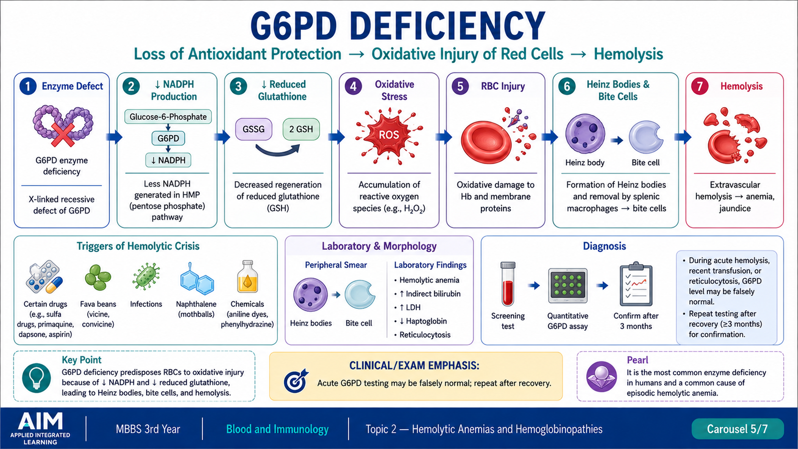
Glucose-6-phosphate dehydrogenase deficiency is an inherited RBC enzyme defect that reduces the cell’s ability to protect itself against oxidative injury. Red cells depend strongly on the pentose phosphate pathway because they lack mitochondria. G6PD is therefore essential for generating NADPH, which keeps glutathione in its reduced protective form.
Classification
G6PD variants can be classified according to the degree of enzyme deficiency and clinical severity. Traditional undergraduate classifications describe a spectrum ranging from severe deficiency with chronic nonspherocytic hemolytic anemia, through severe or moderate deficiency causing episodic oxidant-induced hemolysis, to variants with near-normal or increased activity.
For practical clinical understanding, affected individuals can be considered in two major patterns:
- Episodic hemolysis: the patient is usually well but develops hemolysis after significant oxidative stress.
- Chronic nonspherocytic hemolytic anemia: uncommon severe variants produce ongoing hemolysis even without a major external trigger.
Pathogenesis: Oxidative Injury to RBCs
Reduced glutathione normally neutralizes reactive oxygen species and protects hemoglobin and the RBC membrane from oxidation. G6PD-deficient cells cannot generate adequate NADPH during oxidative stress, so reduced glutathione cannot be maintained efficiently.
Oxidized hemoglobin denatures and precipitates inside the RBC to form Heinz bodies. When affected cells pass through the spleen, macrophages remove these precipitates together with part of the membrane. This produces the characteristic bite cell.
Common forms of oxidative stress include infections, ingestion of fava beans and exposure to certain oxidant drugs. Infection itself can be a powerful trigger because activated inflammatory cells generate oxidants.
Characteristic RBC Morphology
- Heinz bodies: precipitated denatured hemoglobin demonstrated with appropriate supravital staining.
- Bite cells: RBCs from which splenic macrophages have removed Heinz-body-containing portions.
- Reticulocytosis and polychromasia during recovery from hemolysis.
Diagnostic Approach
The diagnosis is suspected from an appropriate clinical trigger together with acute hemolysis and characteristic morphology. Confirmation is obtained by measuring G6PD enzyme activity.
- CBC and reticulocyte count demonstrate anemia with marrow response.
- Hemolysis markers support increased RBC destruction.
- Peripheral smear may show bite cells.
- Supravital staining can demonstrate Heinz bodies.
- Quantitative or screening tests of G6PD activity confirm the enzyme deficiency.
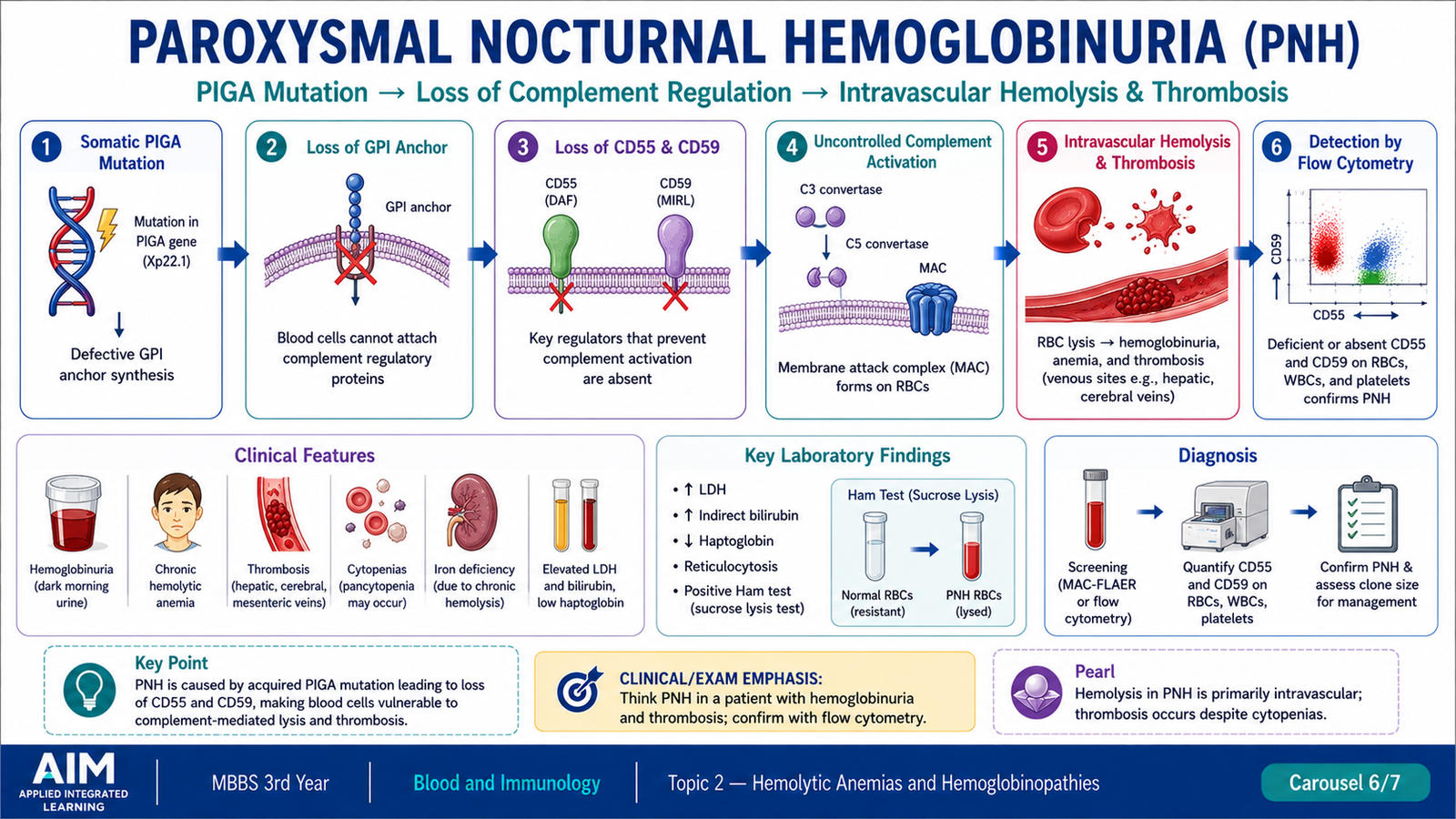
F. Paroxysmal Nocturnal Hemoglobinuria
Paroxysmal nocturnal hemoglobinuria, or PNH, is an acquired clonal hematopoietic stem-cell disorder. Unlike hereditary membrane disorders, the abnormality develops in a stem-cell clone during life. The affected clone produces red cells, white cells and platelets that lack certain protective surface proteins, making them unusually sensitive to complement.
Pathophysiology
The fundamental abnormality is an acquired mutation involving the PIGA gene in a hematopoietic stem cell. This interferes with synthesis of glycosylphosphatidylinositol, or GPI anchors. GPI anchors normally attach several proteins to the cell surface.
Two particularly important GPI-linked proteins are CD55 and CD59. They normally protect blood cells from complement-mediated injury. When these proteins are deficient, complement can damage and lyse RBCs more readily.
PNH is therefore an important cause of acquired intravascular hemolysis. Free hemoglobin may appear in plasma and urine. Despite the historical name, clinically important hemolysis is not limited to the night.
The disorder is also important because the abnormal stem-cell clone can involve multiple blood-cell lines. Patients may have cytopenias associated with marrow dysfunction, and PNH has a particularly important association with venous thrombosis.
Diagnosis
The modern diagnostic approach is based on demonstration of cells lacking GPI-linked proteins.
- Laboratory evidence of intravascular hemolysis may include increased LDH, reduced haptoglobin and hemoglobinuria.
- CBC may reveal anemia and sometimes abnormalities in other blood-cell lines.
- Flow cytometry can demonstrate reduced or absent CD55 and CD59 on affected cells.
- Tests using reagents that bind directly to the GPI anchor, such as FLAER-based flow cytometric analysis, can help identify the abnormal clone.
G. Immune Hemolytic Anemias
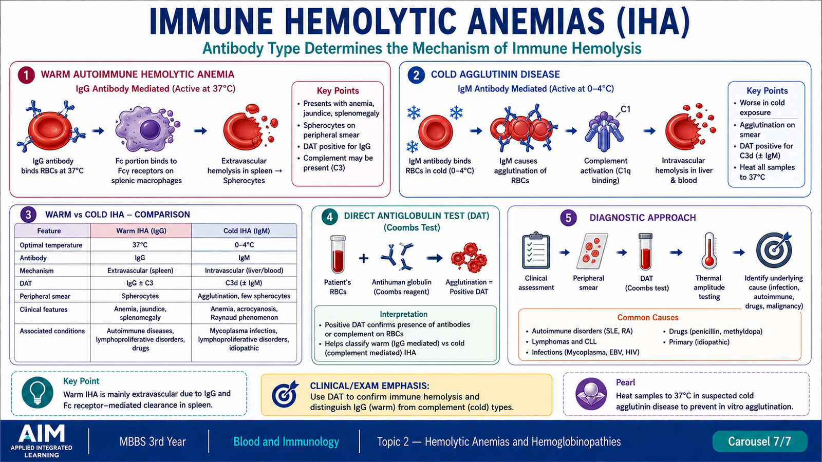
Immune hemolytic anemias occur when antibodies recognize antigens on the surface of red blood cells and promote their destruction. Depending on the antibody involved and the degree of complement activation, hemolysis may occur mainly in the spleen and liver or within the circulation.
Classification
- Autoimmune hemolytic anemia
- Warm-antibody type
- Cold-antibody type
- Paroxysmal cold hemoglobinuria
- Drug-associated immune hemolytic anemia
- Alloimmune hemolytic anemia, including immune destruction after incompatible transfusion or maternal antibody-mediated destruction of fetal/neonatal RBCs.
Warm-Antibody Immune Hemolysis
Warm autoimmune hemolytic anemia is usually mediated by IgG antibodies that react efficiently with RBCs at body temperature. IgG-coated red cells are recognized by Fc receptors on splenic macrophages.
The macrophage may completely remove the RBC or may remove only part of its membrane. Partial membrane loss converts the remaining RBC into a spherocyte. These less-deformable spherocytes are then trapped and destroyed in the spleen.
Cold-Antibody Immune Hemolysis
Cold-antibody hemolysis is usually associated with IgM antibodies that bind more effectively at lower temperatures, particularly in cooler peripheral parts of the body. IgM is very effective at activating complement.
When blood returns to warmer central circulation, IgM may detach, but complement components can remain on the RBC surface. Complement-coated cells may then be removed by macrophages, particularly in the liver. If complement activation proceeds sufficiently, intravascular lysis can also occur.
Diagnostic Workup
The diagnostic approach first establishes hemolysis and then determines whether the process is immune mediated.
- CBC and reticulocyte count: demonstrate anemia and marrow response.
- Hemolysis markers: bilirubin and LDH rise, while haptoglobin may fall.
- Peripheral smear: may show spherocytes in warm immune hemolysis or RBC agglutination in cold-antibody disease.
- Direct antiglobulin test: detects immunoglobulin and/or complement attached to the patient’s RBCs and is the central test for immune-mediated hemolysis.
- The pattern of IgG and complement detected by the direct antiglobulin test helps distinguish warm from cold immune mechanisms.
| Feature | Warm-antibody hemolysis | Cold-antibody hemolysis |
|---|---|---|
| Main antibody | IgG | Usually IgM |
| Important mechanism | Fc-mediated macrophage removal | Complement activation |
| Major site | Predominantly spleen | Complement-mediated removal, often hepatic; intravascular lysis may occur |
| Smear clue | Spherocytes | RBC agglutination may occur |
Integrated Mechanism Flow
↓
2. RBCs become rigid, unstable, oxidatively injured, complement-sensitive or antibody-coated.
↓
3. Abnormal RBCs are removed by splenic/hepatic macrophages or rupture within the circulation.
↓
4. Hemoglobin falls while the marrow increases erythropoiesis, producing reticulocytosis if marrow reserve is adequate.
↓
5. Bilirubin and LDH rise and haptoglobin falls, while the peripheral smear provides the first disease-specific clue.
↓
6. Disease-specific testing identifies the cause: membrane testing, hemoglobin analysis, G6PD assay, PNH flow cytometry or direct antiglobulin testing.
Important Comparison — Major Diagnostic Clues
| Disorder | Core abnormality | Important smear clue | Dominant mechanism | Key diagnostic test/clue |
|---|---|---|---|---|
| Hereditary spherocytosis | Membrane-protein defect | Spherocytes | Splenic extravascular hemolysis | EMA binding / osmotic fragility; DAT negative |
| Sickle cell anemia | HbS β-globin mutation | Sickle cells | Hemolysis + vaso-occlusion | Hemoglobin electrophoresis/HPLC |
| Thalassemia | Reduced globin-chain synthesis | Microcytosis, hypochromia, target cells | Ineffective erythropoiesis + hemolysis | Hemoglobin analysis; genetic testing when needed |
| G6PD deficiency | Reduced antioxidant protection | Bite cells; Heinz bodies on special stain | Oxidative RBC injury | G6PD enzyme assay |
| PNH | Acquired PIGA mutation with deficient GPI-linked proteins | No single pathognomonic smear pattern | Complement-mediated intravascular hemolysis | Flow cytometry for GPI-linked proteins/FLAER |
| Immune hemolytic anemia | Antibody ± complement on RBCs | Spherocytes or agglutination | Immune destruction | Direct antiglobulin test |
⭐ AIM High-Yield Review
- Hemolytic anemia means shortened RBC survival; an appropriate marrow response produces reticulocytosis.
- ⭐ Hereditary spherocytosis: membrane loss → spherocytes → reduced deformability → splenic extravascular hemolysis.
- Spherocytes occur in both hereditary spherocytosis and warm immune hemolysis; the direct antiglobulin test helps distinguish them.
- ⭐ Sickle cell anemia: β-globin Glu→Val substitution produces HbS, which polymerizes during deoxygenation.
- Sickle disease produces two major pathological consequences: hemolysis and vaso-occlusion.
- Hydroxyurea reduces sickling-related complications mainly by increasing HbF.
- ⭐ β-Thalassemia: reduced β-globin synthesis leaves excess α chains, producing ineffective erythropoiesis and hemolysis.
- Thalassemia characteristically produces microcytic hypochromic RBCs and target cells.
- In β-thalassemia trait, increased HbA2 is an important diagnostic clue; mild α-thalassemia may have normal adult hemoglobin electrophoresis.
- ⭐ G6PD deficiency: reduced NADPH/glutathione protection causes oxidative hemoglobin injury, Heinz bodies and bite cells.
- A G6PD assay may be misleadingly normal during an acute hemolytic episode because the most deficient older RBCs have already been destroyed.
- ⭐ PNH: acquired PIGA mutation → deficient GPI-linked CD55/CD59 → complement-mediated intravascular hemolysis.
- PNH should be considered when intravascular hemolysis occurs with cytopenia or unusual venous thrombosis.
- Warm autoimmune hemolysis is mainly IgG-mediated and predominantly extravascular; cold-antibody hemolysis is usually IgM-mediated with prominent complement activation.
- ⭐ The direct antiglobulin test is the central laboratory investigation for confirming immune-mediated RBC destruction.
Hemolytic Anemias and Hemoglobinopathies
Use these videos after reading the AIM Learning Material to reinforce pathogenesis, morphology, diagnosis and the required clinical principles.
Normocytic Anemia — Ninja Nerd
Recommended sections cover intravascular versus extravascular hemolysis, hereditary spherocytosis, G6PD deficiency, PNH, autoimmune hemolytic anemia and sickle cell anemia.
Microcytic Anemia — Ninja Nerd
The linked section begins at the thalassemia discussion and reinforces globin-chain abnormalities, clinical findings, diagnostic approach and treatment principles.
G6PD Deficiency — JJ Medicine
Useful reinforcement for the pentose-phosphate pathway, NADPH and glutathione protection, oxidative hemolysis, triggers, morphology and diagnosis.
Sickle Cell Disease — Osmosis
Reinforces HbS formation, deoxygenation-induced sickling, hemolysis, vaso-occlusion and the major clinical consequences of sickle cell disease.