
This chapter follows the supplied KMU learning outcomes and explains the disorders in a logical sequence. First understand which nervous-system structure is affected and how that produces the characteristic clinical pattern; then use the AIM High-Yield Review for revision.
Huntington Disease, Spinocerebellar Ataxias, Motor Neuron Disease and Friedreich Ataxia
A focused study of selected hereditary and degenerative neurological disorders, linking the main pathological changes with their characteristic movement, motor and coordination abnormalities.
Topic Introduction
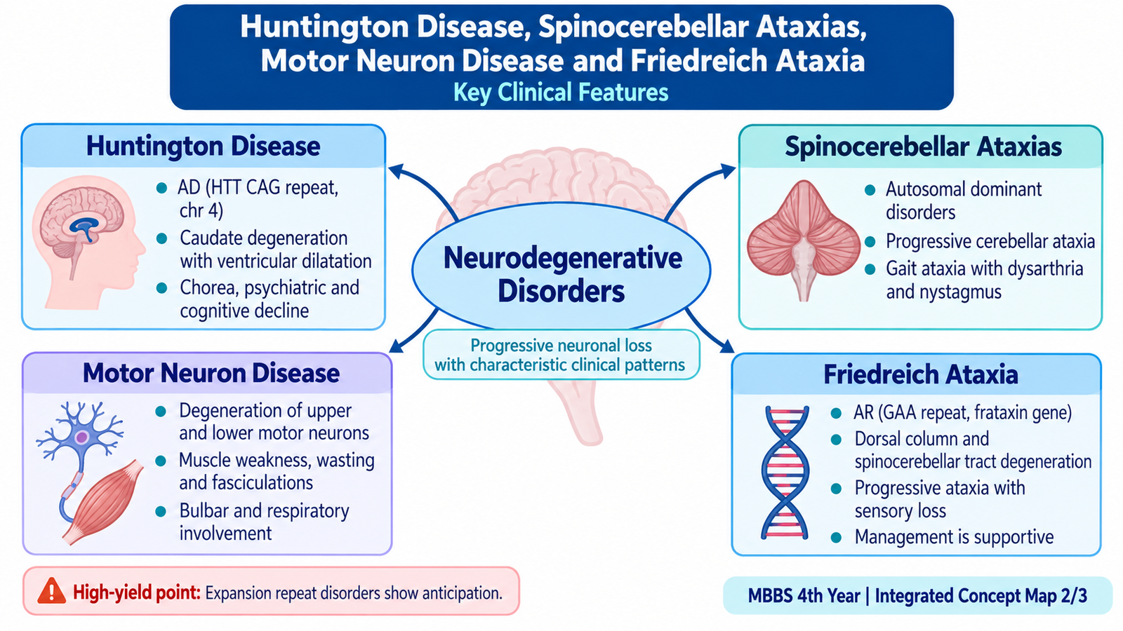
The disorders in this chapter affect different parts of the nervous system but share one important idea: selective degeneration of particular neuronal systems produces recognizable patterns of neurological dysfunction. Huntington disease mainly affects the basal ganglia and produces chorea together with cognitive and behavioral change. Spinocerebellar ataxias mainly disturb cerebellar coordination. Motor neuron disease damages upper and/or lower motor neurons and produces progressive weakness. Friedreich ataxia is an inherited disorder beginning commonly in childhood or adolescence and combines ataxia with sensory and pyramidal abnormalities. Understanding which neuronal pathway is damaged makes the apparently different clinical features much easier to remember.
A. Huntington Disease — Etiology, Pathogenesis, Morphology and Clinical Features
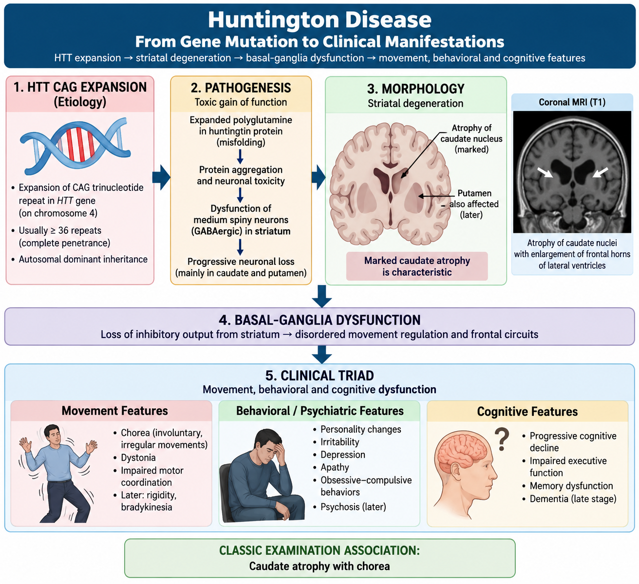
Huntington disease is a progressive inherited neurodegenerative disorder characterized mainly by involuntary choreiform movements, behavioral or psychiatric disturbance and progressive cognitive decline. The major pathological abnormality is degeneration of neurons in the striatum, particularly the caudate nucleus and putamen. Because these structures are important components of the basal ganglia motor circuits, their degeneration interferes with normal control of movement.
Etiology and Risk Factors
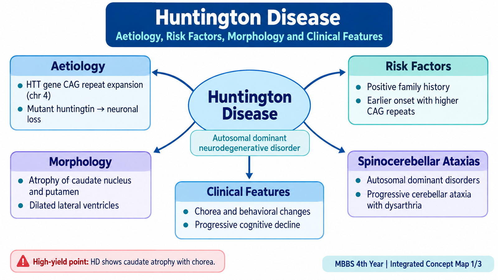
Huntington disease is caused by an expanded CAG trinucleotide repeat in the HTT gene on chromosome 4. The disease is inherited in an autosomal dominant manner. Therefore, an affected heterozygous parent can transmit the abnormal gene to either sons or daughters.
The expanded sequence encodes an abnormally long polyglutamine tract in the huntingtin protein. The abnormal protein interferes with normal neuronal function and promotes progressive neuronal injury. Larger repeat expansions are generally associated with earlier disease expression. The repeat may enlarge during transmission to the next generation, producing anticipation, in which disease appears at a younger age in later generations. This is especially recognized with paternal transmission.
HTT CAG-repeat expansion → abnormal huntingtin protein → neuronal dysfunction and degeneration → marked striatal neuronal loss → disturbed basal-ganglia motor control → chorea and progressive neurological dysfunction.
Because this is primarily a genetic disease, the most important identifiable risk is an affected family line or inherited pathogenic HTT expansion. Lifestyle factors are not the primary cause of Huntington disease.
Morphology
The pathological changes are most striking in the caudate nucleus and putamen. As neuronal loss progresses, the caudate becomes visibly smaller. This produces enlargement of the adjacent lateral ventricles. Cortical atrophy may also occur in advanced disease.
- Gross: atrophy of the caudate nucleus and putamen, often with secondary enlargement of the lateral ventricles.
- Microscopy: loss of striatal neurons with reactive gliosis.
- Medium spiny neurons of the striatum are particularly vulnerable.
- Abnormal huntingtin protein may accumulate within affected neurons.
Clinical Features
The disease usually progresses over years. The typical clinical picture combines movement disorder, behavioral change and cognitive decline. These manifestations arise because the pathological process involves interconnected basal-ganglia and cortical circuits rather than a single isolated motor pathway.
- Chorea: irregular, rapid, involuntary movements that seem to flow from one body region to another.
- Motor incoordination: voluntary movements become progressively less controlled.
- Behavioral and psychiatric change: irritability, altered personality, depression or other psychiatric manifestations may occur.
- Cognitive decline: progressive impairment of planning, judgment and other cognitive functions eventually leads to dementia.
- Speech, swallowing and mobility become increasingly impaired as disease advances.
Why chorea occurs: early degeneration within striatal inhibitory pathways reduces normal suppression of unwanted movement. Motor activity therefore becomes excessive and poorly controlled, producing the characteristic involuntary movements.
B. Spinocerebellar Ataxias — Clinical Pattern
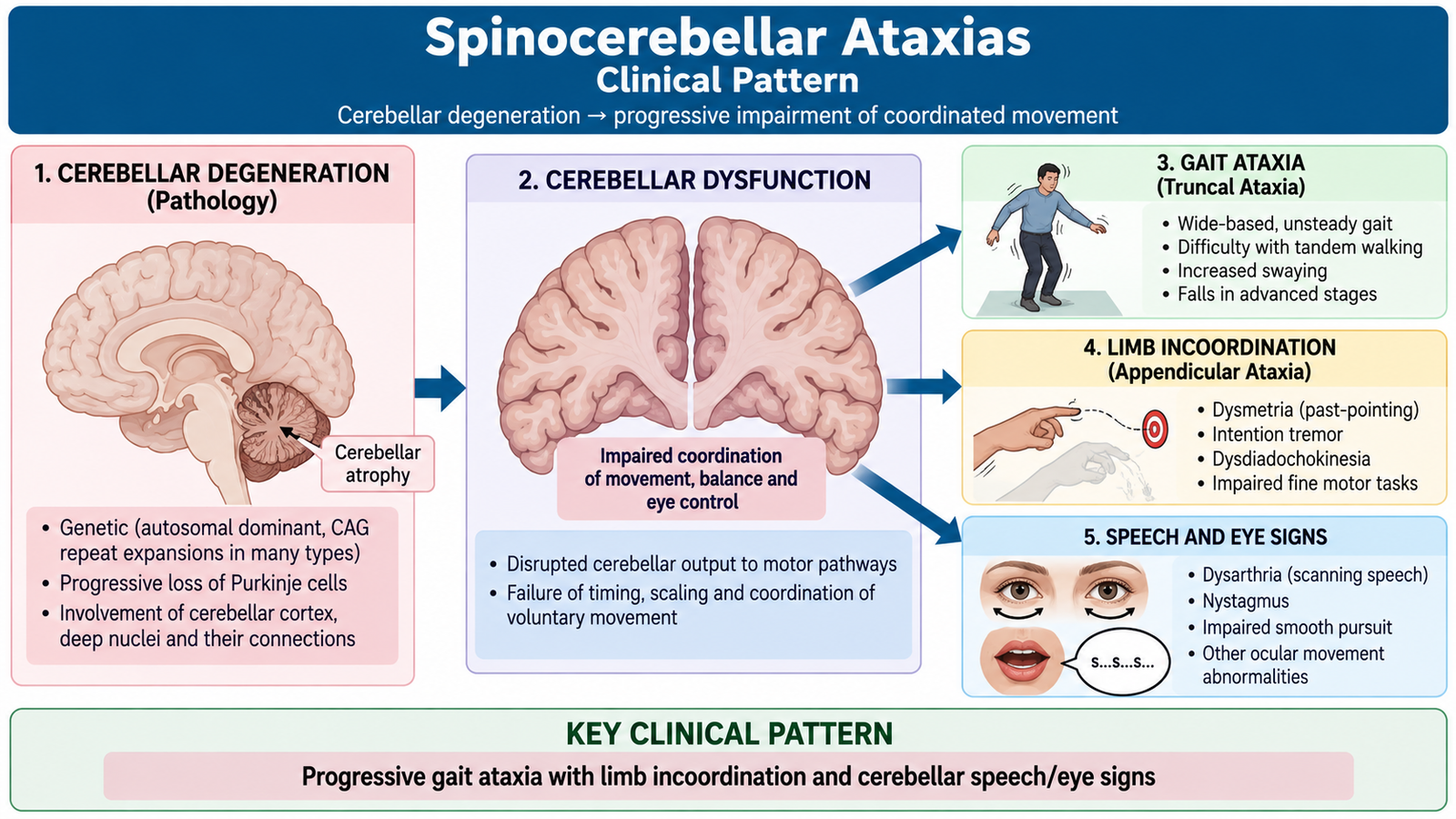
Spinocerebellar ataxias (SCAs) are a heterogeneous group of inherited neurodegenerative disorders in which degeneration involving the cerebellum and its connected pathways produces progressive loss of coordination. Many important forms are inherited in an autosomal dominant pattern, although the exact manifestations vary between individual SCA types.
The cerebellum normally coordinates the timing, range and accuracy of movement. When cerebellar pathways degenerate, muscle power may initially remain relatively preserved, but movements become inaccurate and poorly coordinated. This difference helps distinguish ataxia from weakness caused by motor-system disease.
Clinical Features
- Progressive gait ataxia: the patient develops an unsteady, broad-based gait.
- Limb ataxia: reaching and other purposeful limb movements become inaccurate.
- Dysarthria: speech becomes slow, irregular or scanning because speech muscles are poorly coordinated.
- Nystagmus or abnormal ocular movements may occur because cerebellar connections contribute to control of eye movements.
- Impaired coordination: abnormalities such as dysmetria and intention tremor may be present.
- Some SCA types may additionally show pyramidal, extrapyramidal or peripheral-nerve manifestations.
The exact combination of findings differs between SCA syndromes. The central clinical clue, however, is a gradually progressive cerebellar syndrome, commonly with a relevant family history.
C. Motor Neuron Disease — Etiology, Morphology and Major Types
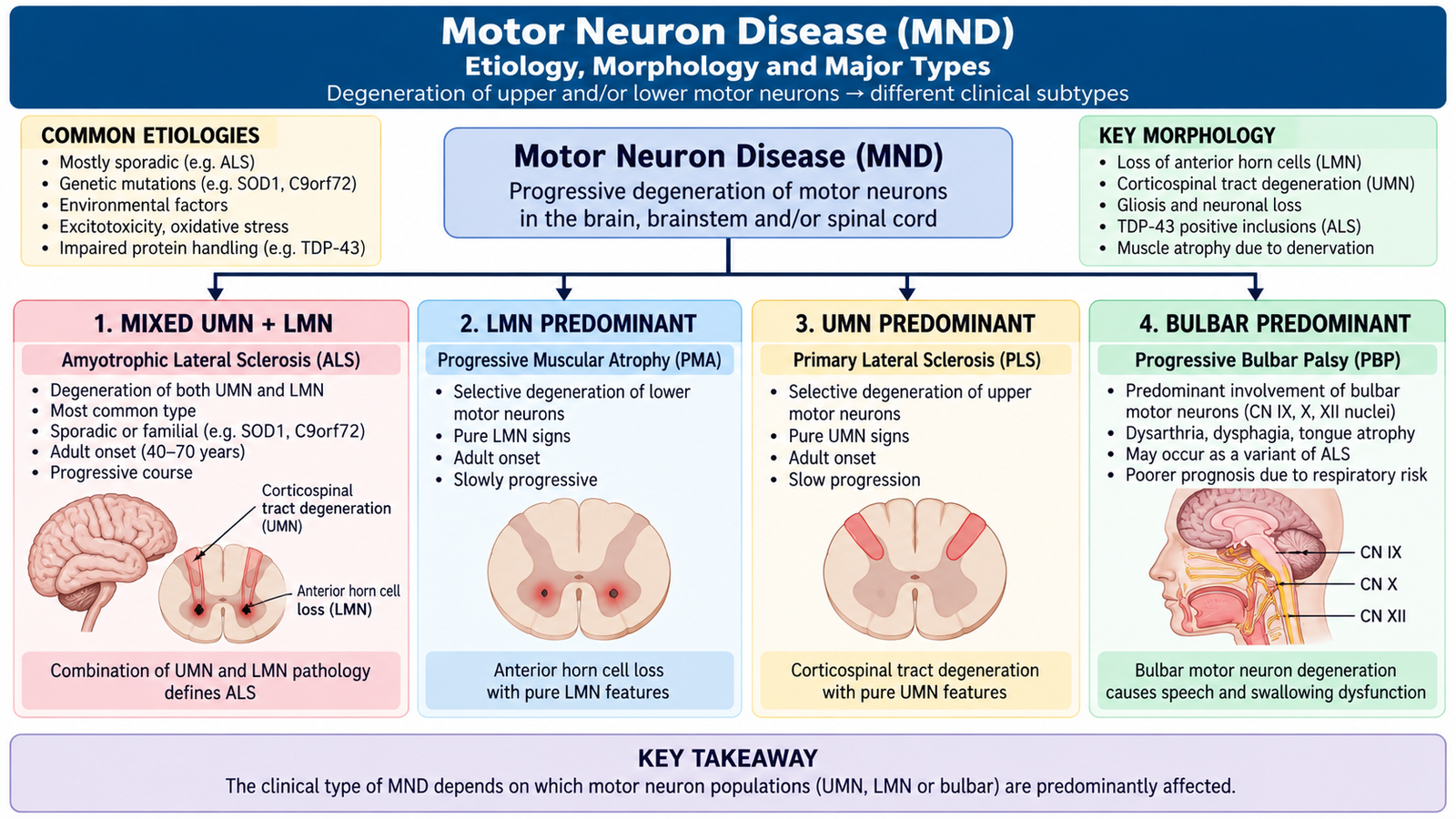
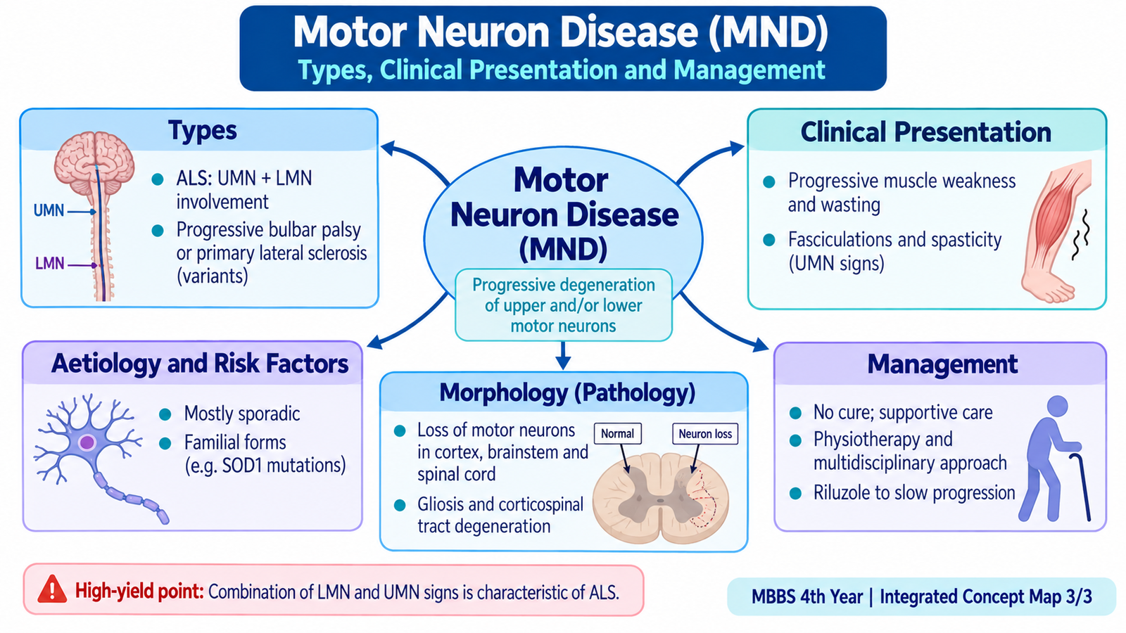
Motor neuron disease (MND) refers to a group of progressive neurodegenerative disorders that selectively damage motor neurons. Depending on the particular form, degeneration may affect upper motor neurons, lower motor neurons or both. Because motor neurons control voluntary skeletal muscle activity, their loss produces progressive weakness rather than primary sensory loss.
Etiology and Risk Factors
Most patients with motor neuron disease have no single identifiable cause and are considered to have sporadic disease. A smaller group has familial disease related to inherited genetic abnormalities. Several abnormalities of protein processing, RNA metabolism, oxidative injury and excitotoxicity have been implicated in motor-neuron degeneration.
The most recognizable predisposing factors are increasing age and inherited susceptibility. A positive family history is particularly important in familial MND. In most sporadic patients, however, a definite external initiating factor cannot be demonstrated.
Morphology
The morphological pattern depends on which motor-neuron populations are affected. In the common mixed upper- and lower-motor-neuron form, degeneration affects anterior horn cells in the spinal cord together with descending corticospinal pathways.
- Anterior horn: loss of lower motor neurons with reactive gliosis.
- Brainstem motor nuclei: degeneration may explain bulbar weakness.
- Corticospinal tracts: degeneration produces upper motor neuron manifestations.
- Skeletal muscle: denervation leads to groups of atrophic muscle fibers and progressive muscle wasting.
- Characteristic cytoplasmic inclusions may occur in affected neurons; abnormal TDP-43 accumulation is important in many cases.
- Bunina bodies are small eosinophilic neuronal inclusions classically associated with amyotrophic lateral sclerosis.
Anterior horn-cell loss → denervation → muscle weakness, wasting and fasciculations.
Corticospinal-tract degeneration → loss of descending motor control → spasticity, hyperreflexia and extensor plantar responses.
Major Types of Motor Neuron Disease
The major clinical types are classified according to whether upper motor neurons, lower motor neurons or bulbar motor neurons are predominantly affected.
- Amyotrophic lateral sclerosis (ALS): combines upper and lower motor neuron degeneration.
- Progressive muscular atrophy (PMA): predominantly lower motor neuron disease.
- Primary lateral sclerosis (PLS): predominantly upper motor neuron disease.
- Progressive bulbar palsy: predominantly affects bulbar motor neurons controlling speech and swallowing.
D. Motor Neuron Disease — Clinical Presentation
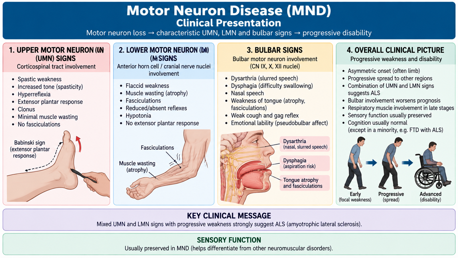
The clinical presentation of motor neuron disease follows directly from the neuronal populations being lost. A patient may initially develop focal weakness in a hand, arm or leg, or may present with difficulty speaking or swallowing. The weakness then progressively spreads. The coexistence of upper motor neuron and lower motor neuron signs in different body regions is particularly characteristic of ALS.
Lower Motor Neuron Features
Lower motor neurons directly supply skeletal muscle. When they degenerate, the muscle loses its neural input and undergoes denervation.
- Progressive muscle weakness
- Muscle wasting
- Fasciculations
- Muscle cramps
- Reduced muscle tone in predominantly lower-motor-neuron regions
- Reduced reflexes when the lower motor neuron lesion dominates
Upper Motor Neuron Features
Upper motor neurons normally regulate spinal motor activity. Loss of corticospinal control makes spinal reflex activity excessively active.
- Spasticity
- Increased muscle tone
- Brisk deep-tendon reflexes
- Extensor plantar responses
Bulbar Features
Degeneration of motor neurons supplying bulbar muscles affects speech, swallowing and control of oral secretions.
- Dysarthria: speech becomes weak, slow or poorly articulated.
- Dysphagia: swallowing becomes difficult and increases the risk of aspiration and poor nutrition.
- Weakness and wasting of the tongue may occur with lower motor neuron bulbar involvement.
- Emotional lability may occur with corticobulbar involvement.
Progression and Characteristic Clinical Pattern
The disease is progressive. Limb weakness may eventually impair walking, hand function and activities of daily living. Weakness of respiratory muscles can later compromise ventilation and is a major cause of serious deterioration.
An important diagnostic concept is that motor function is affected much more than sensory function. Sensory loss is not a typical dominant feature because the primary pathological process involves motor neurons rather than sensory pathways. Bladder control is also usually preserved until very advanced disease. These preserved functions help distinguish MND from several other neurological disorders.
E. Motor Neuron Disease — Management Principles
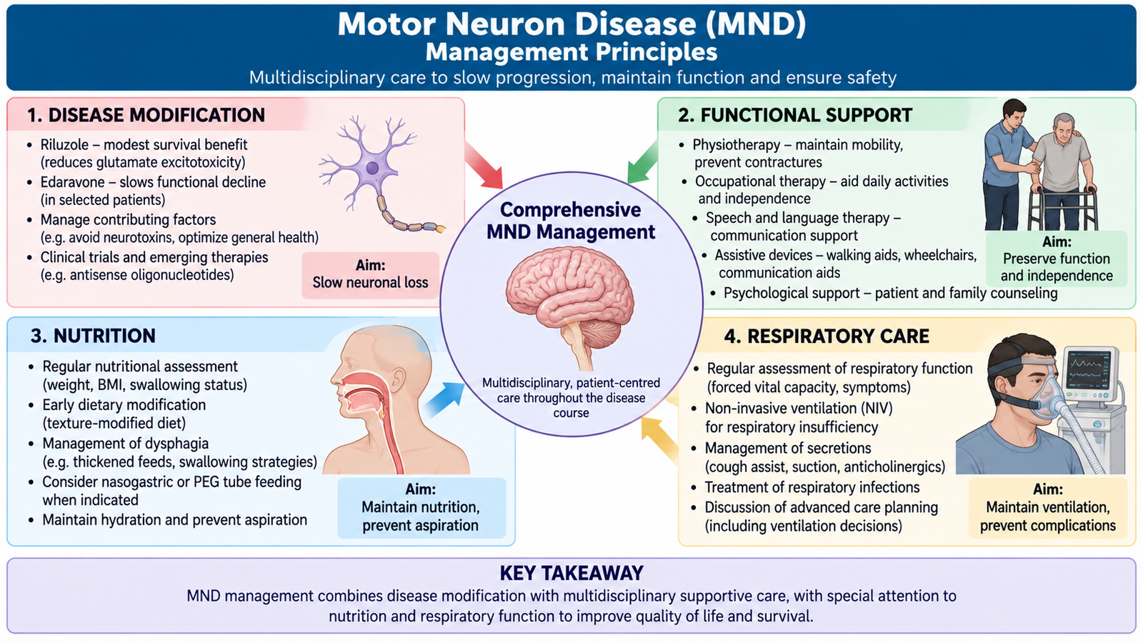
Motor neuron disease is a progressive neurodegenerative disorder for which treatment is aimed at slowing disease progression where possible, maintaining function, controlling symptoms and anticipating nutritional and respiratory complications. Management is therefore multidisciplinary rather than based on a single drug.
Disease-Modifying Treatment
Riluzole is an established disease-modifying treatment for ALS. It reduces glutamate-related excitotoxic effects and can modestly slow disease progression or prolong survival in appropriate patients. Edaravone is another disease-modifying therapy used in selected patients; its proposed benefit relates to reduction of oxidative neuronal injury.
These treatments do not restore neurons that have already been lost. This is why supportive care remains central even when disease-modifying therapy is used.
Supportive and Multidisciplinary Management
- Physiotherapy: maintains mobility, joint range and comfort for as long as possible.
- Occupational support: helps the patient adapt daily activities as weakness progresses.
- Speech and communication support: becomes important when dysarthria interferes with communication.
- Nutritional support: swallowing difficulty should be recognized early because it can lead to weight loss, dehydration and aspiration.
- Enteral feeding: may be considered when safe oral intake becomes inadequate.
- Respiratory assessment and support: respiratory-muscle weakness should be monitored and non-invasive ventilatory support may be required as function declines.
- Symptom control: spasticity, cramps, excessive salivation, pain and emotional symptoms should be treated appropriately.
- Psychological and palliative support: should accompany disease management because progressive loss of function has major effects on both the patient and family.
Progressive motor-neuron loss cannot be reversed → preserve remaining function → anticipate swallowing and respiratory failure → provide coordinated multidisciplinary support.
Respiratory and swallowing deterioration are especially important. Increasing breathlessness, weak cough, recurrent aspiration or marked dysphagia indicate progression and require specialist reassessment because they directly affect survival and safety.
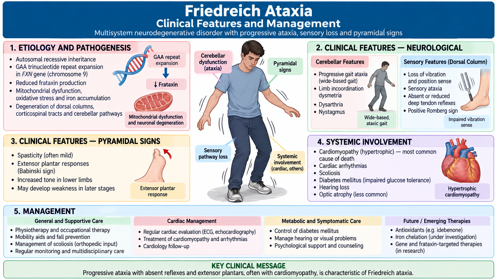
F. Friedreich Ataxia — Clinical Features and Management
Friedreich ataxia is an inherited progressive neurological disorder that usually begins in childhood or adolescence. It primarily affects pathways responsible for coordination, proprioception and motor control. The combination of cerebellar dysfunction, loss of deep sensation and corticospinal-tract involvement produces a characteristic mixed neurological picture.
The disorder is usually inherited in an autosomal recessive pattern and is associated with abnormalities of frataxin. A GAA repeat expansion reduces production of this mitochondrial protein. The resulting cellular dysfunction particularly affects vulnerable neurons and other tissues with high metabolic demands. Detailed molecular mechanisms are less important than recognizing how the affected neural pathways explain the clinical presentation.
Clinical Features
The usual presenting problem is progressively worsening gait and balance. Unlike a purely cerebellar disorder, Friedreich ataxia also damages sensory pathways and corticospinal tracts, so the examination contains features from several neurological systems.
- Progressive gait ataxia: unsteadiness commonly appears early.
- Limb ataxia: voluntary limb movements become poorly coordinated.
- Dysarthria: speech becomes slow and poorly coordinated.
- Loss of vibration and joint-position sense: occurs because posterior-column and sensory pathways are affected.
- Reduced or absent lower-limb tendon reflexes: reflects peripheral sensory or reflex-arc involvement.
- Extensor plantar responses: may coexist because corticospinal tracts are also affected.
- Muscle weakness develops as disease progresses.
- Pes cavus and scoliosis may develop as chronic neuromuscular imbalance affects growth and posture.
- Cardiomyopathy is an important systemic association.
- Abnormal glucose metabolism or diabetes may occur in some patients.
The apparently unusual combination of absent tendon reflexes with extensor plantar responses becomes understandable when both peripheral/reflex pathways and corticospinal tracts are affected at the same time.
Management
Management is mainly multidisciplinary and supportive. The aim is to preserve mobility and independence, recognize systemic complications and provide appropriate support throughout progression.
- Physiotherapy and rehabilitation help maintain mobility, balance, joint movement and functional independence.
- Mobility aids are introduced when required for safety and independence.
- Speech therapy may help when dysarthria interferes with communication.
- Orthopedic assessment is important when scoliosis or significant foot deformity develops.
- Cardiac surveillance and treatment are important because cardiomyopathy may contribute substantially to morbidity.
- Glucose abnormalities should be identified and managed when present.
- Psychological, educational and family support should be incorporated into long-term care.
- Genetic counseling is relevant because the disorder is inherited.
Integrated Mechanism Flow
Genetic mutation or degenerative process
Specific neuronal populations degenerate
Basal ganglia, cerebellar or motor pathways fail
Chorea, ataxia or progressive weakness appears
Mobility, speech, swallowing or respiration may deteriorate
Important Comparison — Major Motor Neuron Disease Patterns
| Type | Predominant Neurons Affected | Typical Pattern | Key Distinction |
|---|---|---|---|
| ALS | Upper + lower motor neurons | Weakness, wasting, fasciculations, spasticity and brisk reflexes | Mixed UMN and LMN signs |
| Progressive muscular atrophy | Lower motor neurons | Weakness, wasting and fasciculations | LMN-predominant pattern |
| Primary lateral sclerosis | Upper motor neurons | Progressive stiffness, spasticity and hyperreflexia | UMN-predominant pattern |
| Progressive bulbar palsy | Bulbar motor neurons | Dysarthria and dysphagia | Bulbar dysfunction dominates |
⭐ AIM High-Yield Review
Neurodegenerative & Hereditary Neurological Disorders
Use these focused videos after reading the learning material to reinforce the major disease mechanisms and clinical patterns.
1. Huntington Disease
Genetics, pathophysiology and major clinical manifestations.
2. Spinocerebellar Ataxia
Inherited cerebellar degeneration and the characteristic ataxic clinical pattern.
3. Motor Neuron Disease / Amyotrophic Lateral Sclerosis
Upper and lower motor neuron involvement, clinical presentation and management.
4. Friedreich Ataxia
Genetic basis, neurological manifestations and important systemic associations.