
This chapter follows the supplied KMU learning outcomes and builds the topic in a logical sequence. First understand where peripheral nerve function is disturbed and why the resulting clinical pattern develops; then use the final AIM High-Yield Review for rapid revision.
Peripheral Neuropathies and Neuromuscular Junction Disorders
GBS, Hereditary Neuropathies, Entrapment and Myasthenia Gravis
Neuroscience Module — understanding peripheral nerve injury, patterns of neuropathy, demyelinating disease, hereditary neuropathy, nerve entrapment and disorders of neuromuscular transmission.
Topic Introduction
Peripheral nerves carry motor, sensory and autonomic signals between the central nervous system and the rest of the body. Disease may damage the axon, its myelin sheath, an individual nerve at a compression site, or the neuromuscular junction where a motor nerve communicates with skeletal muscle. The site and type of damage determine the clinical pattern. In this chapter, you will learn the major pathological patterns of peripheral nerve injury, how peripheral neuropathies are classified and recognized, the mechanisms and management of Guillain–Barré syndrome and myasthenia gravis, the basis of chronic demyelinating neuropathy, important hereditary neuropathies in children, and common entrapment neuropathies including carpal tunnel syndrome.
A. Peripheral Nerve Injury: Pathological Patterns and Orthopedic Classification
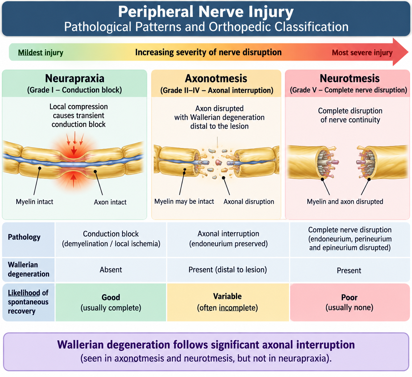
A peripheral nerve contains axons surrounded by Schwann-cell myelin and connective-tissue supporting layers. Injury may primarily damage the axon, primarily damage the myelin sheath, or physically disrupt the whole nerve. This distinction matters because conduction failure, recovery and the need for surgical intervention depend on which structures remain intact.
Axonal injury and Wallerian degeneration
When an axon is severely damaged or divided, the portion of the axon distal to the injury becomes disconnected from its cell body. Because the distal segment can no longer receive proteins and other materials transported from the neuron, it degenerates. This process is called Wallerian degeneration.
Schwann cells and macrophages remove degenerating axon and myelin. If the connective-tissue pathways remain preserved, Schwann cells help guide regenerating axonal sprouts toward the original target. If the nerve has been completely disrupted and the ends are no longer aligned, useful regeneration becomes much more difficult.
Segmental demyelination
In segmental demyelination, the myelin sheath is damaged at portions of the nerve while the underlying axon initially remains intact. Myelin permits rapid saltatory conduction, so its loss causes slowing of nerve conduction or complete conduction block. Schwann cells can remyelinate surviving axons, although newly formed myelin is initially thinner than normal.
Reaction of the neuronal cell body
Significant axonal injury also produces a reaction in the parent neuron. The cell body may enlarge and show central chromatolysis, reflecting increased protein synthesis required for repair. The functional outcome still depends mainly on the severity of axonal damage and whether an appropriate pathway for regeneration is preserved.
Orthopedic classification of peripheral nerve injury
A useful clinical classification divides peripheral nerve injury into three major types:
- Neurapraxia: temporary conduction block, usually related to focal myelin dysfunction, without anatomical disruption of the axon. Recovery is generally expected because the axon remains continuous.
- Axonotmesis: the axon is disrupted and Wallerian degeneration occurs distally, but some supporting connective-tissue structures remain preserved. Regeneration is therefore possible.
- Neurotmesis: the nerve is completely divided or severely disrupted. Spontaneous useful recovery is poor because regenerating axons may not reach their correct distal targets.
Clinical assessment and management principles
Peripheral nerve injury is assessed by identifying the affected nerve and documenting its motor, sensory and, where relevant, autonomic function. Associated fractures, vascular injury and soft-tissue damage must also be considered.
- Neurapraxia: usually managed conservatively while recovery is observed.
- Axonotmesis: requires observation for regeneration, protection of weak muscles and joints, rehabilitation and specialist assessment when recovery is uncertain.
- Suspected neurotmesis: requires early specialist evaluation because surgical repair may be necessary.
- Rehabilitation: physiotherapy, appropriate splintage and maintenance of joint movement help prevent contractures and functional loss while the nerve recovers.
B. Peripheral Neuropathy: Causes, Types, Distribution and Clinical Features
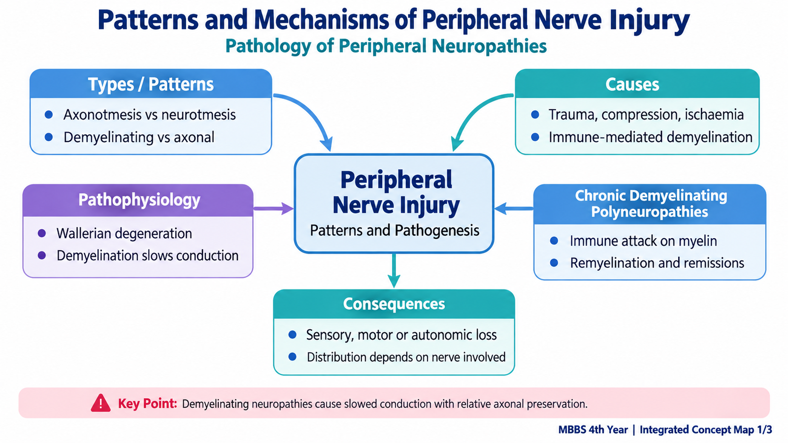
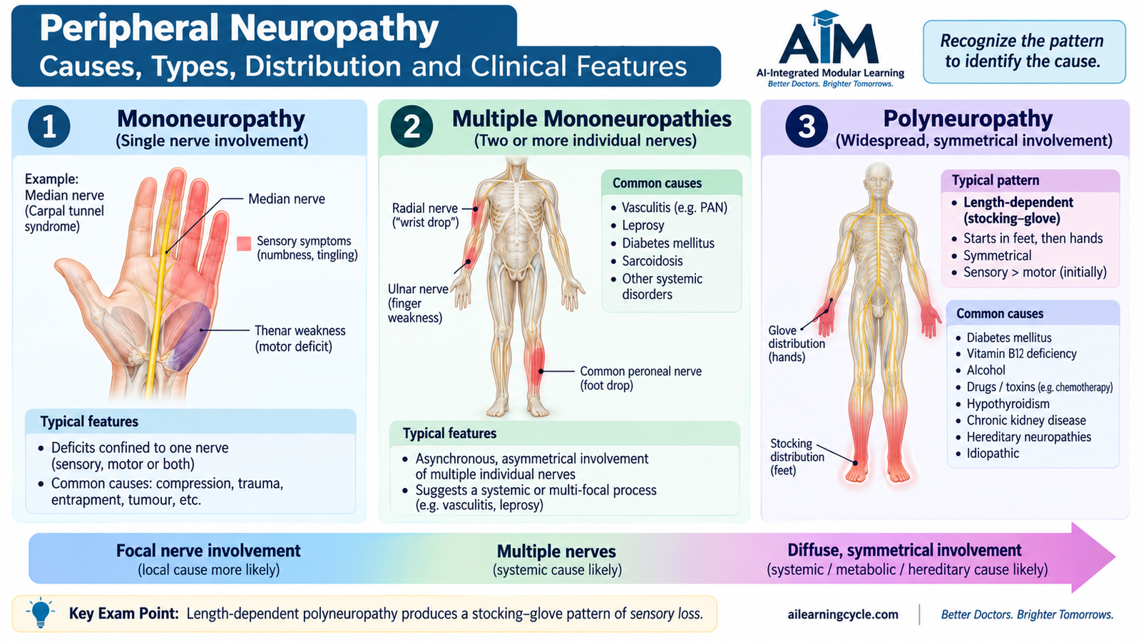
Peripheral neuropathy means dysfunction of peripheral nerves. It is not a single disease. Different disorders may preferentially affect axons or myelin, sensory or motor fibers, one nerve or many nerves. Recognizing the distribution is therefore one of the most useful steps in identifying the underlying process.
Important causes
Peripheral nerves may be damaged by metabolic disease, nutritional deficiency, toxins, immune mechanisms, hereditary abnormalities, infection, trauma or compression. Common clinically important groups include:
- Metabolic: particularly diabetes mellitus and advanced renal disease.
- Nutritional: deficiency of nutrients required for normal nerve function, including vitamin B12.
- Toxic or drug-related: alcohol and neurotoxic medications or chemicals.
- Immune-mediated: acute or chronic inflammatory demyelinating neuropathies.
- Hereditary: inherited disorders such as Charcot–Marie–Tooth disease.
- Mechanical: trauma or focal nerve compression.
Types according to pathological process
In an axonal neuropathy, the axon itself is the main target. Long axons are especially vulnerable, so symptoms often begin in the feet. In a demyelinating neuropathy, myelin is primarily damaged, causing marked slowing or block of conduction. Some diseases eventually produce both axonal and myelin damage.
Distribution of neuropathy
| Pattern | Meaning | Typical clinical pattern |
|---|---|---|
| Mononeuropathy | One peripheral nerve is affected. | Deficit follows the motor and sensory territory of that nerve. |
| Multiple mononeuropathies | Several individual nerves are affected in an asymmetric pattern. | Patchy motor and sensory deficits in different nerve territories. |
| Polyneuropathy | Many peripheral nerves are affected diffusely. | Usually bilateral and symmetrical, often beginning distally. |
Why does a stocking–glove pattern occur?
Many generalized axonal neuropathies are length dependent. The longest axons are affected first because maintaining a very long axon requires continuous transport of proteins and other materials over a large distance. Symptoms therefore usually begin in the toes and feet, then ascend in the legs. When the disease becomes sufficiently advanced, the fingertips and hands become involved, producing the classic stocking–glove distribution.
Clinical features
- Sensory: numbness, tingling, burning pain, impaired vibration or position sense, and reduced distal sensation.
- Motor: weakness, muscle wasting and, in distal neuropathies, difficulty with ankle dorsiflexion or foot drop.
- Reflexes: reduced or absent deep-tendon reflexes, particularly ankle reflexes in length-dependent neuropathy.
- Autonomic: some neuropathies disturb sweating, blood-pressure regulation, gastrointestinal function or other autonomic functions.
C. Guillain–Barré Syndrome
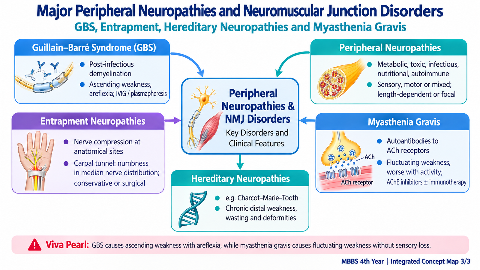
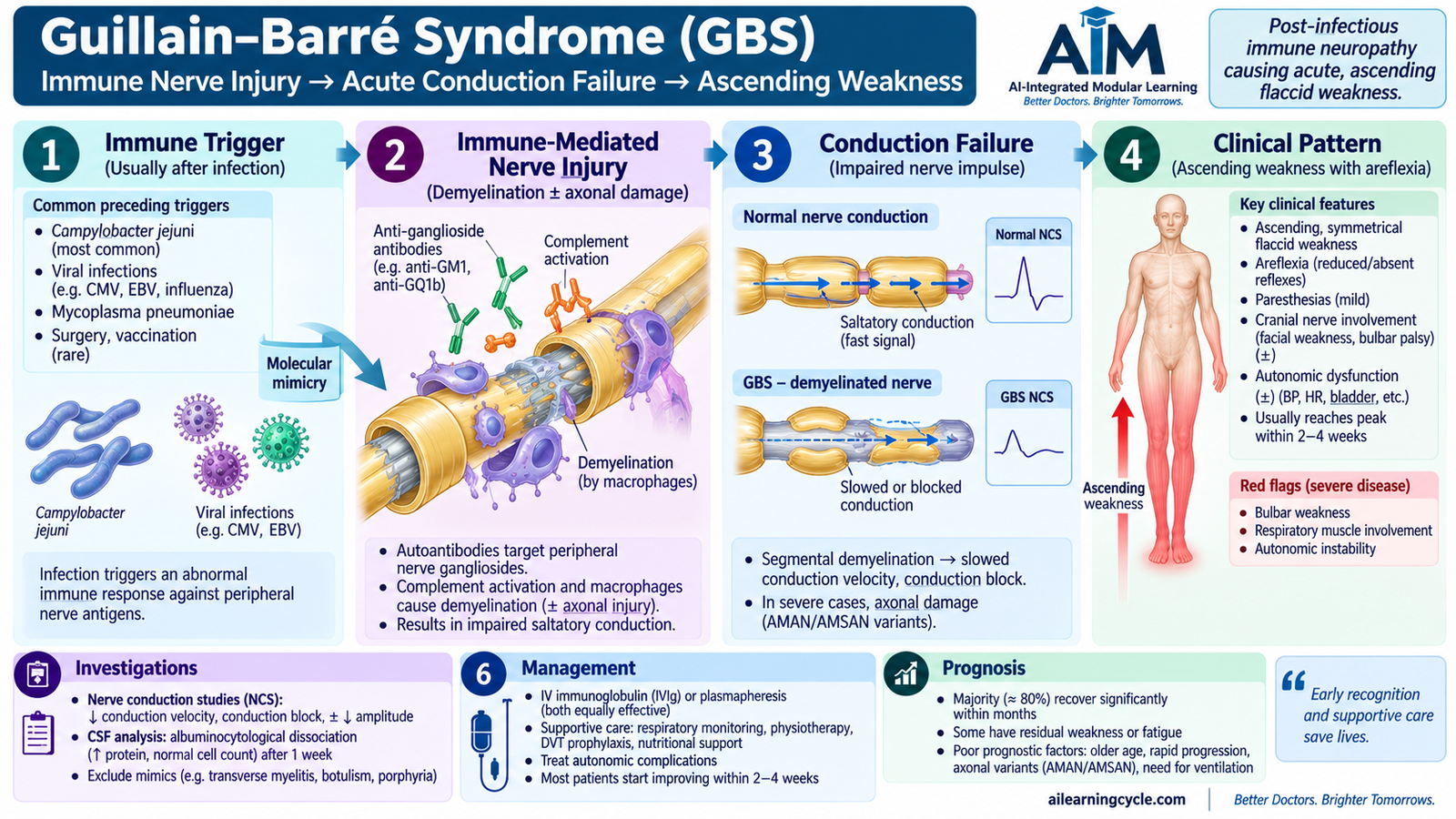
Guillain–Barré syndrome (GBS) is an acute immune-mediated polyradiculoneuropathy. The immune response attacks peripheral nerves and nerve roots, producing rapidly evolving weakness and reduced reflexes. In the common demyelinating form, myelin is the major initial target; axonal forms also occur.
Pathophysiology
GBS frequently follows an infection. The preceding immune response may recognize microbial antigens that resemble components of peripheral nerves. This phenomenon, called molecular mimicry, allows antibodies and immune cells to cross-react with nerve components.
Loss of myelin prevents rapid impulse conduction. When many motor fibers are affected, muscles receive inadequate neural stimulation and weakness develops. Reflex arcs also depend on intact peripheral sensory and motor fibers, which explains the characteristic reduction or loss of deep-tendon reflexes.
Clinical features
The classic presentation is rapidly progressive, relatively symmetrical weakness, usually beginning in the lower limbs and spreading upward. Sensory symptoms such as tingling may occur, but motor weakness is generally more prominent.
- Symmetrical limb weakness, often ascending.
- Reduced or absent deep-tendon reflexes.
- Paresthesia or mild sensory symptoms.
- Facial or other cranial nerve weakness in some patients.
- Bulbar weakness with difficulty swallowing in severe disease.
- Respiratory muscle weakness that may progress to ventilatory failure.
- Autonomic dysfunction, including unstable heart rate or blood pressure.
Diagnostic support
GBS is primarily recognized from its clinical pattern. Nerve-conduction studies can demonstrate features of demyelination or axonal damage. Cerebrospinal fluid classically develops an increased protein concentration with relatively few cells, known as albuminocytologic dissociation. A normal early cerebrospinal fluid result does not by itself exclude the clinical diagnosis.
Management
Management has two priorities: detect life-threatening deterioration early and modify the immune process. Patients with significant or progressing weakness require close monitoring of respiratory, bulbar and autonomic function.
- Respiratory monitoring and support: ventilatory assistance is required if respiratory muscle weakness becomes severe.
- Intravenous immunoglobulin (IVIG): reduces the harmful immune response and is an established disease-modifying treatment.
- Plasma exchange: removes circulating pathogenic immune factors and is another established treatment.
- Supportive care: includes prevention of complications of immobility, pain management, nutritional support and protection against aspiration when bulbar function is impaired.
- Rehabilitation: physiotherapy and gradual functional rehabilitation are important as nerve function recovers.
D. Chronic Demyelinating Polyneuropathies
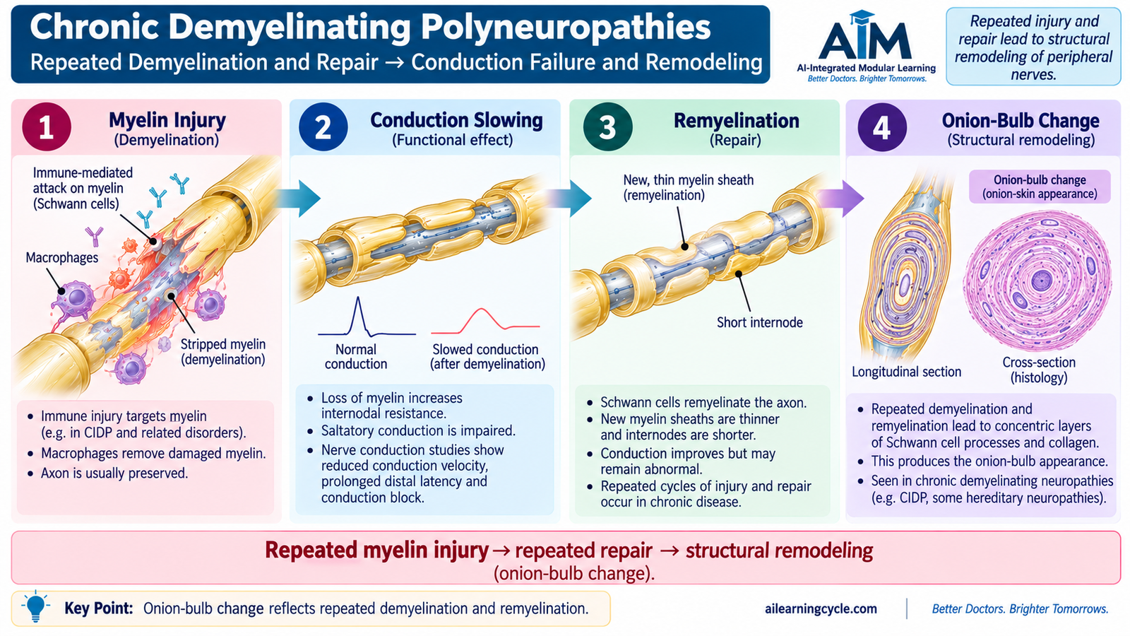
Chronic demyelinating polyneuropathies are disorders in which peripheral nerve myelin is repeatedly or persistently damaged over a longer period. The major pathological consequence is failure of saltatory conduction. An important acquired example is chronic inflammatory demyelinating polyneuropathy (CIDP), while inherited disorders can also produce chronic demyelination.
Pathophysiology of chronic immune-mediated demyelination
In an acquired chronic inflammatory demyelinating neuropathy, immune mechanisms injure myelin and Schwann cells along peripheral nerves and roots. Because myelin normally allows the impulse to jump rapidly between nodes of Ranvier, loss of myelin slows conduction and may produce conduction block.
Repeated cycles of demyelination and repair can stimulate Schwann-cell processes to accumulate around axons. On microscopy this may produce concentric layers called onion-bulb formations. These formations indicate repeated demyelination and remyelination rather than a single acute episode.
When motor fibers are affected, conduction failure causes weakness and reduced reflexes. Sensory fiber involvement produces numbness, paresthesia and loss of sensation. Long-standing demyelinating disease may eventually be accompanied by secondary axonal loss, which makes functional recovery less complete.
Acute versus chronic demyelinating disease
GBS and CIDP share the concept of immune-mediated peripheral nerve demyelination, but their clinical time course differs. GBS is an acute illness that progresses rapidly, whereas CIDP develops more gradually or follows a relapsing or progressive course over a longer period.
E. Hereditary Neuropathies in Children
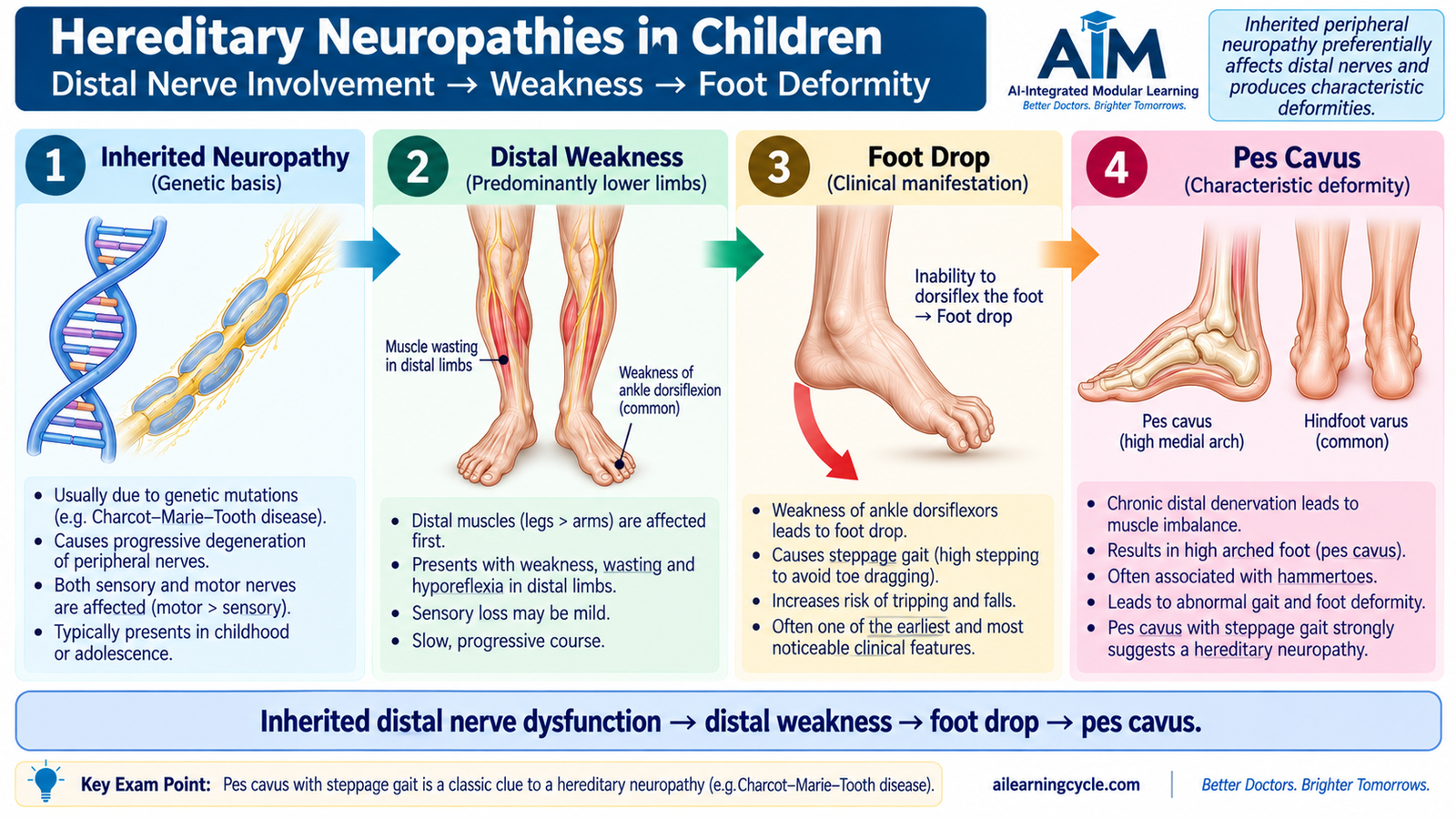
Hereditary neuropathies are genetically determined disorders of peripheral nerves. Because the abnormality is inherited, symptoms often begin during childhood, adolescence or early adult life and usually progress slowly. Many affect both motor and sensory fibers. The best-known group is Charcot–Marie–Tooth disease (CMT).
Major types
Hereditary neuropathies can be grouped according to the main function or nerve component affected. At undergraduate level, the important patterns include:
- Hereditary motor and sensory neuropathies: represented particularly by Charcot–Marie–Tooth disease.
- Demyelinating hereditary neuropathies: myelin dysfunction is prominent, as in common forms of CMT type 1.
- Axonal hereditary neuropathies: axonal degeneration predominates, as in CMT type 2.
- Hereditary neuropathy with liability to pressure palsies: peripheral nerves are unusually susceptible to focal compression.
- Hereditary sensory and autonomic neuropathies: sensory and autonomic functions are affected to varying degrees.
Clinical features of Charcot–Marie–Tooth disease
CMT usually produces a slowly progressive, length-dependent neuropathy. Long nerves supplying the distal lower limbs are affected early. Weakness and wasting of the small muscles of the feet and lower legs therefore appear before more proximal weakness.
- Distal leg weakness and muscle wasting.
- Difficulty with ankle dorsiflexion, producing foot drop.
- A high-stepping or steppage gait as the patient lifts the foot higher to prevent the toes dragging.
- Pes cavus and other long-standing foot deformities.
- Reduced or absent ankle reflexes.
- Variable distal sensory loss.
- Later involvement of distal upper-limb muscles in more advanced disease.
Because the disorder often develops gradually, a child may initially be brought because of frequent tripping, difficulty running, unusual footwear wear or progressive foot deformity rather than an obvious complaint of “neuropathy.” A family history can provide an important clue, although its absence does not exclude an inherited disorder.
Management principles
There is no single treatment that reverses all forms of hereditary neuropathy, so management is directed toward maintaining mobility, preventing deformity and maximizing function.
- Physiotherapy to preserve flexibility, strength and mobility.
- Appropriate footwear and orthoses for foot drop or instability.
- Prevention and treatment of contractures and progressive deformity.
- Orthopedic assessment when significant structural deformity interferes with function.
- Occupational support when hand weakness affects daily activities.
- Genetic assessment and counseling where appropriate for the child and family.
F. Entrapment Neuropathies and Carpal Tunnel Syndrome
An entrapment neuropathy occurs when a peripheral nerve is compressed at a predictable anatomical site, often where it passes through a narrow canal or around a bony prominence. Persistent pressure can disturb intraneural blood flow and myelin function, producing focal conduction failure. Longer-standing severe compression can eventually damage axons.
Examples of entrapment neuropathy
- Median nerve at the carpal tunnel.
- Ulnar nerve around the elbow.
- Common fibular nerve near the fibular neck.
The resulting weakness and sensory loss follow the anatomical distribution of the compressed nerve. This is different from a generalized polyneuropathy, in which multiple nerves are affected.
Carpal tunnel syndrome
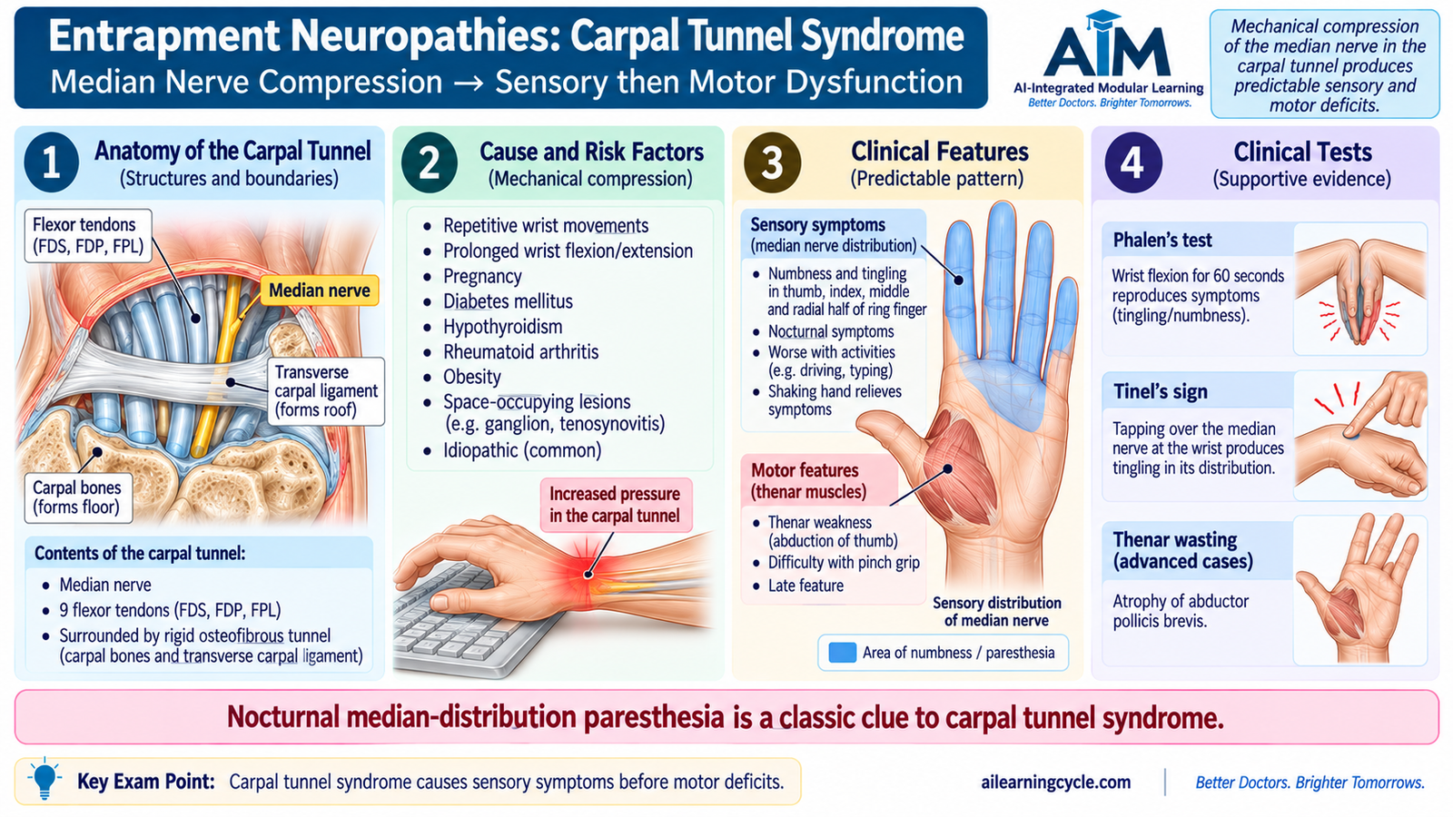
Carpal tunnel syndrome is compression of the median nerve as it passes beneath the flexor retinaculum through the carpal tunnel at the wrist. Increased pressure within this confined space interferes first with nerve conduction and, if prolonged, may lead to axonal injury.
Risk factors
Any factor that reduces available space in the tunnel, increases the volume of its contents or makes the nerve more vulnerable can contribute to carpal tunnel syndrome.
- Repetitive or sustained wrist loading.
- Pregnancy and other states associated with tissue swelling.
- Obesity.
- Diabetes mellitus.
- Hypothyroidism.
- Inflammatory joint disease such as rheumatoid arthritis.
- Previous wrist trauma or structural alteration.
Clinical features
Sensory symptoms commonly appear before obvious weakness. Patients often describe tingling, numbness or burning in the median-nerve distribution, particularly at night. Symptoms may wake the patient from sleep.
- Paresthesia involving the thumb, index finger, middle finger and radial side of the ring finger.
- Nocturnal numbness or pain.
- Difficulty with fine hand activity as motor involvement progresses.
- Weakness of median-innervated thenar muscles.
- Thenar wasting in advanced disease.
Sensation over the skin of the thenar eminence may remain relatively preserved because the palmar cutaneous branch of the median nerve usually leaves the nerve before it enters the carpal tunnel and therefore does not pass through the compressed tunnel.
Clinical examination
Phalen testing may reproduce median-nerve paresthesia when the wrist is maintained in flexion. Tinel testing may produce tingling in the median-nerve distribution when the nerve is tapped over the carpal tunnel. These findings support the diagnosis but should be interpreted together with the history and neurological examination.
Management
Management depends on severity, duration and evidence of motor or axonal impairment.
- Reduce or modify aggravating wrist activity when possible.
- A neutral-position wrist splint, particularly at night, can reduce pressure on the nerve.
- Local corticosteroid injection may provide symptomatic improvement in selected patients.
- Underlying contributing conditions should be addressed when present.
- Surgical decompression is considered when symptoms persist despite conservative treatment or when there is significant weakness, thenar wasting or evidence of severe nerve impairment.
G. Myasthenia Gravis
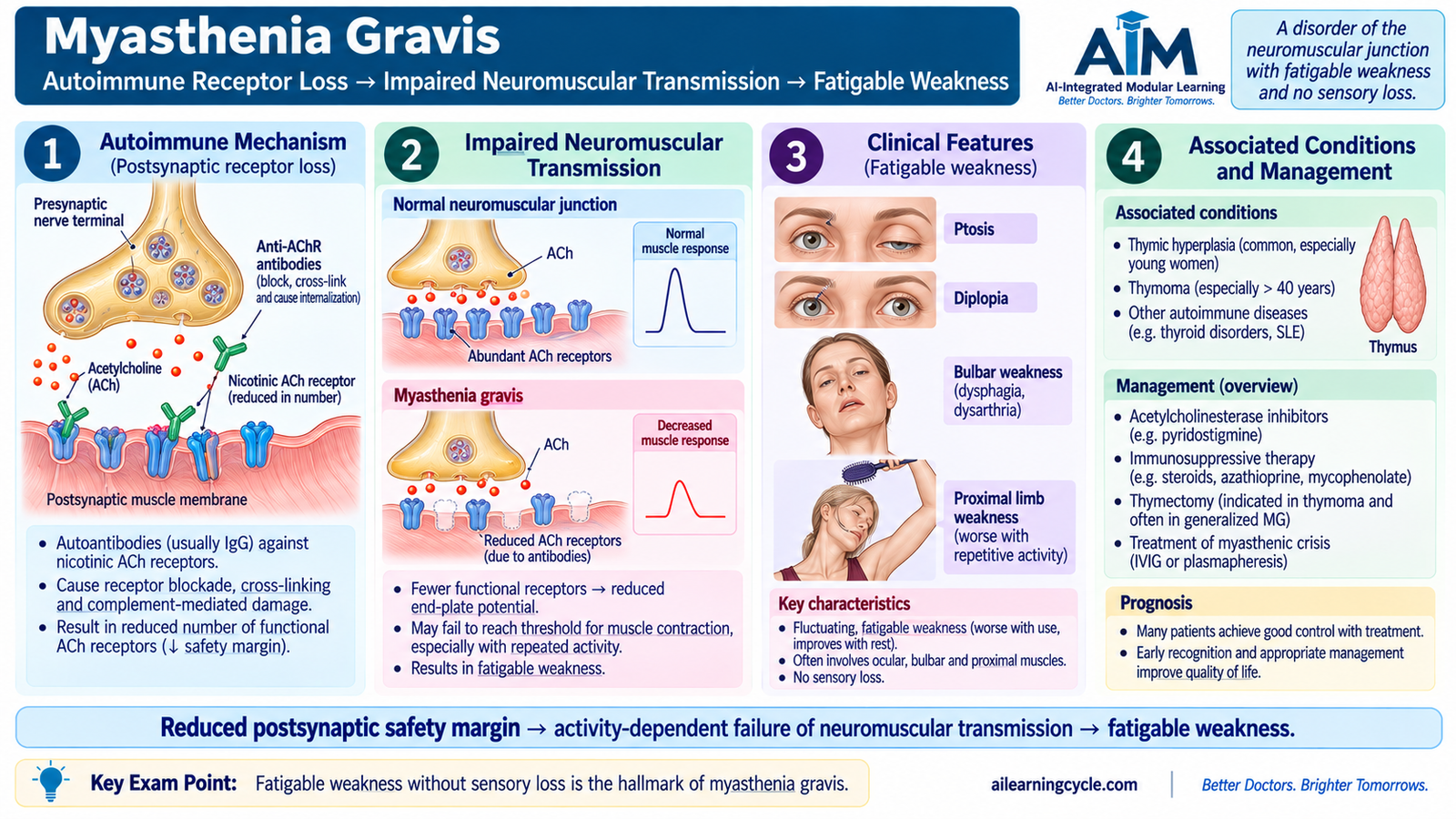
Myasthenia gravis (MG) is an autoimmune disorder of the postsynaptic neuromuscular junction. The peripheral nerve and skeletal muscle may be structurally capable of functioning, but transmission of the nerve impulse from nerve to muscle becomes unreliable. The hallmark clinical result is fluctuating, fatigable skeletal-muscle weakness.
Normal neuromuscular transmission
When a motor nerve impulse reaches the nerve terminal, acetylcholine is released into the neuromuscular junction. Acetylcholine binds to nicotinic acetylcholine receptors on the muscle end plate and produces depolarization. If the end-plate potential is sufficient, a muscle action potential is generated and the muscle contracts.
Pathophysiology
In most patients with autoimmune MG, antibodies are directed against the postsynaptic acetylcholine receptor. These antibodies reduce functional receptor availability and contribute to structural damage at the postsynaptic membrane. Some patients instead have antibodies against other proteins important for neuromuscular junction organization, such as MuSK.
At the beginning of activity, enough acetylcholine may still activate sufficient receptors for contraction. With repeated activity, the reduced safety margin becomes more important, so muscle power falls. After rest, transmission temporarily becomes more effective again. This explains the characteristic fatigability.
Clinical features
- Ocular weakness: fluctuating ptosis and diplopia are common presentations.
- Bulbar weakness: dysarthria, difficulty chewing or dysphagia may occur.
- Facial weakness: facial expression may become weak.
- Limb weakness: usually affects proximal muscles more than distal muscles.
- Fatigability: weakness becomes more apparent with sustained or repeated activity and improves after rest.
Because MG affects the neuromuscular junction rather than sensory nerves, sensation remains normal. Deep-tendon reflexes are usually preserved. Pupillary function is also generally spared.
Diagnostic approach
The diagnosis is supported by the characteristic pattern of fatigable weakness together with tests demonstrating autoimmune or neuromuscular-junction dysfunction. Antibodies against the acetylcholine receptor are important in many patients, while other antibodies such as MuSK may be relevant when acetylcholine-receptor antibodies are absent. Electrophysiological testing can demonstrate abnormal neuromuscular transmission.
When ptosis is present, temporary improvement after cooling the eyelid can support the clinical suspicion because lower temperature can improve neuromuscular transmission for a short period.
Management
Treatment aims to improve neuromuscular transmission, reduce the autoimmune attack and recognize situations requiring thymic treatment or emergency respiratory support.
- Acetylcholinesterase inhibition: pyridostigmine reduces breakdown of acetylcholine, allowing acetylcholine to remain longer at the neuromuscular junction and increasing the chance that the reduced number of receptors will be activated.
- Immunosuppression: corticosteroids and other immunosuppressive treatments may be used to reduce autoimmune activity when required.
- Thymectomy: is required when a thymoma is present and may also have a therapeutic role in selected patients with generalized autoimmune MG.
- Acute severe deterioration: IVIG or plasma exchange can produce relatively rapid improvement and are particularly useful in myasthenic crisis or serious exacerbation.
- Respiratory and bulbar support: airway protection and ventilatory support take priority when respiratory muscle weakness is severe.
Integrated Mechanism Flow
Important Comparison: GBS and Chronic Inflammatory Demyelinating Polyneuropathy
Both disorders can produce demyelination, weakness and reduced reflexes. The major undergraduate distinction is the time course.
| Feature | GBS | Chronic inflammatory demyelinating neuropathy |
|---|---|---|
| Course | Acute, rapidly evolving illness | More gradual, progressive or relapsing illness |
| Basic mechanism | Acute immune-mediated peripheral nerve/root injury | Persistent or recurrent immune-mediated demyelination |
| Reflexes | Usually reduced or absent | Usually reduced or absent |
| Pathological emphasis | Acute demyelination or axonal injury | Repeated demyelination/remyelination; onion-bulb change may develop |
⭐ AIM High-Yield Review
Peripheral Neuropathies & Neuromuscular Junction Disorders
Use these videos after reading the AIM Learning Material to reinforce the major mechanisms and clinical patterns.
1. Guillain–Barré Syndrome
Focus on pathophysiology, ascending weakness, areflexia, investigations and management.
2. Myasthenia Gravis
Focus on neuromuscular-junction dysfunction, fatigable weakness, diagnosis and treatment.