
25. Multiple Sclerosis, Transverse Myelitis and Other CNS Demyelinating Disorders
A disease-centered introduction to CNS demyelination, linking the pathological process to neurological dysfunction, characteristic morphology and the major principles of clinical management.
Topic Introduction
Demyelinating disorders damage the myelin that surrounds nerve fibers in the central nervous system. Because myelin allows rapid and efficient conduction of electrical impulses, its loss can produce weakness, sensory disturbance, visual symptoms, imbalance and other neurological deficits. Multiple sclerosis is the most important chronic CNS demyelinating disease in this topic. Transverse myelitis mainly affects the spinal cord and produces a characteristic combination of motor, sensory and autonomic dysfunction. Acute demyelinating encephalomyelitis and acute necrotizing hemorrhagic encephalitis are important pathological patterns of acute inflammatory demyelination. The key to understanding these disorders is to connect inflammation and myelin injury with the site of the lesion and the resulting neurological deficit.
A. Multiple Sclerosis: Core Concept and Pathogenesis
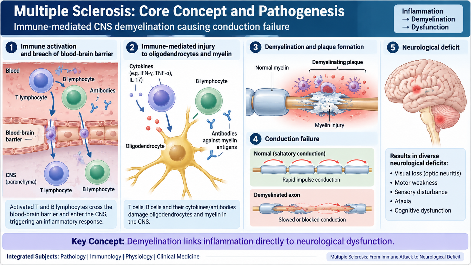
Multiple sclerosis (MS) is a chronic inflammatory demyelinating disease of the central nervous system. It affects the brain, optic nerves and spinal cord. The disease produces multiple areas of myelin loss, called plaques, which occur at different sites and may develop at different times.
The basic problem is an immune-mediated attack against CNS myelin and oligodendrocytes. Oligodendrocytes are the cells responsible for producing myelin in the central nervous system. Myelin normally insulates axons and allows impulses to travel rapidly by saltatory conduction. When myelin is damaged, conduction becomes slow or may fail completely. This explains why a demyelinating plaque can cause a neurological deficit even when many axons initially remain structurally intact.
How the disease develops
MS develops in a genetically susceptible person in whom environmental influences are believed to contribute to abnormal immune activation. Activated lymphocytes cross the blood-brain barrier and initiate inflammation within the CNS. T lymphocytes, B lymphocytes, macrophages and their inflammatory mediators participate in damage to myelin and oligodendrocytes.
Immune activation → lymphocytes enter the CNS → inflammatory injury to oligodendrocytes and myelin → focal demyelination → impaired impulse conduction → neurological deficit.
Early inflammatory lesions may show substantial demyelination while many axons are still preserved. This relative axonal preservation helps explain why neurological function can improve after an acute episode when inflammation settles and surviving axons regain some ability to conduct impulses. With repeated or severe disease, however, irreversible axonal loss and gliosis may occur, producing persistent disability.
Different plaques affect different CNS pathways. For example, involvement of the optic nerve can disturb vision, a spinal cord plaque can produce weakness or sensory dysfunction, and lesions involving cerebellar pathways can impair coordination.

B. Multiple Sclerosis: Morphology and Clinicopathological Correlation
The characteristic pathological lesion of MS is the demyelinating plaque. Plaques are well-demarcated areas in which myelin has been damaged or lost. Their appearance changes according to whether the lesion is actively inflamed or has become an old inactive scar.
Distribution of plaques
MS plaques commonly occur in CNS white matter, particularly in locations where relatively small lesions can disturb important neurological pathways. Characteristic sites include:
- periventricular white matter;
- optic nerves and optic pathways;
- brainstem;
- cerebellar pathways;
- spinal cord.
The presence of lesions in different anatomical locations explains why MS may produce a mixture of visual, motor, sensory, cerebellar and other neurological manifestations.
Gross morphology
On gross examination, established plaques are typically sharply defined areas of discoloration within the CNS. Older lesions may appear firm because of astrocytic gliosis. Their distribution is often more diagnostically informative than their individual size.
Microscopic morphology
An active plaque is an area of ongoing inflammation and myelin breakdown. Macrophages remove damaged myelin, and lymphocytes are commonly present around small blood vessels. Axons may initially be relatively preserved despite extensive myelin loss.
- loss and breakdown of myelin;
- macrophages containing myelin debris;
- perivascular lymphocytic inflammation;
- injury or loss of oligodendrocytes;
- relative preservation of many axons in early lesions.
As inflammation becomes less active, the lesion evolves into an inactive plaque. Macrophages and inflammatory cells decrease, myelin remains lost, and astrocytic gliosis becomes prominent. Variable axonal loss may be present, particularly in longstanding disease.

C. Multiple Sclerosis: Clinical Features and Management
The clinical manifestations of MS depend on where demyelinating plaques occur. A patient therefore does not have one fixed neurological pattern. Different parts of the CNS may be affected during different episodes, and symptoms may improve, recur or become persistent.
Important clinical features
Visual symptoms are particularly important. Demyelination of the optic nerve may cause optic neuritis, typically producing reduced vision associated with pain on eye movement. Damage to other CNS pathways can produce motor, sensory, cerebellar or brainstem manifestations.
- Motor: weakness, stiffness and upper motor neuron features such as spasticity and exaggerated reflexes.
- Sensory: numbness, tingling or altered sensation.
- Visual: optic neuritis, reduced visual acuity or other visual disturbance.
- Brainstem: diplopia or other cranial-nerve-related symptoms depending on lesion location.
- Cerebellar: imbalance, gait disturbance, tremor or incoordination.
- Spinal/autonomic: bladder dysfunction may occur when relevant CNS pathways are involved.
The important clinical principle is dissemination of neurological dysfunction within the CNS. Symptoms arising from anatomically separate pathways strongly support a multifocal CNS process rather than a single peripheral nerve lesion.
Clinical assessment
Diagnosis is based on the neurological pattern together with evidence that demyelinating lesions are present in characteristic CNS locations. MRI is particularly useful because it can demonstrate plaques in the brain and spinal cord. Additional investigations may be used by clinicians when needed to support the diagnosis or exclude alternative disorders.
Management principles
Management has three broad aims: treat acute inflammatory relapses, reduce future disease activity and manage the functional consequences of neurological damage.
1. Acute relapse
Corticosteroids are used for significant acute inflammatory relapses because they suppress inflammation and can speed recovery. Severe attacks that fail to respond adequately may require specialist escalation such as plasma exchange.
2. Disease-modifying treatment
Disease-modifying therapies reduce inflammatory disease activity and the frequency of future relapses in appropriate patients. Available therapies act at different parts of the immune response. Examples include interferon-beta, glatiramer acetate, selected oral immunomodulatory drugs and monoclonal-antibody therapies. Selection requires specialist assessment because efficacy, route of administration and adverse-effect risks differ between agents.
3. Symptomatic treatment and rehabilitation
Persistent problems such as spasticity, pain, bladder dysfunction, mobility limitation and fatigue require symptom-directed care. Physiotherapy, occupational therapy and rehabilitation help maintain independence and function. Psychological and social support may also be important in a chronic neurological disease.

D. Transverse Myelitis: Etiology, Pathophysiology and Clinical Features
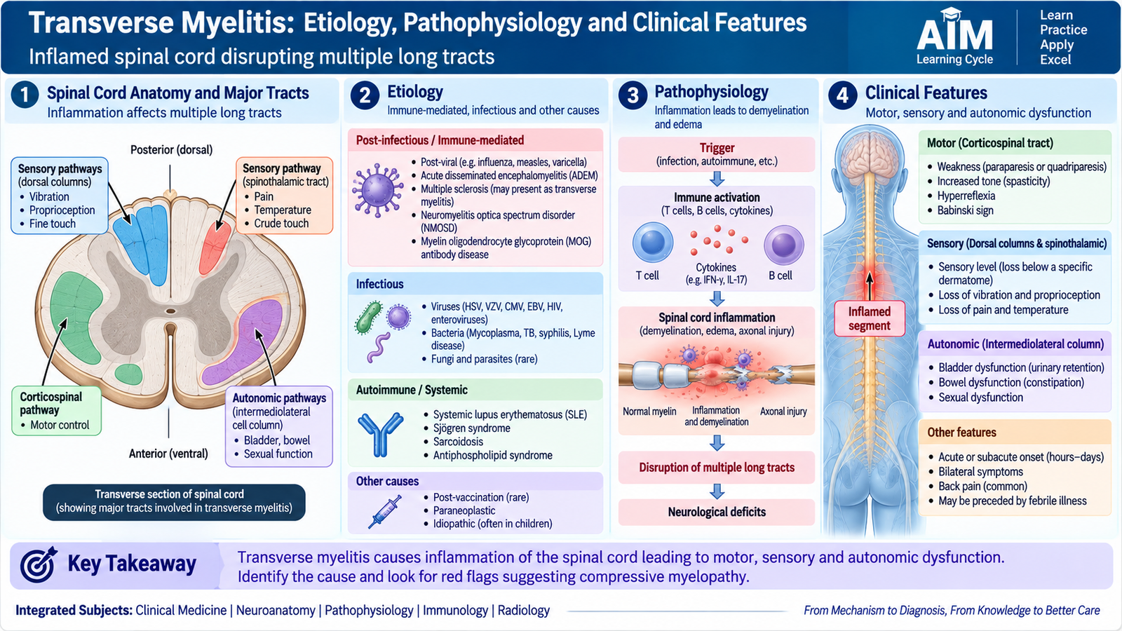
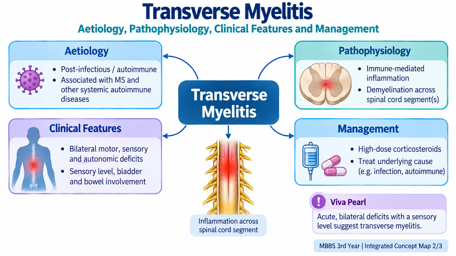
Transverse myelitis is an inflammatory disorder of the spinal cord that produces neurological dysfunction below the affected level. The word myelitis means inflammation of the spinal cord. The term transverse reflects involvement across the cord sufficient to disturb several functional pathways at approximately the same spinal level.
This produces the characteristic combination of motor, sensory and autonomic abnormalities. Unlike a single peripheral nerve lesion, the neurological findings involve functions carried by several long spinal tracts.
Etiology
Transverse myelitis may occur as an isolated inflammatory disorder or in association with another disease. Important etiological groups include:
- idiopathic inflammatory transverse myelitis;
- post-infectious or infection-associated inflammation;
- CNS demyelinating disease, including multiple sclerosis;
- other immune-mediated demyelinating disorders;
- systemic autoimmune inflammatory disease.
The cause matters because transverse myelitis is a syndrome of spinal cord inflammation, not a single disease with one universal mechanism.
Pathophysiology
Inflammation within a segment of the spinal cord damages myelin and may also injure axons. Damage to multiple long tracts interrupts communication between the brain and structures below the lesion.
Spinal cord inflammation → myelin injury and edema → conduction failure in motor, sensory and autonomic pathways → neurological deficits below the lesion.
Damage to descending corticospinal pathways produces weakness. Interruption of ascending sensory pathways produces altered or lost sensation. Involvement of autonomic pathways can impair bladder, bowel and sexual function.
Clinical features
Symptoms usually develop over a relatively short period and reflect the spinal cord level involved. The most important diagnostic clue is the simultaneous involvement of several spinal-cord functions.
- Motor dysfunction: weakness affecting the limbs below the lesion.
- Sensory dysfunction: numbness, tingling or altered sensation, often with a clinically identifiable sensory level.
- Autonomic dysfunction: urinary retention or other bladder disturbance, bowel dysfunction and sometimes sexual dysfunction.
- Pain: back pain or radicular-type discomfort may occur.
A sensory level is especially useful because it points toward a spinal cord lesion. The patient may describe relatively normal sensation above a particular level and altered sensation below it.
E. Transverse Myelitis: Management and Red Flags
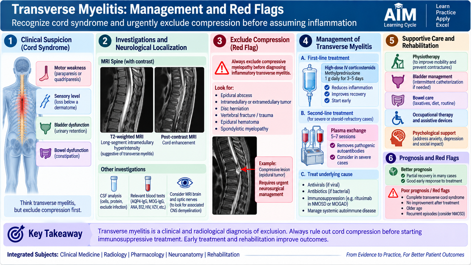
Transverse myelitis requires prompt assessment because similar symptoms can result from mechanical compression of the spinal cord, which may require urgent surgical treatment. The first management priority is therefore not simply to label the illness as inflammatory, but to identify the spinal-cord syndrome and exclude dangerous alternative causes.
Initial assessment
Neurological examination helps identify the level and severity of cord dysfunction. MRI of the spinal cord is important because it can demonstrate an inflammatory lesion and, crucially, identify or exclude a compressive lesion. Depending on the clinical setting, brain imaging, cerebrospinal-fluid examination and targeted investigations may help determine whether the episode is associated with infection, MS or another inflammatory disorder.
Treatment principles
When an acute inflammatory transverse myelitis is established, treatment aims to suppress spinal-cord inflammation, prevent complications and preserve function.
- Anti-inflammatory treatment: corticosteroids are commonly used for significant acute inflammatory disease.
- Escalation: severe disease with inadequate response may require specialist consideration of plasma exchange.
- Treat an identified cause: management must be adapted when a specific infection, systemic autoimmune disease or underlying demyelinating disorder is found.
- Bladder and bowel care: autonomic dysfunction needs active assessment and supportive management.
- Rehabilitation: physiotherapy and rehabilitation are important when weakness, mobility limitation or functional impairment persists.
Recovery varies because neurological dysfunction may arise from reversible inflammation and conduction block or from more permanent axonal injury. Early recognition and appropriate treatment therefore matter clinically.
F. Acute Demyelinating Encephalomyelitis and Acute Necrotizing Hemorrhagic Encephalitis: Morphology
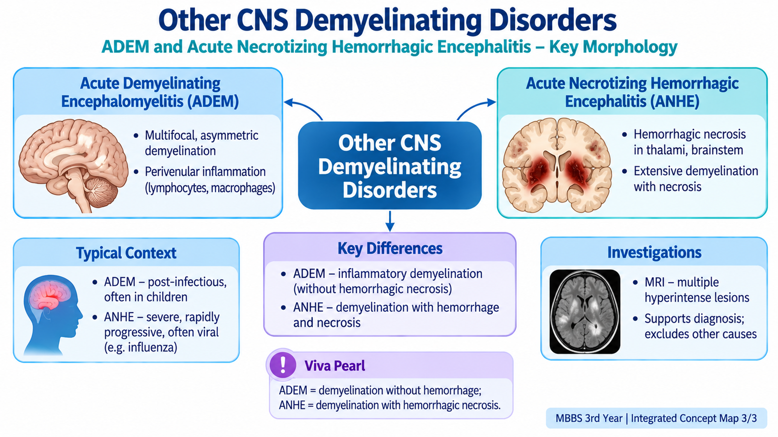
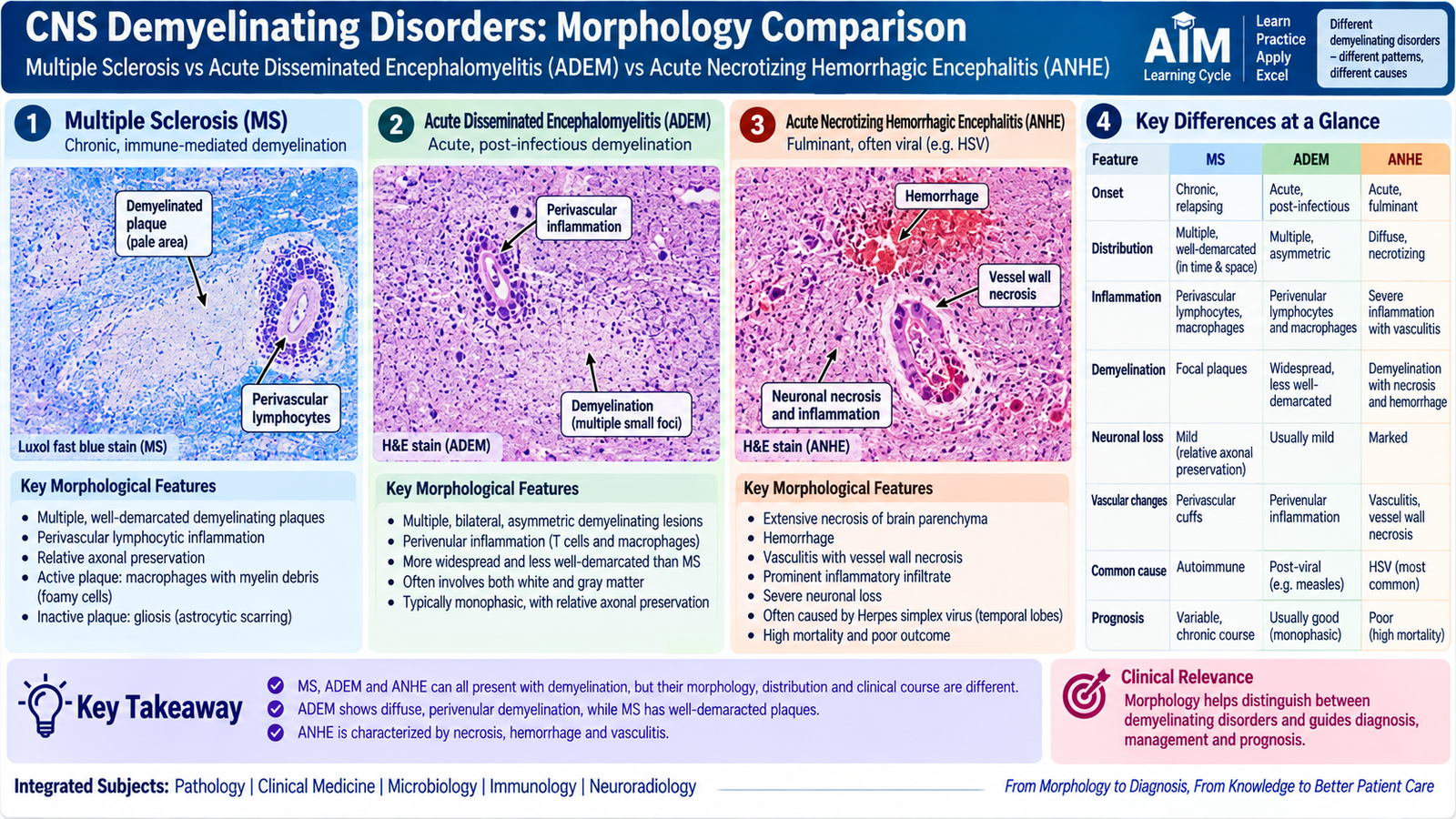
Acute demyelinating encephalomyelitis (ADEM) and acute necrotizing hemorrhagic encephalitis represent acute inflammatory demyelinating processes. In this curriculum, their most important requirement is recognition of their characteristic morphological pattern.
Acute demyelinating encephalomyelitis
The characteristic lesion of ADEM is perivenular demyelination. Small veins and venules are surrounded by inflammatory cells, and myelin is lost in sleeves around these vessels.
Microscopically, the important features are:
- perivascular inflammatory infiltrates, particularly around small veins and venules;
- perivenular zones of demyelination;
- macrophages associated with areas of myelin breakdown;
- multifocal involvement of CNS white matter.
The distribution is useful diagnostically: rather than the sharply defined established plaques typical of MS, ADEM characteristically shows areas of demyelination centered around small vessels.
Acute necrotizing hemorrhagic encephalitis
Acute necrotizing hemorrhagic encephalitis, also called acute hemorrhagic leukoencephalitis, is a severe fulminant inflammatory demyelinating process. Morphologically, it resembles an aggressive hemorrhagic and necrotizing form of acute demyelinating encephalomyelitis.
Important morphological findings include:
- widespread or multifocal demyelination;
- prominent tissue necrosis;
- hemorrhage within affected areas;
- marked perivascular inflammation;
- severe vascular injury, which contributes to hemorrhage and tissue destruction.
Integrated Mechanism Flow
Important Comparison: Demyelinating Morphology
| Feature | Multiple Sclerosis | ADEM | Acute Necrotizing Hemorrhagic Encephalitis |
|---|---|---|---|
| Basic lesion | Focal demyelinating plaque | Perivenular demyelination | Severe demyelination with hemorrhage and necrosis |
| Inflammation | Prominent in active plaques | Centered around small veins and venules | Marked perivascular inflammation |
| Hemorrhage | Not the defining feature | Not the defining feature | Prominent |
| Necrosis | Not a typical defining feature | Not the dominant feature | Prominent tissue necrosis |
| High-yield clue | Sharply defined plaques | Sleeves of demyelination around venules | Hemorrhagic, necrotizing demyelinating lesion |
AIM High-Yield Review
- ⭐ Multiple sclerosis is an immune-mediated inflammatory demyelinating disease of the central nervous system.
- The central lesion of MS is the demyelinating plaque.
- Active MS plaques show myelin breakdown, macrophages and perivascular lymphocytes, with relative preservation of many axons early in the lesion.
- Inactive plaques show persistent myelin loss and prominent astrocytic gliosis, with variable axonal loss.
- MS plaques commonly affect periventricular white matter, optic pathways, brainstem, cerebellar pathways and spinal cord.
- The neurological manifestation of MS depends on the anatomical pathway affected by demyelination.
- Acute significant MS relapses are treated with anti-inflammatory therapy, while disease-modifying therapies aim to reduce future inflammatory disease activity.
- ⭐ Transverse myelitis produces a spinal cord syndrome involving motor, sensory and autonomic pathways.
- A sensory level combined with weakness and bladder or bowel dysfunction is an important clue to spinal cord disease.
- Spinal-cord compression must be excluded urgently when evaluating suspected transverse myelitis.
- Management of inflammatory transverse myelitis includes suppression of inflammation, treatment of an identified cause, supportive care and rehabilitation.
- ⭐ ADEM characteristically shows perivenular demyelination with perivascular inflammation.
- Acute necrotizing hemorrhagic encephalitis is distinguished by severe inflammatory demyelination associated with hemorrhage, vascular injury and necrosis.
- Persistent neurological disability in demyelinating disease becomes more likely when inflammation is accompanied by irreversible axonal injury.
🎥 Recommended Video
Multiple Sclerosis — Etiology, Pathophysiology, Clinical Features, Diagnosis & Treatment
Ninja Nerd