
Tumours of the Nervous System: Brain, Spinal Cord and Peripheral Nerve Tumours
Topic Introduction
Tumours of the nervous system arise from several different cell types and may involve the brain, meninges, spinal canal or peripheral nerves. Their clinical importance depends not only on whether they are histologically benign or malignant, but also on where they grow. Even a slowly growing benign tumour can become dangerous when it compresses the brain, spinal cord or an important cranial nerve. In this chapter, you will learn the major brain tumour groups and their morphology, understand the presentation and surgical principles of spinal tumours, and distinguish peripheral nerve sheath tumours. The final section connects these tumours with neurofibromatosis, an inherited group of disorders in which particular nerve-system tumours occur with increased frequency.
A. How Nervous System Tumours Are Classified and Why Location Matters
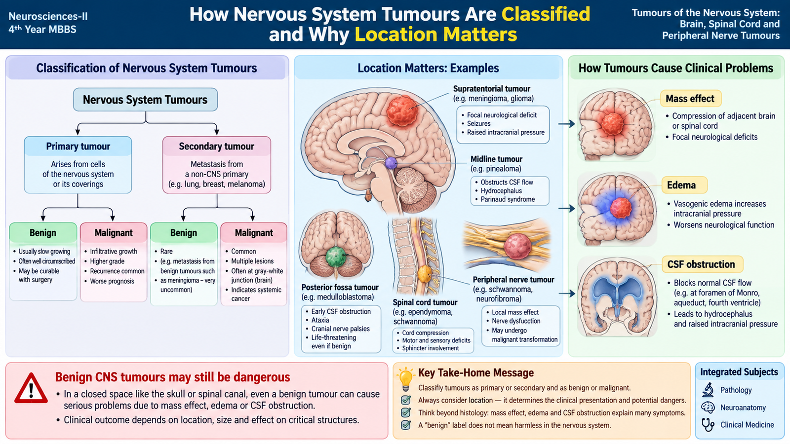
Nervous-system tumours can be classified according to their site of origin and their biological behaviour. This provides a simple framework before individual tumour types are studied.
Primary and secondary tumours
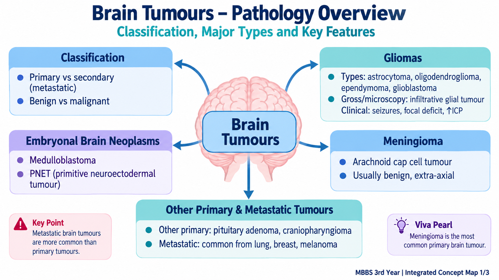
Primary tumours arise from structures within or closely associated with the nervous system. They include gliomas arising from glial-lineage cells, meningiomas arising from meningothelial cells and nerve-sheath tumours such as schwannomas.
Secondary tumours are metastases from a cancer elsewhere in the body. Brain metastases are particularly important in adults and may be single or multiple.
Benign and malignant behaviour
Histologically benign tumours usually grow slowly, show limited cellular atypia and tend to remain localized. Malignant tumours grow more aggressively and may show infiltration, marked cellular atypia, mitotic activity, vascular proliferation or necrosis, depending on the tumour type.
How a tumour produces symptoms
The clinical picture is strongly determined by anatomical location. A growing mass may directly destroy or compress neural tissue, produce surrounding vasogenic edema, obstruct cerebrospinal-fluid pathways or increase intracranial pressure.
Mass + edema or CSF obstruction → increased intracranial pressure → headache, vomiting, reduced consciousness and, in severe cases, herniation.
Therefore, the same histological tumour can present differently when it occurs in different anatomical locations.
B. Gliomas: Major Types, Morphology and Clinical Features
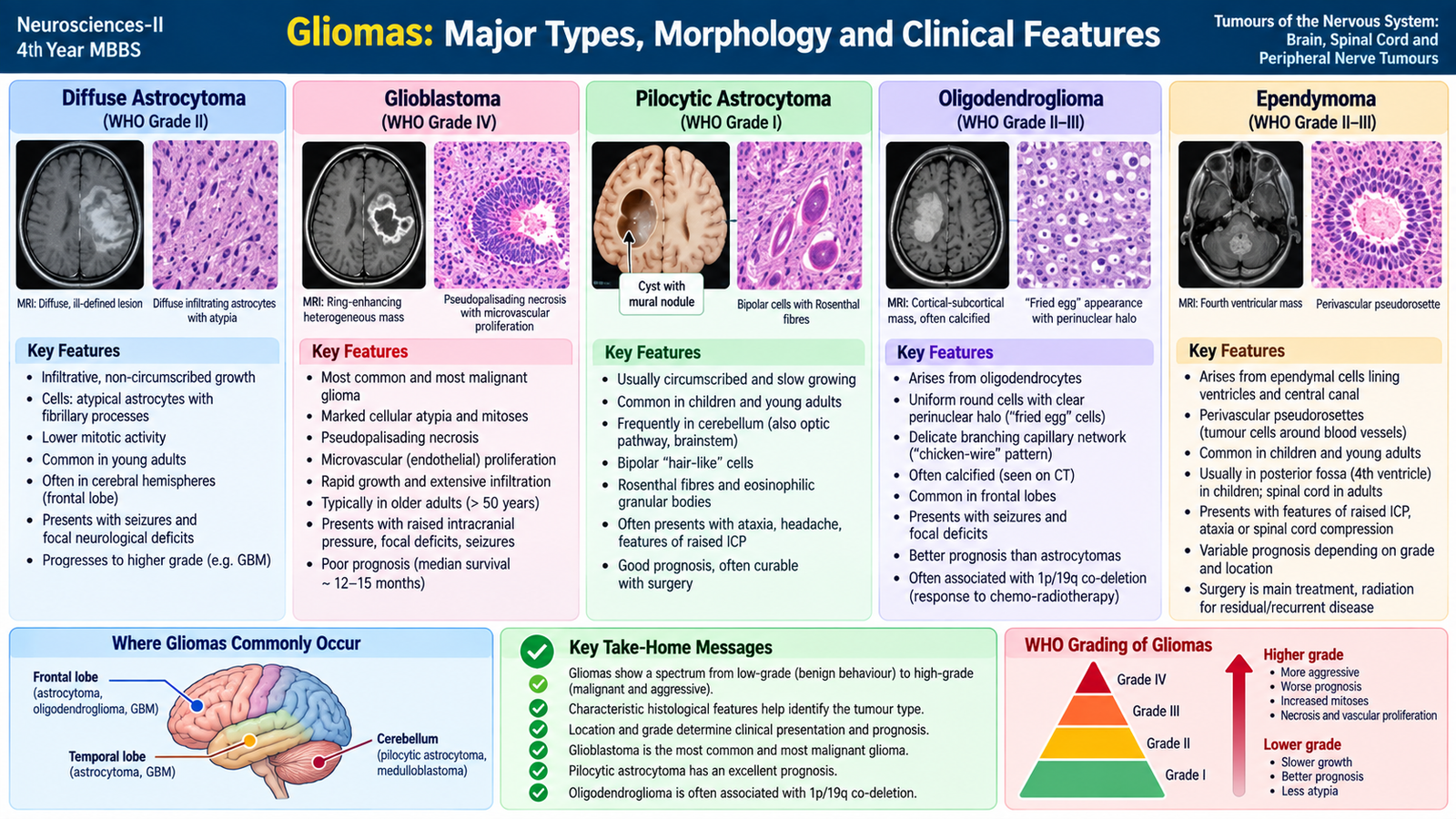
Gliomas are primary central nervous system tumours showing differentiation toward glial-lineage cells. Important undergraduate groups include astrocytic tumours, oligodendroglioma and ependymoma. Their behaviour ranges from relatively circumscribed, slowly growing lesions to highly infiltrative malignant tumours.
Astrocytic tumours
Diffuse astrocytoma
Diffuse astrocytomas infiltrate the surrounding brain rather than forming a sharply separated mass. They commonly involve the cerebral hemispheres of adults. Lower-grade tumours may initially produce seizures or slowly progressive focal deficits.
Grossly, the affected area may appear poorly defined and expand the involved cerebral tissue. Microscopically, neoplastic astrocytic cells infiltrate between normal neural structures and show variable nuclear atypia. Modern classification also uses molecular features, especially IDH status, to define important diffuse astrocytic tumour categories.
Glioblastoma
Glioblastoma is a highly malignant, rapidly infiltrating astrocytic tumour, usually occurring in adults. It commonly arises in the cerebral hemispheres and can extend through white-matter pathways; involvement of both hemispheres through the corpus callosum may produce the classic “butterfly” pattern.
Gross morphology: the tumour is poorly circumscribed and often shows areas of hemorrhage, softening and necrosis.
Microscopic morphology: marked cellular atypia, mitotic activity, pseudopalisading necrosis and microvascular proliferation are characteristic high-grade features.
Rapid growth and surrounding edema produce progressive headache, focal deficits, cognitive change, seizures or raised intracranial pressure.
Pilocytic astrocytoma
Pilocytic astrocytoma is a comparatively circumscribed, usually low-grade astrocytic tumour seen particularly in children and younger patients. The cerebellum is a common site.
Grossly, it may form a cyst with a mural nodule. Microscopically, elongated bipolar cells with hair-like processes may be associated with Rosenthal fibres and eosinophilic granular bodies. Cerebellar lesions commonly produce gait disturbance, limb incoordination and other signs of cerebellar dysfunction.
Oligodendroglioma
Oligodendroglioma is an infiltrating glioma that commonly affects adults and often develops in the cerebral hemispheres, particularly the frontal region. Seizures are an important presentation.
These tumours are defined by characteristic molecular abnormalities including IDH mutation with combined deletion of chromosome arms 1p and 19q. Grossly, the lesion may contain calcification. On routine histology, tumour cells often show round nuclei surrounded by clear halos, producing the familiar “fried-egg” appearance, together with a delicate branching capillary network sometimes described as “chicken-wire” vasculature.
Ependymoma
Ependymomas show differentiation toward ependymal cells that normally line the ventricular system and central canal. In children they frequently occur around the fourth ventricle, where they may obstruct CSF flow and cause hydrocephalus. In adults, the spinal cord is an important site.
Microscopically, tumour cells characteristically arrange themselves around blood vessels, forming perivascular pseudorosettes. True ependymal rosettes may also occur.
| Tumour | Typical pattern | Gross clue | Microscopic clue | Clinical clue |
|---|---|---|---|---|
| Diffuse astrocytoma | Infiltrating cerebral tumour | Poorly defined expansion | Infiltrating atypical astrocytic cells | Seizures or focal deficits |
| Glioblastoma | Highly malignant infiltrative tumour | Necrosis and hemorrhage | Pseudopalisading necrosis + vascular proliferation | Rapid neurological deterioration |
| Pilocytic astrocytoma | Often circumscribed; younger patients | Cyst with mural nodule | Rosenthal fibres | Often cerebellar signs |
| Oligodendroglioma | Adult cerebral tumour | May calcify | Clear halos + delicate capillaries | Seizures |
| Ependymoma | Ventricular in children; spinal in adults | Related to ventricular or spinal canal structures | Perivascular pseudorosettes | Hydrocephalus or cord symptoms |
C. Embryonal Brain Neoplasms
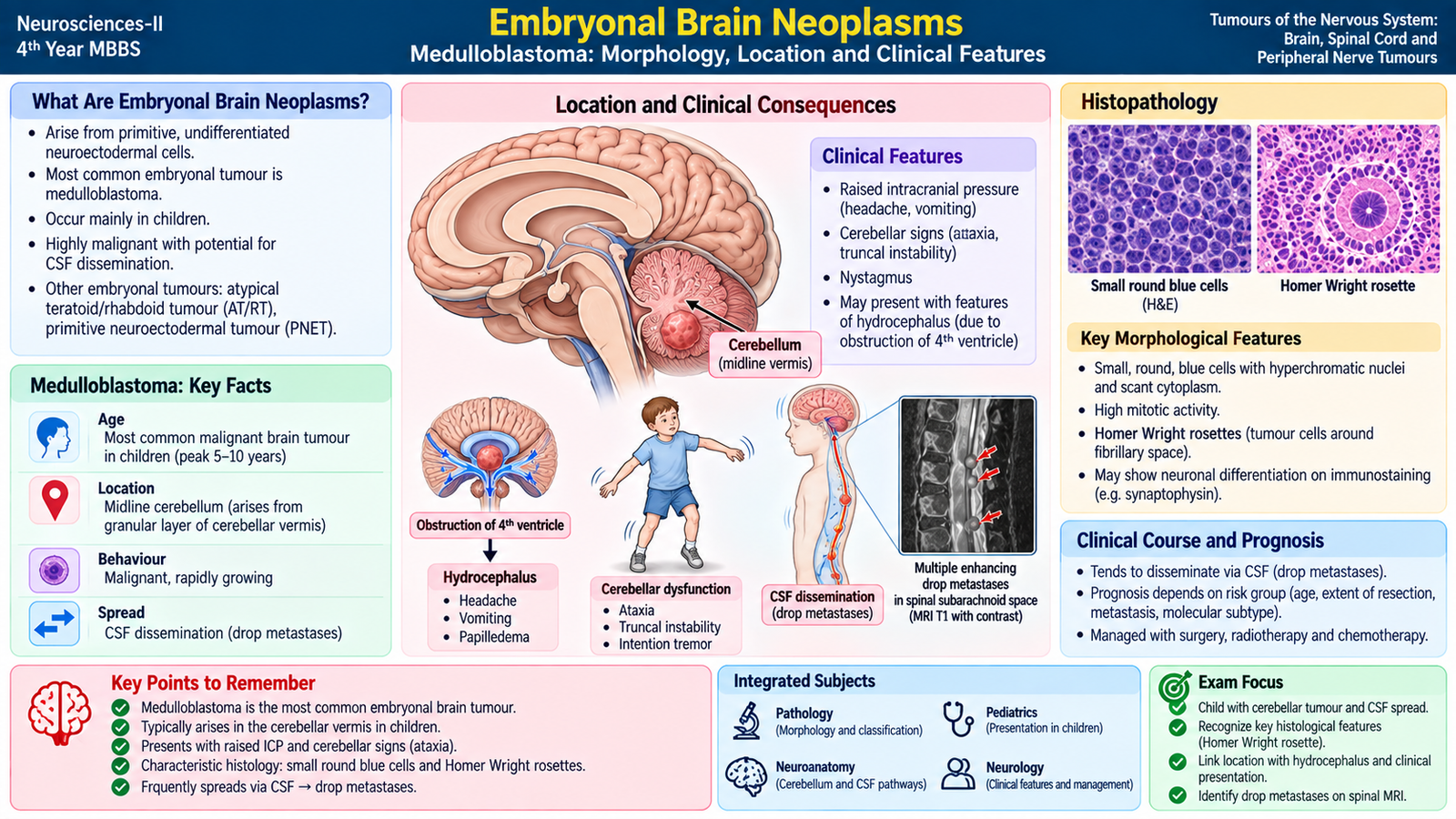
Embryonal tumours are malignant central nervous system tumours composed of poorly differentiated primitive cells. They occur predominantly in children. For undergraduate pathology, the most important example is medulloblastoma.
Medulloblastoma
Medulloblastoma is a highly malignant embryonal tumour that most often develops in the cerebellum. Its cerebellar location explains why a child may present with gait unsteadiness, poor coordination or truncal ataxia. A posterior fossa mass can also obstruct CSF pathways, producing hydrocephalus, headache and vomiting.
Morphology
Grossly, medulloblastoma is typically a soft, relatively well-demarcated cerebellar mass. Microscopically, it is a highly cellular tumour composed of small primitive cells with dark nuclei and scant cytoplasm—the classic small round blue cell appearance. Tumour cells may arrange around central neuropil to form Homer Wright rosettes.
Spread
Unlike many primary brain tumours, medulloblastoma has a significant tendency to disseminate through cerebrospinal fluid. Tumour deposits may therefore appear at other sites along the neuraxis, including the spinal subarachnoid space. These are often referred to clinically as drop metastases.
D. Meningioma, Other Primary Brain Tumours and Metastatic Tumours
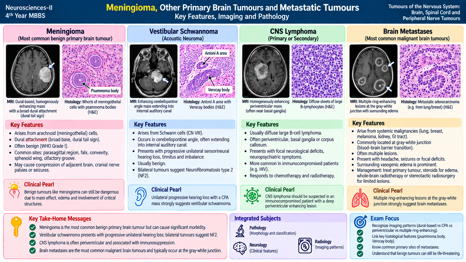
Not every intracranial tumour arises from glial cells. Some arise from the meninges or cranial nerve sheaths, while others originate from non-neural cell populations. Metastases from systemic malignancies form another important group and must always be distinguished from primary brain tumours.
Meningioma
Meningiomas are tumours derived from meningothelial or arachnoid-cap cells. Most are slow-growing and histologically benign, but their location can still produce significant neurological disease through compression of the underlying brain.
They are typically extra-axial and attached to the dura. Common sites include the parasagittal region, cerebral convexities and skull base.
Gross morphology: a well-circumscribed, firm, dura-based mass that compresses rather than diffusely infiltrates the adjacent brain. The cut surface may show a whorled appearance.
Microscopic morphology: meningothelial cells often form characteristic whorls. Calcification within these whorls may produce laminated structures called psammoma bodies.
Clinical features depend on location. A convexity lesion may cause seizures or focal weakness, whereas skull-base lesions may affect cranial nerves. Symptoms usually develop gradually because many meningiomas grow slowly.
Selected other primary intracranial tumours
Schwannoma
Schwannomas arise from Schwann cells of peripheral or cranial nerves. Intracranially, the vestibular portion of cranial nerve VIII is an important site. A vestibular schwannoma may present with progressive unilateral hearing impairment, tinnitus or imbalance. The detailed nerve-sheath morphology is discussed later with peripheral nerve tumours.
Primary central nervous system lymphoma
Primary CNS lymphoma is a malignant lymphoid tumour arising within the central nervous system, most often of B-cell origin. Lesions may involve deep cerebral or periventricular structures. Clinical manifestations include focal neurological deficits, cognitive or behavioural changes and other features related to lesion location.
Metastatic brain tumours
Metastatic tumours reach the brain from a primary cancer outside the nervous system. Important primary sources include cancers of the lung and breast, as well as melanoma, renal and gastrointestinal malignancies.
Metastases are often multiple, well-demarcated lesions and commonly occur near the junction between cerebral gray and white matter, where circulating tumour emboli may lodge in small vessels. Marked surrounding vasogenic edema can make a relatively small metastasis produce substantial symptoms.
On microscopy, metastatic tumour cells usually resemble the original primary malignancy rather than normal CNS tissue. This can help the pathologist identify or suggest the systemic source.
Patients may present with headache, seizures, focal neurological deficits or altered cognition. Occasionally, neurological symptoms from a brain metastasis reveal an underlying systemic cancer that has not yet been diagnosed.
E. Spinal Tumours: Types, Presentation and Surgical Principles
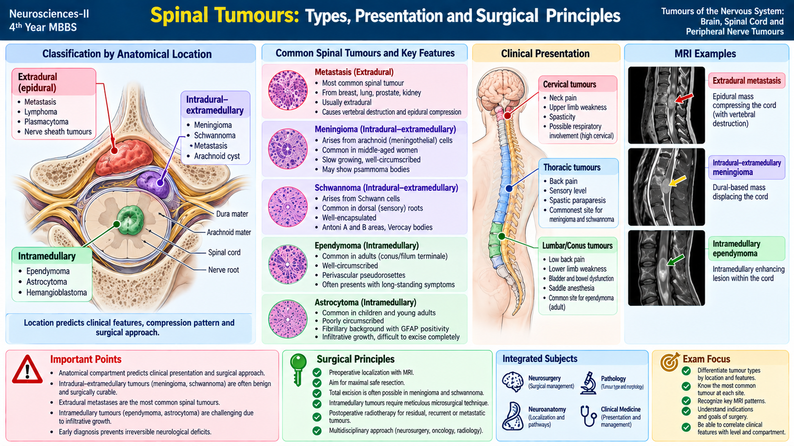
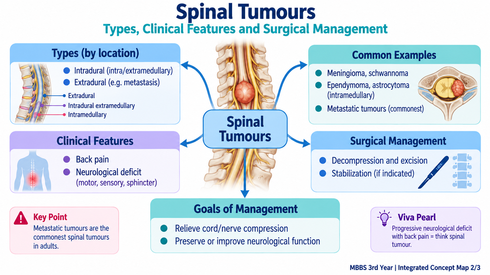
Spinal tumours are best understood according to their anatomical relationship to the spinal cord and dura. This classification is clinically useful because the compartment in which the tumour grows helps predict the pattern of compression, neurological findings and surgical approach.
Types according to anatomical location
These lie outside the dura. Vertebral metastases are an important cause. Expansion into the spinal canal may compress the cord or cauda equina.
These lie inside the dura but outside the spinal cord substance. Common examples include meningiomas and schwannomas. Because they compress the cord from outside, they may produce progressive myelopathy.
These arise within the spinal cord itself. Important examples include ependymomas and astrocytomas. Their growth expands and disrupts the cord from within.
Clinical features
The presentation depends on the level of the lesion, the direction of compression and the rate of tumour growth. Symptoms usually become progressively more obvious as neural structures are compressed.
- Local or back pain, sometimes persistent or progressive.
- Radicular pain when a spinal nerve root is irritated or compressed.
- Motor weakness below the level of significant cord compression.
- Upper motor neuron signs below the lesion, such as increased tone and exaggerated reflexes, when corticospinal tracts are affected.
- Segmental lower motor neuron findings may occur at the level where anterior horn cells or roots are damaged.
- Sensory disturbance, including numbness, paresthesia or a definable sensory level.
- Gait difficulty due to motor, sensory or proprioceptive pathway involvement.
- Bladder or bowel dysfunction in advanced cord or cauda-equina compression.
A clearly identifiable sensory level is particularly useful because it helps localize a spinal cord lesion.
Assessment before surgery
MRI is the key imaging investigation because it demonstrates the level of the lesion, whether it is extradural or intradural, its relationship to the spinal cord, and the degree of neural compression. Imaging also helps the neurosurgical team judge whether the lesion appears resectable and whether vertebral stability is threatened.
Principles of surgical management
Surgery is planned according to tumour location, pathology, neurological deficit, resectability and overall patient condition. The objective is not simply to “remove a mass”; it is to obtain disease control while preserving as much neurological function as possible.
- Decompress the spinal cord or nerve roots when significant compression is present.
- Obtain tissue for histopathological diagnosis when the nature of the tumour is not already established.
- Achieve maximal safe resection when the tumour can be separated from important neural structures.
- Preserve neurological function; aggressive removal is inappropriate if it would cause unacceptable cord injury.
- Restore or maintain spinal stability when vertebral destruction or the required decompression makes instability a concern.
- Use additional oncological treatment when indicated by tumour type and biological behaviour.
Intradural-extramedullary tumours such as many meningiomas and schwannomas are often well-defined and may be amenable to surgical excision. Intramedullary tumours require particularly careful surgery because the tumour lies within functioning spinal cord tissue. Extradural metastatic disease may require decompression and stabilization in selected patients, together with treatment directed at the underlying malignancy.
F. Peripheral Nerve Sheath Tumours
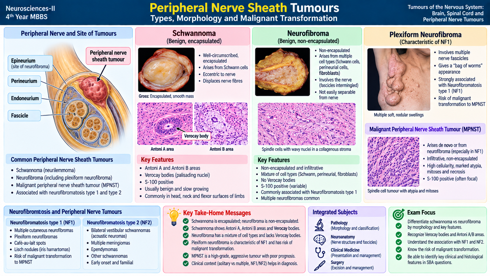
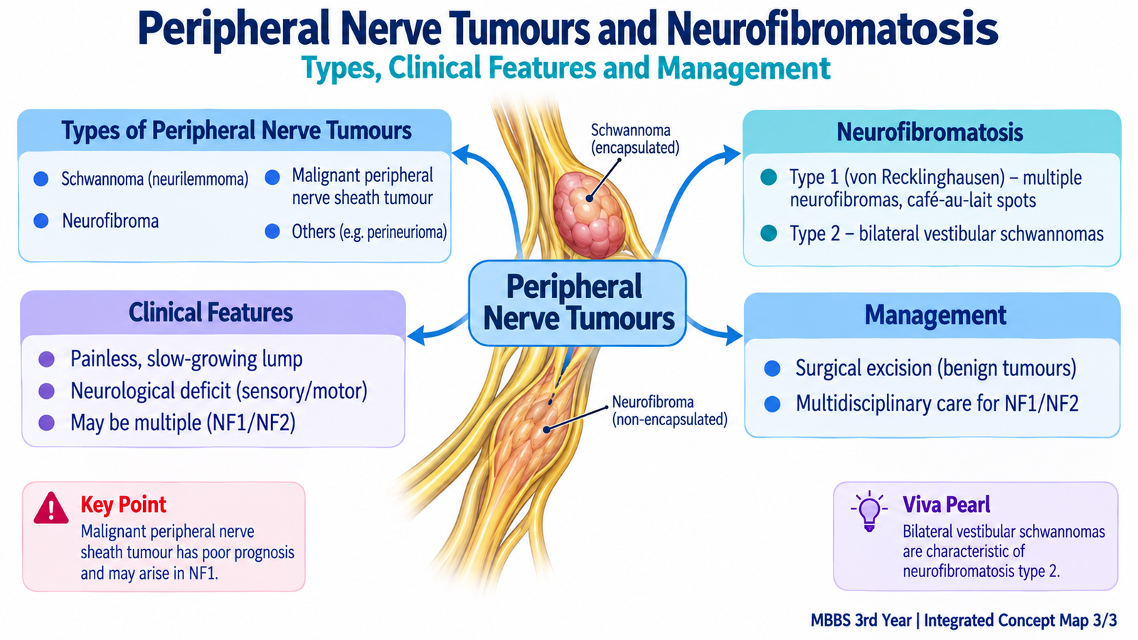
Peripheral nerves contain Schwann cells, fibroblasts, perineurial cells and axons. Tumours arising from nerve-sheath components therefore have different patterns of growth and morphology. The three major lesions students should recognize are schwannoma, neurofibroma and malignant peripheral nerve sheath tumour.
Schwannoma
A schwannoma is a usually benign tumour composed of Schwann cells. It tends to be well circumscribed and encapsulated. It commonly grows eccentrically from the involved nerve, meaning the tumour lies to one side rather than diffusely expanding the entire nerve.
Microscopically, schwannomas classically show two patterns:
- Antoni A areas: more cellular regions containing spindle Schwann cells.
- Antoni B areas: less cellular, loosely arranged areas.
In Antoni A regions, aligned nuclei may palisade around acellular zones, producing Verocay bodies.
Symptoms depend on the nerve involved and include a slowly enlarging mass, pain, paresthesia or functional loss from nerve compression. Vestibular schwannoma is the important intracranial example.
Neurofibroma
A neurofibroma is a benign peripheral nerve sheath tumour containing a mixture of cell types, with Schwann cells forming an important component. Unlike the typical schwannoma, neurofibromas are generally unencapsulated and may incorporate the involved nerve fibres within the lesion.
Microscopically, spindle cells with elongated, wavy nuclei are arranged in a collagenous or myxoid background. Neurofibromas may occur as solitary lesions or as multiple tumours in neurofibromatosis type 1.
Plexiform neurofibroma is particularly important. It expands multiple nerve fascicles and may produce a tortuous, multinodular enlargement of a nerve. This lesion is strongly associated with NF1 and carries clinical importance because malignant transformation can occur.
Malignant peripheral nerve sheath tumour
Malignant peripheral nerve sheath tumour is an aggressive malignant spindle-cell tumour arising from a peripheral nerve or a pre-existing nerve-sheath lesion. Risk is increased in patients with NF1, particularly in association with plexiform neurofibromas.
A previously stable nerve-related mass that begins to show rapid enlargement, persistent increasing pain or new neurological deficit raises concern for malignant transformation and requires specialist evaluation.
G. Neurofibromatosis: Types, Clinical Features and Management
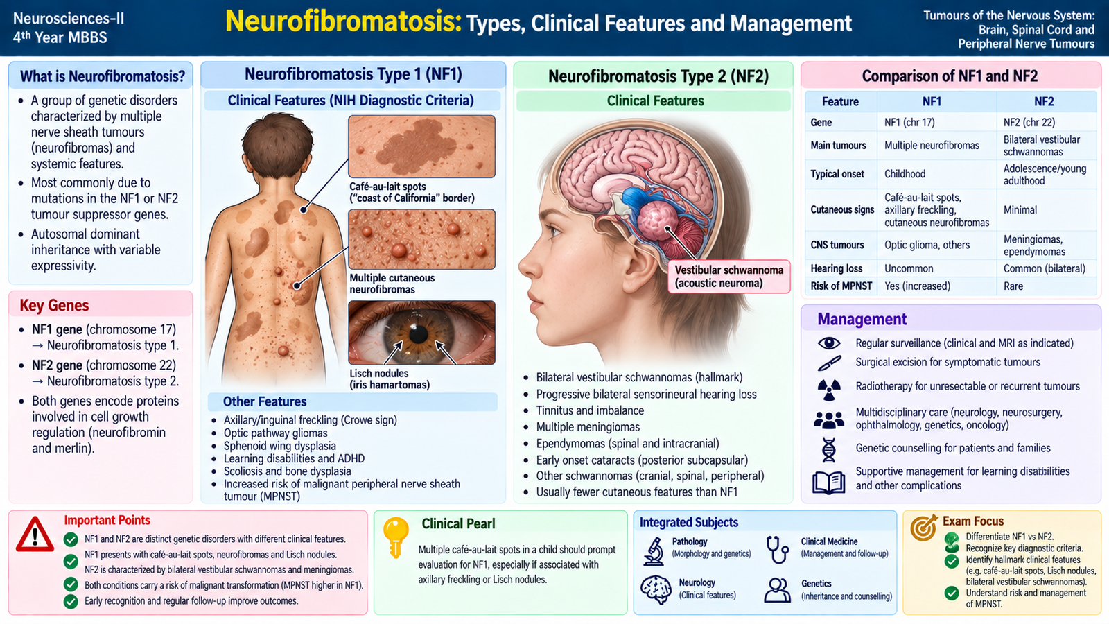
Neurofibromatosis refers to inherited tumour-predisposition disorders in which patients develop characteristic nervous-system tumours together with other clinical manifestations. For undergraduate learning, the two classic forms are neurofibromatosis type 1 (NF1) and neurofibromatosis type 2 (NF2).
Neurofibromatosis type 1
NF1 is an autosomal dominant disorder caused by abnormal function of the NF1 tumour-suppressor gene, which encodes neurofibromin. Loss of normal tumour-suppressor control increases the tendency of affected cells to form particular tumours.
Important clinical features include:
- Multiple café-au-lait macules.
- Freckling in characteristic intertriginous areas such as the axilla.
- Multiple cutaneous and subcutaneous neurofibromas.
- Plexiform neurofibromas, which may involve large segments of a nerve or nerve plexus.
- Lisch nodules of the iris.
- Association with optic pathway glioma.
- Characteristic skeletal abnormalities may occur in some patients.
- Increased risk of malignant peripheral nerve sheath tumour.
The wide range of manifestations occurs because NF1 affects tumour suppression in several tissues rather than producing only one isolated tumour type.
Neurofibromatosis type 2
NF2 is also an autosomal dominant tumour-predisposition disorder. It results from abnormal function of the NF2 tumour-suppressor gene, which encodes the protein merlin.
The hallmark clinical association is bilateral vestibular schwannomas. Because these tumours affect cranial nerve VIII, progressive hearing impairment, tinnitus and balance disturbance are important manifestations.
Other associated nervous-system tumours include:
- other schwannomas;
- multiple meningiomas;
- ependymomas and other central nervous system tumours.
Ocular abnormalities such as early cataract may also occur.
Management of neurofibromatosis
Neurofibromatosis is a genetic tumour-predisposition disorder, so management is based on surveillance, early recognition of complications and treatment of clinically important tumours. There is no single operation that removes the underlying inherited tendency to develop tumours.
- Regular clinical follow-up to identify new neurological, skin, visual, hearing or skeletal problems.
- Targeted imaging when symptoms or clinical findings suggest an important internal tumour.
- Visual assessment when optic pathway involvement is a concern.
- Hearing and neurological assessment in patients with NF2 and suspected vestibular schwannomas.
- Surgical treatment for selected symptomatic, enlarging, compressive or function-threatening tumours.
- Urgent assessment of suspicious tumour change, especially rapid enlargement, persistent pain or new deficit suggesting malignant transformation.
- Genetic counselling because both classic neurofibromatosis syndromes are inherited disorders with important implications for patients and families.
- Rehabilitation and functional support when neurological, visual, hearing or musculoskeletal disability develops.
Management should be individualized because tumour number, location and functional impact differ greatly between patients.
Integrated Mechanism Flow
⭐ AIM High-Yield Review
- Primary nervous-system tumours arise within nervous-system structures; secondary tumours are metastases from cancers elsewhere.
- A histologically benign CNS tumour can still cause serious disease because the skull and spinal canal provide little room for expansion.
- Glioblastoma: aggressive infiltrating tumour with necrosis, pseudopalisading tumour cells and microvascular proliferation.
- Pilocytic astrocytoma: often occurs in younger patients; may be cystic with a mural nodule and shows Rosenthal fibres.
- Oligodendroglioma: IDH-mutant, 1p/19q-codeleted glioma; seizures, calcification and clear-cell “fried-egg” histology are useful clues.
- Ependymoma: ventricular tumours are important in children, spinal tumours in adults; perivascular pseudorosettes are characteristic.
- Medulloblastoma: malignant cerebellar embryonal tumour with small round blue cells, possible Homer Wright rosettes and CSF drop metastases.
- Meningioma: usually a dura-based extra-axial mass showing whorls and psammoma bodies.
- Brain metastases are often multiple and well demarcated with marked surrounding edema; they commonly occur near the gray-white junction.
- Spinal tumours are classified as extradural, intradural-extramedullary or intramedullary.
- Progressive weakness, a sensory level and sphincter dysfunction suggest significant spinal cord compression and require urgent assessment.
- A schwannoma is usually encapsulated and eccentric to the nerve; Antoni A/B areas and Verocay bodies are classic morphological findings.
- A neurofibroma is generally unencapsulated and incorporates nerve fibres; plexiform neurofibroma is strongly associated with NF1.
- NF1: café-au-lait macules, freckling, neurofibromas, plexiform neurofibromas, Lisch nodules and optic pathway glioma are important associations.
- NF2: bilateral vestibular schwannomas are the hallmark; management of neurofibromatosis centers on surveillance and treatment of symptomatic or threatening tumours.
🎥 Brain Tumours
Classification, major brain tumours, clinical presentation, pathology, diagnosis and management.
🎥 Spinal Tumours
Location-based understanding of intramedullary, intradural-extramedullary and extradural spinal tumours.
🎥 Peripheral Nerve Sheath Tumours & Neurofibromatosis
Schwannoma, neurofibroma, malignant peripheral nerve sheath tumour, NF1 and NF2.