
Head Injury: Skull Fractures, Cerebral Injury and Medicolegal Evaluation
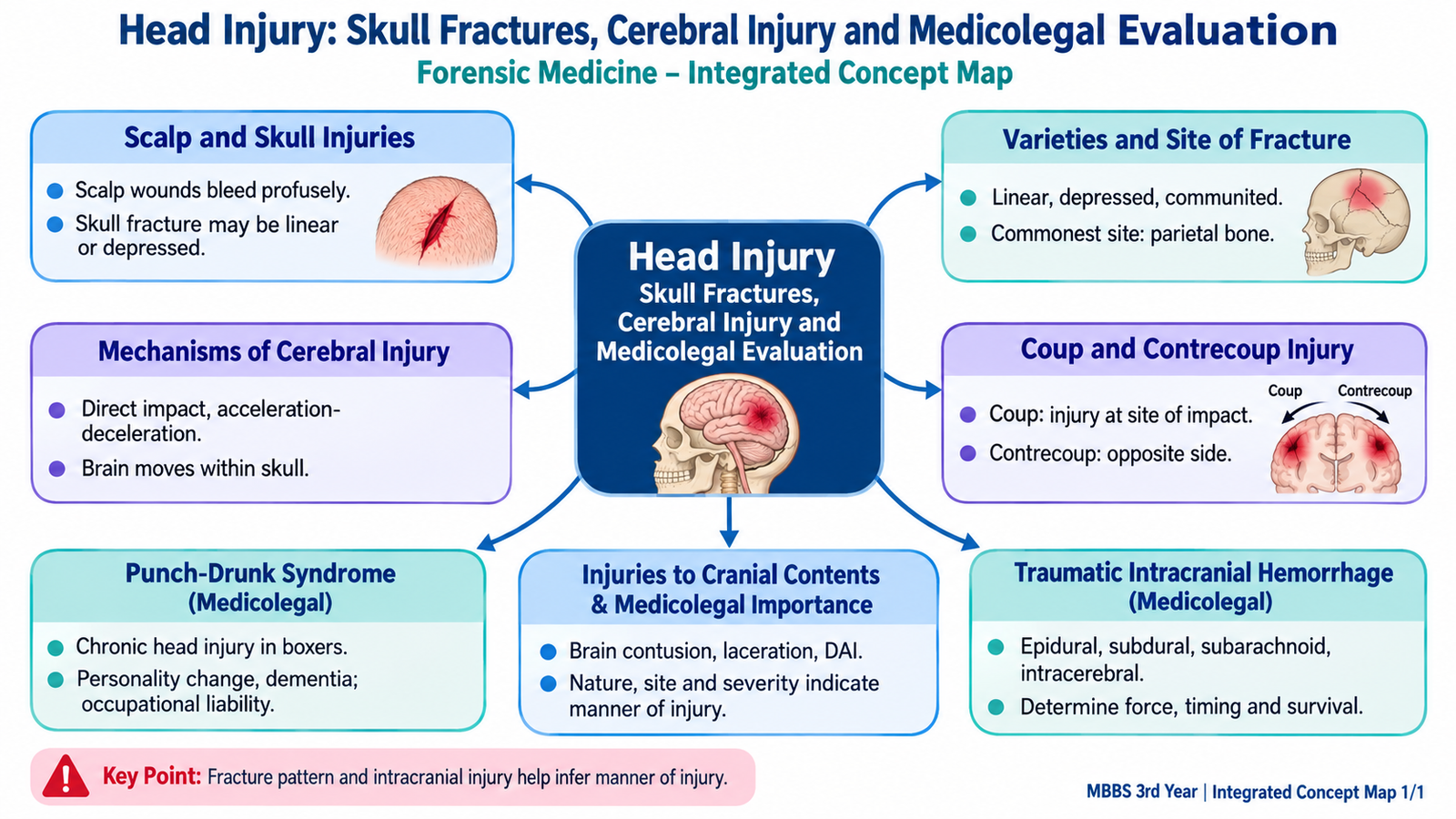
Head trauma may leave evidence in the scalp, skull, meninges, blood vessels and brain. Understanding how external force produces these different injuries is essential for both clinical recognition and careful medico-legal interpretation.
Topic Introduction
Head injury ranges from a superficial scalp wound to severe disruption of the skull and intracranial contents. The external appearance does not always predict the damage underneath: serious cerebral injury or intracranial hemorrhage may occur with little external injury, while an impressive scalp wound may occur without major brain damage. For forensic interpretation, the important task is therefore to examine the injuries as a connected pattern. This chapter explains scalp injury, major varieties of skull fracture, mechanisms of cerebral damage, coup and contrecoup injury, injuries to the cranial contents, traumatic intracranial hemorrhage and the chronic effects of repeated head trauma known historically as punch-drunk syndrome.
A. Scalp Injuries and Their Medico-Legal Meaning
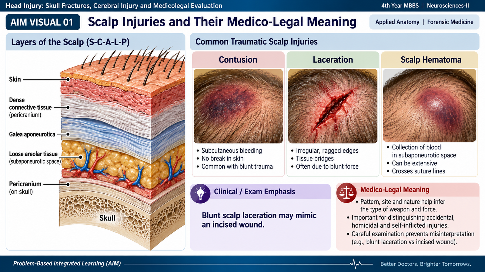
The scalp is the first structure exposed to most blunt impacts on the head. Because it contains a rich vascular supply and its vessels are held open within dense connective tissue, even a relatively small scalp wound may bleed considerably. The underlying skull provides a firm surface against which the scalp can be crushed, so blunt trauma may produce abrasions, bruises and lacerations with characteristic features.
Main forms of scalp injury
- Abrasion: superficial loss of epidermis caused by friction or scraping. Its shape or direction may occasionally provide useful information about the contact surface or movement.
- Contusion: bleeding into scalp tissues caused by rupture of small vessels without disruption of the skin. Swelling may be localized or may spread through tissue planes.
- Laceration: tearing of the scalp by blunt force. When scalp is crushed against underlying bone, the wound may look deceptively clean and resemble an incised wound.
- Scalp hematoma: collection of blood within or beneath scalp layers. Its extent does not necessarily indicate the severity of intracranial injury.
Medico-legal importance
A scalp injury should be interpreted together with the skull and intracranial findings. Its location may identify a possible impact site, while its shape may sometimes reflect the contacting surface. However, the appearance of a single wound should never be used alone to identify a particular weapon or reconstruct an entire event.
- Record the site, size, shape, margins and surrounding bruising or abrasion.
- Look for more than one injury, because multiple impacts may be present.
- Correlate the external injury with the underlying skull fracture and cerebral injury.
- Remember that absence of a major scalp wound does not exclude serious intracranial injury.
🖼 AIM VISUAL 01 — Scalp Injury Recognition Map
B. Skull Fractures: Types, Mechanism and Common Sites
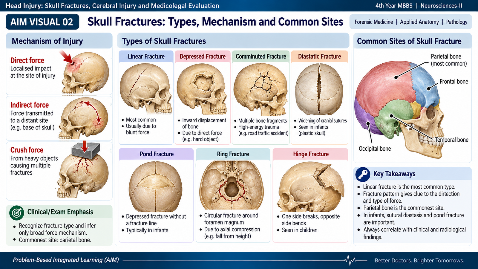
A skull fracture occurs when applied mechanical force exceeds the elastic strength of the cranial bone. The resulting pattern depends on the magnitude of force, the area over which it acts, the direction of impact, the shape of the object and the properties of the skull. A broad impact tends to produce a linear fracture, whereas a concentrated force acting over a smaller area is more likely to produce depression or comminution.
Important varieties of skull fracture
| Type | Core concept | Forensic significance |
|---|---|---|
| Linear / fissured | A crack in the skull without major displacement of bone. | Common after blunt impact over a relatively broad area. |
| Depressed | A portion of skull is driven inward. | Suggests localized application of substantial force over a relatively small area. |
| Comminuted | Bone is broken into several fragments. | Usually reflects greater local deformation or force. |
| Diastatic | A cranial suture is widened or separated. | More relevant in younger individuals in whom sutural relationships differ from adults. |
| Pond fracture | Localized indentation of the pliable skull, classically in infants. | Age and skull flexibility are important when interpreting the lesion. |
| Gutter fracture | A groove-like defect produced by tangential force along the skull. | The direction and circumstances of force require correlation with the complete injury pattern. |
| Basilar fracture | Fracture involving the floor of the cranial cavity. | May be associated with meningeal tears, cranial nerve injury or leakage of cerebrospinal fluid. |
Special basal fracture patterns
- Ring fracture: a fracture encircling or partly encircling the foramen magnum. It may result from severe axial loading transmitted between the skull and vertebral column.
- Hinge fracture: a transverse fracture passing across the base so that the skull is effectively divided into two portions. Severe lateral compression is a classical mechanism.
Commonest site
For undergraduate forensic learning, the parietal-temporoparietal region of the cranial vault is an important and commonly involved fracture region. The lateral skull is vulnerable because parts of the temporal region are relatively thin. The exact distribution of fractures nevertheless depends strongly on the mechanism and direction of trauma, so a fracture site should not be interpreted in isolation. :contentReference[oaicite:1]{index=1}
Medico-legal interpretation
Skull fractures help establish that significant mechanical force reached the head, but the fracture alone does not reveal the complete circumstances. A fatal cerebral injury may occur without fracture, and a fracture may occur without immediately fatal brain injury. Interpretation therefore requires correlation with scalp findings, intracranial lesions, history and scene circumstances.
🖼 AIM VISUAL 02 — Skull Fracture Classification Tree
C. Mechanisms of Cerebral Injury: Coup, Contrecoup and Diffuse Damage
The brain is soft tissue suspended in cerebrospinal fluid inside a rigid, irregular bony cavity. During trauma, the skull and brain do not always move together. Sudden acceleration, deceleration and rotation may therefore cause the brain to strike the internal skull, stretch axons or tear blood vessels. Cerebral injury may consequently occur directly beneath the impact, opposite the impact or diffusely throughout the brain.
Mechanism chain
1. Coup injury
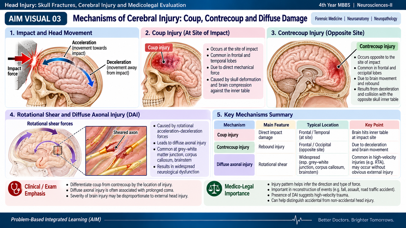
A coup injury is cerebral injury occurring at or immediately beneath the site where the external force strikes the head. Local deformation of the skull can compress and bruise the underlying cortex. It is classically prominent when an object strikes a relatively stationary head.
2. Contrecoup injury
A contrecoup injury occurs away from, usually opposite to, the external impact. It is classically associated with a moving head striking a fixed surface. Sudden deceleration causes relative movement of the brain within the skull, allowing the brain to strike the internal bony surface on the opposite side. The inferior frontal and temporal regions are especially vulnerable because they lie over irregular parts of the skull base. :contentReference[oaicite:2]{index=2}
| Feature | Coup | Contrecoup |
|---|---|---|
| Location | At or beneath impact | Remote from, usually opposite, impact |
| Typical mechanical setting | Blow delivered to the head | Moving head suddenly stopped by a surface |
| Underlying principle | Direct local deformation and compression | Relative brain movement and opposite-side impact |
These are useful mechanical concepts rather than absolute rules. Real head trauma may produce both coup and contrecoup contusions, and the complete injury pattern must be considered before drawing forensic conclusions.
3. Acceleration-deceleration and rotational injury
Rapid change in head velocity can stretch or tear tissues even when no major fracture is present. Rotational forces are particularly important because different parts of the brain move at different velocities. This produces shearing strain within white matter and may damage axons widely.
Severe widespread axonal damage is called diffuse axonal injury. It can cause immediate and prolonged impairment of consciousness despite relatively limited external evidence of trauma.
4. Secondary cerebral injury
Trauma may initiate cerebral edema, impaired perfusion and increasing intracranial pressure after the original mechanical injury. These secondary processes can enlarge the damage and contribute to later neurological deterioration. Thus, the condition of the brain some time after trauma may reflect both the original impact and evolving secondary injury.
🖼 AIM VISUAL 03 — Coup–Contrecoup and Shearing Mechanism
D. Injuries to the Cranial Contents and Medico-Legal Importance
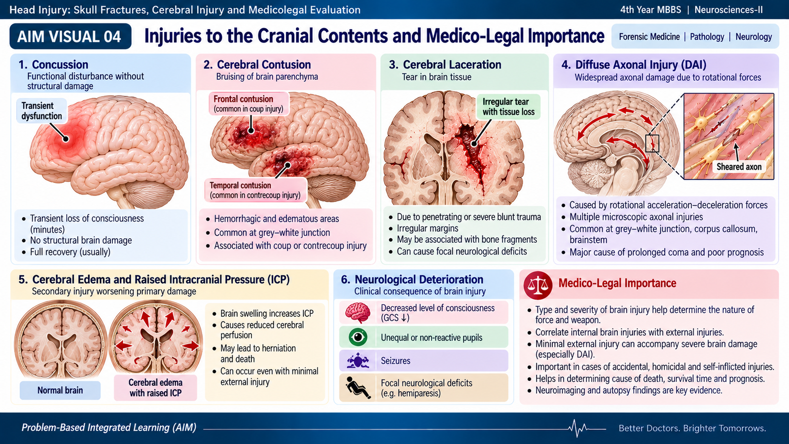
The cranial contents include the meninges, cerebral blood vessels and brain itself. Head trauma may disturb brain function without obvious structural destruction, produce localized bruising or laceration, cause widespread axonal damage, or create an expanding intracranial mass. The forensic significance lies not only in recognizing each lesion but in relating it cautiously to the external injury and circumstances.
Concussion
Cerebral concussion is a transient disturbance of neurological function following trauma. It may produce temporary loss or alteration of consciousness, amnesia or confusion without a major structural lesion visible to the naked eye. Its importance is that significant functional disturbance can exist even when gross anatomical damage is minimal.
Cerebral contusion
A contusion is a bruise of brain tissue. Small vessels rupture and hemorrhage occurs within damaged cortex. Contusions commonly involve surfaces of the brain that strike rough internal skull structures. They may occur as coup or contrecoup lesions and may enlarge after injury.
Cerebral laceration
A laceration represents actual tearing of brain tissue. It is generally a more severe structural injury and may occur with marked skull deformation, depressed fracture or severe movement of the brain against bony structures.
Diffuse axonal injury
Diffuse axonal injury results from widespread shearing of axons, particularly during rapid rotational acceleration or deceleration. The external head injury may appear modest despite severe disturbance of consciousness. This illustrates a major forensic principle: external injury severity does not always equal brain injury severity.
Medico-legal interpretation
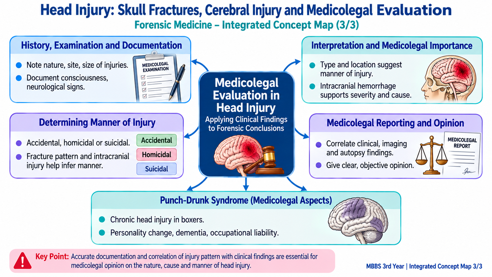
In a medico-legal examination, no single cerebral lesion should be interpreted independently. The pattern must be correlated with the scalp, skull, hemorrhage distribution, clinical course and circumstances.
- Compatibility: Does the internal injury reasonably fit the alleged mechanism?
- Causation: Is the traumatic brain injury sufficient to explain deterioration or death?
- Multiplicity: Are there injuries suggesting more than one impact?
- Survival: Some intracranial lesions may permit a period of apparently preserved function before deterioration.
- Associated factors: Natural disease, anticoagulant use or bleeding disorders may alter the extent of hemorrhage and must be considered where relevant.
The cause of death, mechanism of death and manner of death are related but different concepts. Traumatic head injury may be the cause; brain swelling, intracranial pressure and herniation may contribute to the physiological mechanism; while classification of the manner as accidental, homicidal, suicidal or undetermined requires the total medico-legal evidence rather than the brain lesion alone.
🖼 AIM VISUAL 04 — Cranial Contents Injury Map
E. Traumatic Intracranial Hemorrhage and Forensic Interpretation
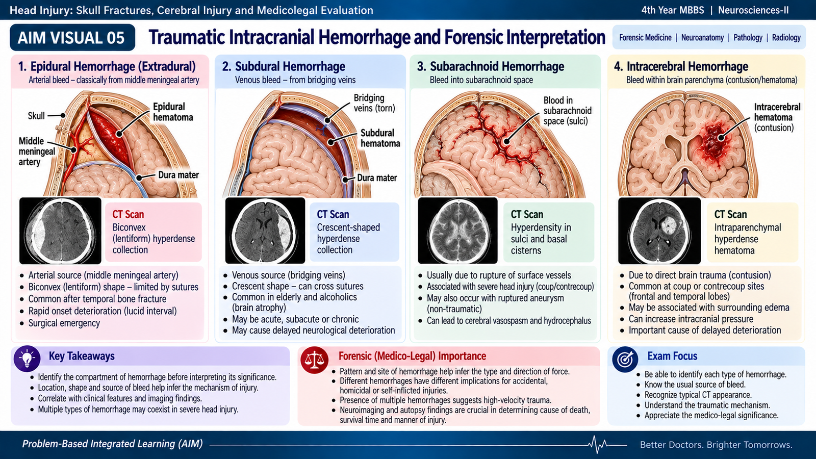
Trauma may rupture arteries, veins or small cerebral vessels, producing blood in different intracranial compartments. The anatomical location of blood gives important clues to the injured vessel and mechanical process. For medico-legal purposes, however, identifying the hemorrhage is only the first step; it must then be distinguished from possible natural hemorrhage and interpreted together with the rest of the injuries.
| Hemorrhage | Location | Typical traumatic mechanism | Important clue |
|---|---|---|---|
| Epidural / extradural | Between skull and dura | Often arterial bleeding associated with skull fracture, classically involving the middle meningeal artery | Localized expanding collection; classically biconvex on CT |
| Subdural | Between dura and arachnoid | Stretching and tearing of bridging veins during acceleration-deceleration | Crescentic collection; may be acute or evolve over time |
| Subarachnoid | Within the CSF-containing subarachnoid space | Damage to vessels accompanying contusion or other traumatic brain injury | Blood tracks within sulci and subarachnoid spaces |
| Intracerebral | Within brain parenchyma | Contusion, laceration or shearing of intraparenchymal vessels | Often accompanies other evidence of cerebral injury |
Epidural hemorrhage
Epidural hemorrhage commonly follows fracture near a meningeal arterial vessel, classically in the temporal or temporoparietal region. Arterial blood can accumulate between skull and dura and compress the brain. A patient may occasionally regain apparent consciousness after the initial impact and then deteriorate as the collection enlarges. This is the classical lucid interval, but it is not present in every case and is not unique to epidural hemorrhage.
Subdural hemorrhage
Subdural hemorrhage most often follows tearing of bridging veins as the brain moves relative to the dura. Because this is mainly an acceleration-deceleration mechanism, a skull fracture is not required. Blood can spread over a relatively broad cerebral surface. The hemorrhage may be acute or may evolve over a longer period, making correlation with history and other findings particularly important.
Subarachnoid hemorrhage
Traumatic subarachnoid hemorrhage results when small vessels in the subarachnoid space are damaged. Because subarachnoid hemorrhage can also occur naturally, for example after rupture of an intracranial aneurysm, the mere presence of subarachnoid blood does not automatically establish trauma as its cause. Distribution of blood, accompanying contusions, external injuries, vascular findings and circumstances all contribute to interpretation.
Intracerebral hemorrhage
Traumatic intracerebral bleeding occurs within damaged brain tissue and may accompany cerebral contusions or lacerations. Natural intracerebral hemorrhage is also possible, so location, associated trauma and the overall pathological pattern must be assessed before attributing the bleed to injury.
Medico-legal questions in traumatic hemorrhage
- Is the hemorrhage traumatic, natural, or potentially influenced by both?
- Does its site correspond with the scalp, skull and cerebral injuries?
- Could an underlying bleeding tendency have increased its severity?
- Is the reported clinical course compatible with an evolving intracranial lesion?
- Can the hemorrhage reasonably explain unconsciousness, neurological deterioration or death?
Precise dating of an intracranial hemorrhage from appearance alone has limitations. The safest forensic conclusion combines morphology with clinical information, imaging, other injuries and circumstances rather than assigning an exact time solely from the color or consistency of blood.
🖼 AIM VISUAL 05 — Intracranial Hemorrhage Comparison
F. Punch-Drunk Syndrome: Repetitive Head Trauma and Medico-Legal Aspects
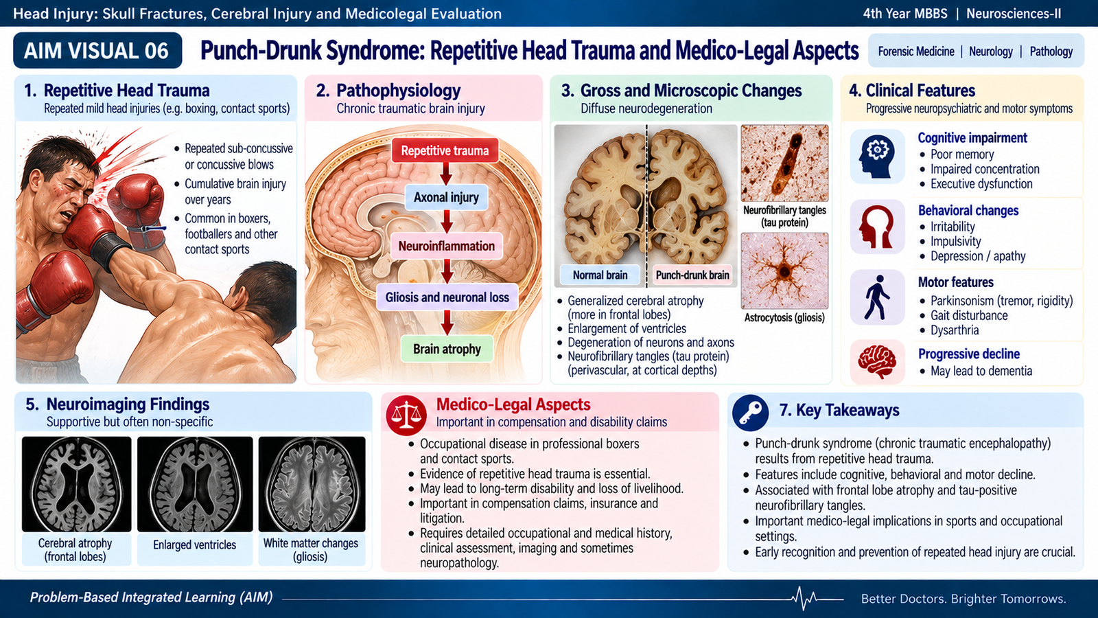
Punch-drunk syndrome is the historical term used for progressive neurological and behavioral abnormalities described in boxers exposed to repeated blows to the head. The condition later became known as dementia pugilistica and is now considered within the broader concept of chronic traumatic encephalopathy (CTE), which is associated with repetitive head impacts. :contentReference[oaicite:3]{index=3}
How repeated injury produces chronic damage
A single blow is not the central idea. Repeated concussive and subconcussive impacts expose the brain to recurrent mechanical strain. Over time, abnormal neurodegenerative changes may develop, including abnormal accumulation of phosphorylated tau protein in characteristic microscopic patterns.
Mechanism concept
Clinical features
The manifestations develop gradually and may involve several functional domains rather than one single characteristic symptom.
- Cognitive: impaired memory, attention, judgment and executive function.
- Behavioral or mood: personality or behavioral change and emotional disturbance may occur.
- Speech and coordination: dysarthria, poor coordination and ataxic features may develop.
- Motor: tremor, rigidity, slowness or parkinsonian features may occur in advanced disease.
- Progression: significant cognitive decline or dementia may occur in severe cases.
Medico-legal importance
Punch-drunk syndrome is important because injury may be cumulative. There may be no single dramatic traumatic event that explains the eventual neurological impairment. A proper medico-legal assessment therefore depends on the history of repeated head impacts, clinical evolution and, where relevant, neuropathological findings.
- A detailed history of participation in boxing or other activities involving repetitive head impacts is important.
- Symptoms may appear or progress after the period of repeated exposure has ended.
- Other neurological and neurodegenerative conditions must be considered before attributing symptoms solely to previous trauma.
- The presence of a history of repetitive impacts does not, by itself, establish the cause of every later neurological symptom.
- In fatal cases where CTE is relevant, careful neuropathological examination may contribute to the final interpretation.
🖼 AIM VISUAL 06 — Repetitive Head Trauma to Chronic Encephalopathy
Integrated Mechanism Flow
Blow, fall or acceleration-deceleration
Deformation, translation or rotation
Coup, contrecoup, shearing or fracture
Epidural, subdural, SAH or intracerebral bleeding
Edema and rising intracranial pressure
Neurological deficit, deterioration or death
⭐ AIM High-Yield Review
- Scalp lacerations can bleed heavily because scalp vessels are held open within dense connective tissue.
- A blunt-force scalp laceration may resemble an incised wound; bruising, abrasion and tissue bridges support laceration.
- Linear or fissured fracture is the common general pattern of skull fracture.
- A depressed fracture suggests concentrated localized force over a relatively small area.
- The parietal-temporoparietal vault is an important commonly involved fracture region in undergraduate forensic teaching.
- Coup injury occurs at or beneath the impact; contrecoup injury occurs away from, usually opposite to, the impact.
- Rotational acceleration-deceleration produces shearing and may cause diffuse axonal injury without a major skull fracture.
- A severe intracranial injury can occur despite relatively little external scalp injury.
- Epidural hemorrhage: classically associated with skull fracture and injury to a meningeal artery.
- Subdural hemorrhage: classically results from tearing of bridging veins during acceleration-deceleration.
- Traumatic subarachnoid hemorrhage must be distinguished from natural causes of subarachnoid hemorrhage by examining the entire injury pattern.
- A lucid interval can occur with an expanding intracranial hematoma but should not be treated as an invariable or exclusive diagnostic sign.
- Punch-drunk syndrome is the historical boxer-associated syndrome now considered within the spectrum of chronic traumatic encephalopathy related to repetitive head impacts.
- Definitive CTE is a postmortem neuropathological diagnosis; symptoms during life are not by themselves proof of CTE.
- Medico-legal interpretation must be cautious: correlate scalp, skull, brain, hemorrhage, clinical course and circumstances rather than drawing conclusions from one lesion alone.
contrecoup injury and diffuse axonal shearing.
Impact at the contact side produces coup injury, movement of the brain
against the opposite skull produces contrecoup injury, and rotational
forces may produce diffuse axonal shearing.