
This chapter follows the supplied KMU Eye learning outcomes and explains each concept in a logical sequence. First understand the pathways, examination findings and investigation principles; then use the AIM High-Yield Review for rapid revision.
Foundations of Vision: Blindness, Pupillary Reflexes, Visual Pathway & Ophthalmic Investigations
Module: EYE — Fundamental visual function, pupil examination, visual-pathway localization, essential ophthalmic drugs and interpretation of OCT, visual fields, fluorescein angiography and ocular ultrasonography.
Topic Introduction
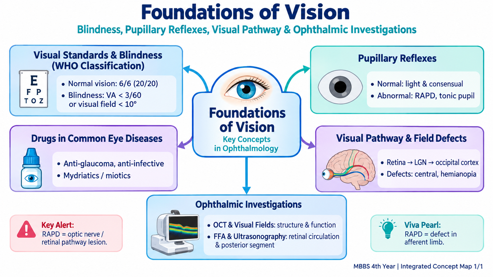
Vision depends on an intact optical system, retina, optic nerve and visual pathway extending from the eye to the occipital cortex. Ophthalmic examination therefore does more than measure how clearly a patient sees: pupillary responses and visual-field patterns can help localize disease, while modern investigations reveal structural and functional changes that may not be visible on routine examination. In this chapter, you will learn how visual acuity and blindness are described, how normal and abnormal pupillary reflexes are interpreted, how the visual pathway produces characteristic field defects, how major ophthalmic drug groups are used, and why OCT, visual-field testing, fundus fluorescein angiography and ultrasonography are selected in different clinical situations.
A. Visual Acuity, Visual Standards and WHO Classification of Blindness
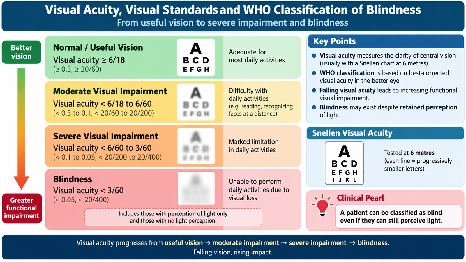
Visual acuity describes the ability of the eye to resolve fine detail. It is commonly measured with a distance chart and recorded as a fraction such as 6/6. The first number represents the testing distance, while the second represents the distance at which a person with standard vision can identify the same optotype. Therefore, a patient with 6/60 vision must be 6 metres from a symbol that a person with standard vision can identify at 60 metres.
Recording reduced vision
When a patient cannot read the largest letter on the distance chart, vision is assessed progressively using increasingly gross visual stimuli. This helps document severe visual loss consistently.
- Counting fingers (CF): the patient can identify the number of fingers shown at a stated distance.
- Hand movements (HM): the patient detects movement of the examiner’s hand but cannot count fingers.
- Perception of light (PL): the patient can detect whether a light is present.
- Projection of rays: the patient can identify the direction from which light is coming.
- No perception of light (NPL): no light can be perceived.
WHO distance-vision impairment
The functional severity of distance-vision impairment is classified using the visual acuity of the better eye. The important principle is that impairment becomes progressively more severe as the best available distance acuity falls.
| Category | Presenting distance visual acuity in better eye | Interpretation |
|---|---|---|
| Mild impairment | Worse than 6/12 to 6/18 | Reduced distance vision but useful vision remains. |
| Moderate impairment | Worse than 6/18 to 6/60 | Significant limitation of distance vision. |
| Severe impairment | Worse than 6/60 to 3/60 | Marked visual disability. |
| Blindness | Worse than 3/60 | Profound loss of distance vision. |
Older undergraduate ophthalmology descriptions may further subdivide profound visual loss into categories extending from less than 3/60 through perception of light to no perception of light. The important examination concept is that blindness is not synonymous with complete absence of light perception; a person may meet the visual-acuity definition of blindness while retaining some useful residual vision.
B. Normal Pupillary Reflexes and Abnormal Pupillary Responses
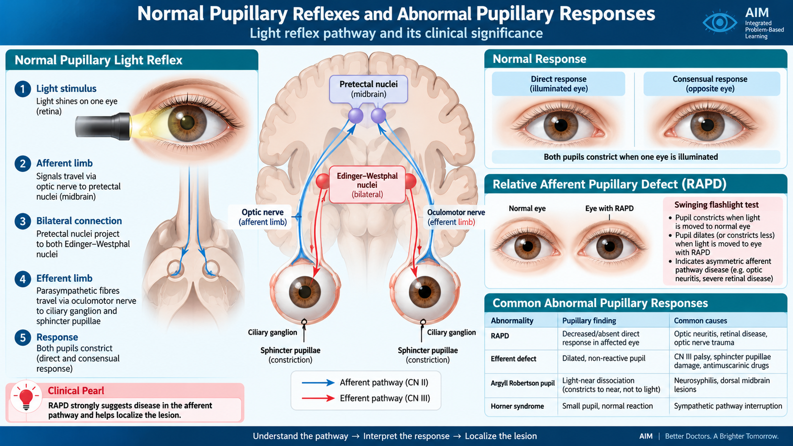
The pupils provide a rapid way of assessing the retina, optic nerve, midbrain and parasympathetic and sympathetic pathways. A normal pupil becomes smaller when exposed to light and during near fixation. Interpretation becomes much easier when the student separates the pathway into an afferent limb, which carries sensory information toward the brain, and an efferent limb, which carries the motor response back to the pupil.
Pupillary light reflex
Light falling on the retina generates impulses that travel through the optic nerve and optic tract to the pretectal region of the midbrain. Each pretectal nucleus communicates with both Edinger-Westphal nuclei. Parasympathetic fibres then travel with the oculomotor nerve to the ciliary ganglion and through the short ciliary nerves to the sphincter pupillae.
Because the pretectal area activates both Edinger-Westphal nuclei, illumination of one normal eye causes two responses:
- Direct light reflex: constriction of the illuminated pupil.
- Consensual light reflex: simultaneous constriction of the opposite pupil.
Near response
When fixation changes from a distant target to a near object, three coordinated actions occur: accommodation, convergence and pupillary constriction. Accommodation increases the refractive power of the lens, convergence aligns both eyes with the near object, and miosis improves optical quality and depth of focus.
Relative afferent pupillary defect
A relative afferent pupillary defect (RAPD or Marcus Gunn pupil) occurs when one eye sends a weaker afferent light signal than the other, usually because of significant optic-nerve disease or severe asymmetric retinal disease. It is detected with the swinging-flashlight test. When the light is moved from the healthier eye to the affected eye, both pupils appear to dilate rather than maintain constriction. They dilate because the brain receives a weaker afferent stimulus from the diseased eye, reducing bilateral parasympathetic output. The pupils themselves may still constrict normally when the healthy eye is illuminated because the efferent pathways remain intact.
Other important abnormal patterns
| Abnormality | Typical pupil finding | Localization / significance |
|---|---|---|
| RAPD | Reduced response when light is moved to affected eye | Asymmetric afferent defect, especially optic nerve |
| Oculomotor parasympathetic defect | Affected pupil is dilated and poorly reactive | Efferent parasympathetic pathway |
| Horner syndrome | Miosis; anisocoria greater in darkness | Sympathetic pathway lesion; may accompany mild ptosis |
| Adie tonic pupil | Usually dilated; poor light response with better near response and slow redilatation | Postganglionic parasympathetic dysfunction |
| Light-near dissociation | Near response is better preserved than light response | Seen in selected neurological pupillary disorders; classically described in Argyll Robertson pupils |
C. Drugs Used in Common Eye Diseases
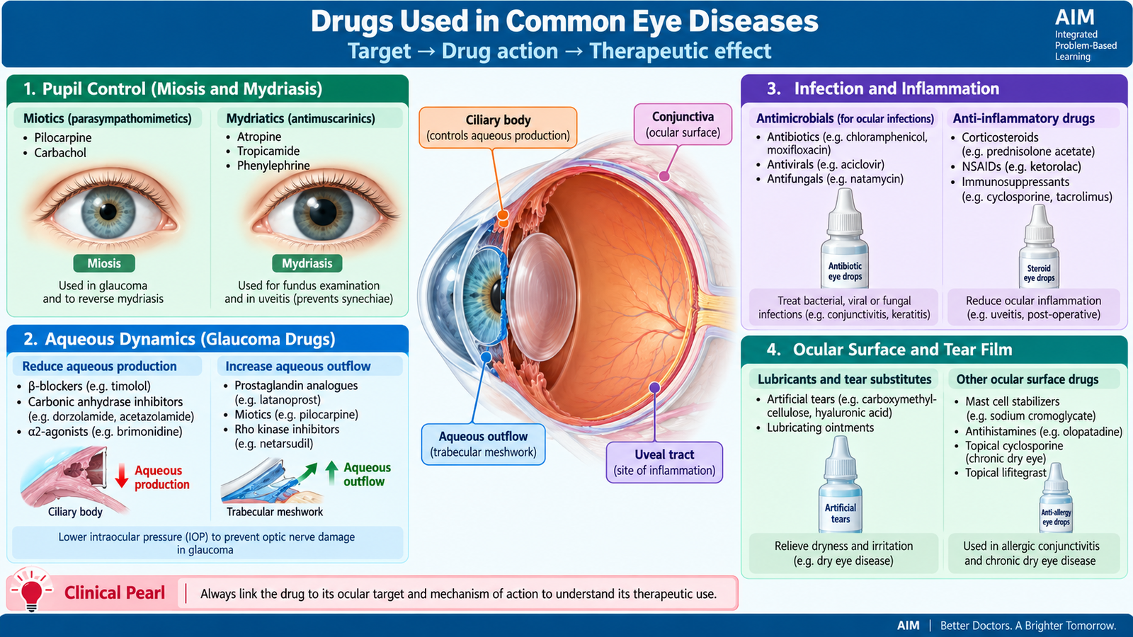
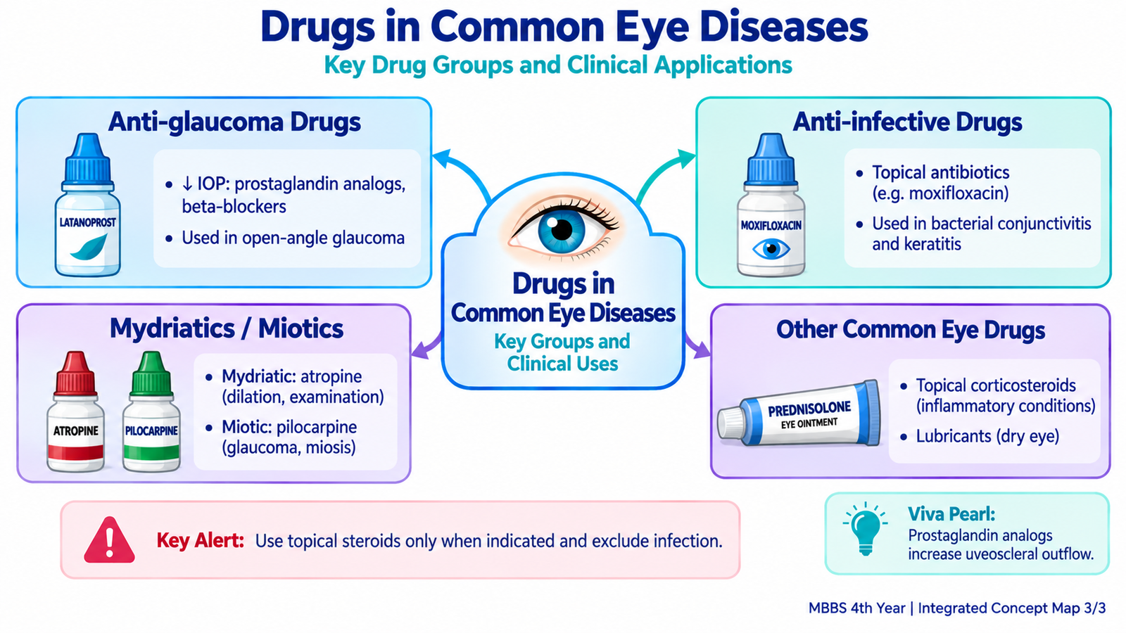
Ophthalmic drugs are selected according to the ocular structure or physiological process that needs to be modified. Some dilate or constrict the pupil, some alter aqueous-humour dynamics to reduce intraocular pressure, and others treat infection, inflammation, allergy or ocular-surface dryness. Because topical eye drops can be absorbed locally and systemically, their adverse effects and contraindications remain clinically important.
Mydriatics and cycloplegics
Antimuscarinic drugs such as tropicamide, cyclopentolate and atropine block muscarinic receptors in the iris sphincter and ciliary muscle. Sphincter paralysis produces mydriasis, while ciliary-muscle paralysis produces cycloplegia, meaning loss of accommodation. Tropicamide is commonly used when short-lasting dilation is required for fundus examination. Cycloplegic agents are useful for cycloplegic refraction, particularly when accommodation would otherwise distort the refractive assessment. Longer-acting atropine also has selected therapeutic ophthalmic uses. Antimuscarinic dilation can precipitate marked intraocular-pressure elevation in a susceptible eye with a narrow anterior-chamber angle. Phenylephrine stimulates alpha-adrenergic receptors in the iris dilator muscle and produces mydriasis without cycloplegia. Systemic cardiovascular effects are possible because sympathomimetic drug can be absorbed from the ocular surface.
Drugs that lower intraocular pressure
In glaucoma, treatment aims to reduce intraocular pressure by either decreasing aqueous-humour production or increasing its outflow. Understanding this mechanism is more useful than memorizing an isolated list of drug names.
| Drug group | Main action | Important ophthalmic use / caution |
|---|---|---|
| Prostaglandin analogues | Increase mainly uveoscleral aqueous outflow | Common long-term glaucoma therapy; may increase iris pigmentation and eyelash growth |
| Beta blockers | Reduce aqueous production | Timolol is an important example; systemic beta blockade may worsen bronchospasm or bradycardia |
| Alpha-2 agonists | Reduce aqueous production and enhance outflow | Brimonidine is a common example; allergy and systemic effects may occur |
| Carbonic anhydrase inhibitors | Reduce bicarbonate-dependent aqueous secretion | Available as topical agents and systemic acetazolamide; systemic therapy can cause metabolic and electrolyte adverse effects |
| Cholinergic agonists | Contract ciliary muscle and facilitate trabecular outflow | Pilocarpine causes miosis and may produce brow ache or accommodative symptoms |
Anti-infective drugs
Topical antimicrobial therapy is used when the infection involves accessible ocular tissues such as the conjunctiva or cornea. The choice depends on whether the suspected cause is bacterial, viral or another organism. Antibiotic drops or ointments are used for appropriate bacterial infections, while antiviral therapy is required for herpetic ocular disease. A major principle is that topical corticosteroids should not be used casually in an undiagnosed red eye. Steroids can suppress local immune responses, worsen certain corneal infections and delay epithelial healing.
Anti-inflammatory and anti-allergic drugs
Corticosteroids are valuable when ocular inflammation requires suppression, such as selected forms of uveitis, but prolonged or inappropriate use can raise intraocular pressure, promote cataract formation and worsen infection. Anti-allergic preparations, including antihistamine or mast-cell-stabilizing agents, reduce itching and allergic inflammation in allergic conjunctival disease.
Lubricants
Artificial tears and ocular lubricants supplement the tear film and reduce friction over the ocular surface. They are widely used in dry-eye disease to relieve irritation, burning and foreign-body sensation.
D. Visual Pathway: From Retina to Visual Cortex
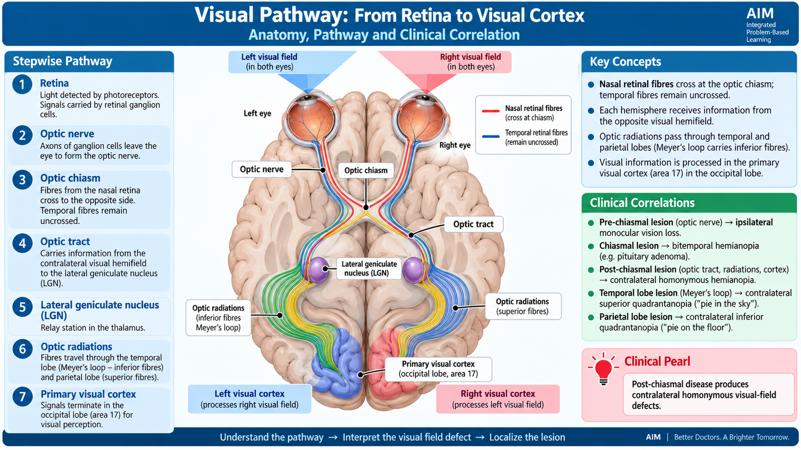
The visual pathway carries information from retinal ganglion cells to the occipital cortex. Its organization is highly systematic, so damage at different sites produces predictable patterns of visual loss. The most important principle is that fibres from the nasal half of each retina cross in the optic chiasm, while fibres from the temporal retina remain uncrossed.
Retina and optic nerve
Light from the visual field is focused onto the retina in an inverted orientation. The temporal visual field falls on the nasal retina, while the nasal visual field falls on the temporal retina. Similarly, the superior visual field is represented on the inferior retina and the inferior visual field on the superior retina. Axons of retinal ganglion cells converge at the optic disc and form the optic nerve. Therefore, a complete lesion of one optic nerve removes visual information from that eye and causes ipsilateral monocular blindness.
Optic chiasm
At the optic chiasm, nasal retinal fibres cross to the opposite side, while temporal retinal fibres remain ipsilateral. Because the nasal retina receives the temporal visual field, a central chiasmal lesion preferentially affects the crossing fibres from both eyes and therefore produces a bitemporal visual-field defect.
Optic tract and lateral geniculate nucleus
After the chiasm, each optic tract contains fibres representing the opposite visual hemifield. The right optic tract therefore carries information from the left visual field of both eyes, and the left optic tract carries information from the right visual field of both eyes. Most fibres synapse in the lateral geniculate nucleus of the thalamus.
Optic radiations
From the lateral geniculate nucleus, visual fibres travel to the occipital cortex through the optic radiations. Fibres carrying information from the superior visual field arise from the inferior retina and sweep anteriorly through the temporal lobe as Meyer’s loop. A temporal-lobe lesion can therefore cause a contralateral superior quadrantanopia. Fibres representing the inferior visual field pass more directly through the parietal lobe. Parietal optic-radiation damage therefore produces a contralateral inferior quadrantanopia.
Visual cortex
The optic radiations terminate around the calcarine fissure of the occipital lobe. The upper bank receives information from the inferior visual field, while the lower bank receives information from the superior visual field. Macular vision occupies a disproportionately large area near the occipital pole because high-acuity central vision requires extensive cortical processing.
E. Common Visual-Field Defects and Anatomical Localization
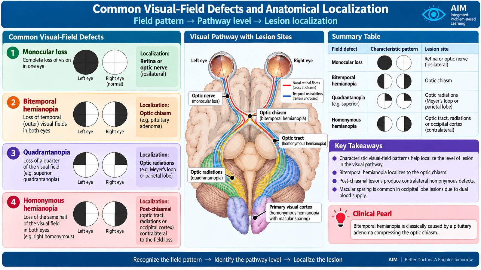
A visual-field defect is an area of reduced or absent vision within the field seen by one or both eyes. Because fibres are arranged predictably throughout the visual pathway, the shape and laterality of a defect can identify the probable site of disease. The first step is to decide whether the defect is monocular, bitemporal or homonymous.
Monocular visual loss
A lesion affecting one retina or optic nerve occurs before visual information from the two eyes is combined. It therefore produces a defect confined to the affected eye. Complete optic-nerve interruption causes ipsilateral monocular blindness, while partial optic-nerve disease may produce defects such as a central scotoma.
Bitemporal hemianopia
A central optic-chiasmal lesion affects crossing nasal retinal fibres from both eyes. Because those fibres represent the temporal visual fields, the patient loses the temporal half of the field in each eye. Compression of the chiasm from a lesion in the sellar region is an important clinical association.
Homonymous visual-field loss
Once fibres leave the optic chiasm, each side of the brain carries information from the opposite visual field of both eyes. Therefore, a lesion of the optic tract, optic radiations or occipital cortex causes a contralateral homonymous defect.
Quadrantanopia
Damage limited to part of the optic radiation may affect only one visual-field quadrant. Meyer’s loop in the temporal lobe carries information from the contralateral superior visual quadrant. Temporal-lobe damage therefore causes a contralateral superior quadrantanopia. Parietal optic-radiation damage affects fibres representing the lower visual field and produces a contralateral inferior quadrantanopia.
Occipital lesions and macular sparing
An occipital-cortex lesion may produce a highly congruous contralateral homonymous hemianopia. Central or macular vision may sometimes be relatively preserved, producing macular sparing. This pattern strongly supports a posterior visual-pathway lesion.
F. Optical Coherence Tomography and Visual-Field Testing
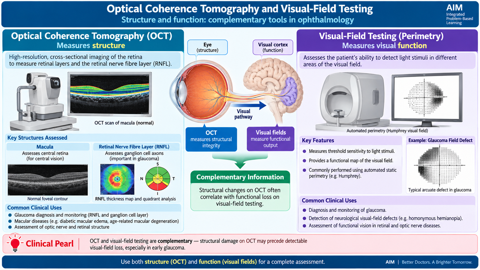
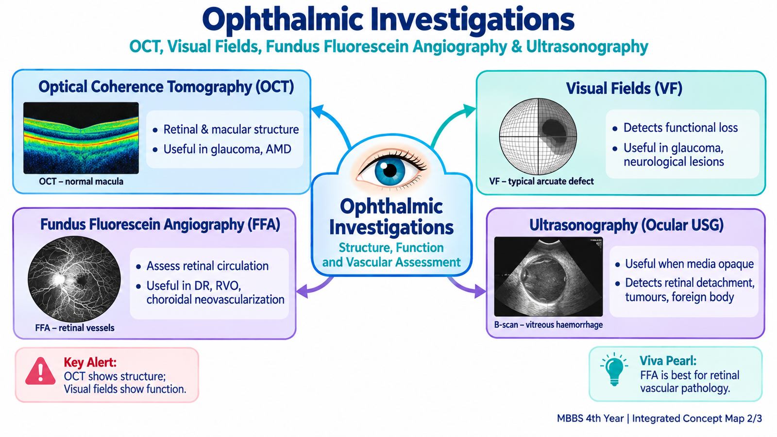
OCT and visual-field testing answer different but complementary questions. OCT primarily assesses structure, while visual-field testing assesses visual function. This structure-function relationship is especially important in diseases such as glaucoma, but the tests also have major roles in retinal and neuro-ophthalmic disease.
Optical Coherence Tomography
Optical coherence tomography (OCT) is a non-contact imaging technique that uses reflected light to create high-resolution cross-sectional images of ocular tissues. It allows the retina to be viewed almost as if its layers had been sectioned optically. Why it is used: OCT objectively assesses retinal architecture, macular thickness, the retinal nerve-fibre layer and selected optic-nerve-head parameters. What important abnormalities look like: Retinal swelling may appear as increased thickness with intraretinal fluid spaces. Loss of retinal nerve-fibre tissue appears as thinning. Subretinal or intraretinal fluid, disruption of retinal layers and other structural abnormalities can be directly demonstrated. What the finding means clinically: OCT helps determine whether reduced vision is associated with structural macular disease, identifies and monitors retinal fluid, and detects or follows structural loss of retinal nerve fibres in glaucoma.
Important clinical uses of OCT
- Macular disease: demonstrates macular thickness, retinal distortion and fluid.
- Diabetic retinal disease: helps detect and quantify diabetic macular edema.
- Age-related macular disease: demonstrates important changes in outer retinal and subretinal anatomy.
- Glaucoma: assesses retinal nerve-fibre-layer and ganglion-cell loss.
- Optic-nerve assessment: provides objective structural information that can be compared with functional visual-field loss.
Visual-field testing
A visual-field test determines which areas of the patient’s field of vision are seen normally and which have reduced sensitivity. A simple confrontation field test is useful for bedside screening of major defects, but automated perimetry provides a more detailed map of visual sensitivity. Why it is used: Perimetry is particularly important when disease may damage the optic nerve or visual pathway, or when glaucoma is suspected or being monitored. Key findings: The pattern may show a central scotoma, arcuate defect, nasal step, hemianopia or quadrantanopia. Clinical meaning: The pattern helps distinguish focal optic-nerve or retinal dysfunction from glaucoma and from lesions affecting the chiasm, optic radiations or occipital cortex.
| Investigation | Main information | Typical question answered |
|---|---|---|
| OCT | Structural | Is the retina or retinal nerve-fibre layer structurally abnormal? |
| Visual field | Functional | Which part of the patient’s vision is functionally impaired? |
In glaucoma, this distinction is especially useful: OCT may demonstrate progressive structural thinning of the retinal nerve-fibre layer, while perimetry demonstrates the corresponding loss of visual-field function. Neither test should be interpreted in isolation from the clinical examination.
G. Fundus Fluorescein Angiography and Ocular Ultrasonography
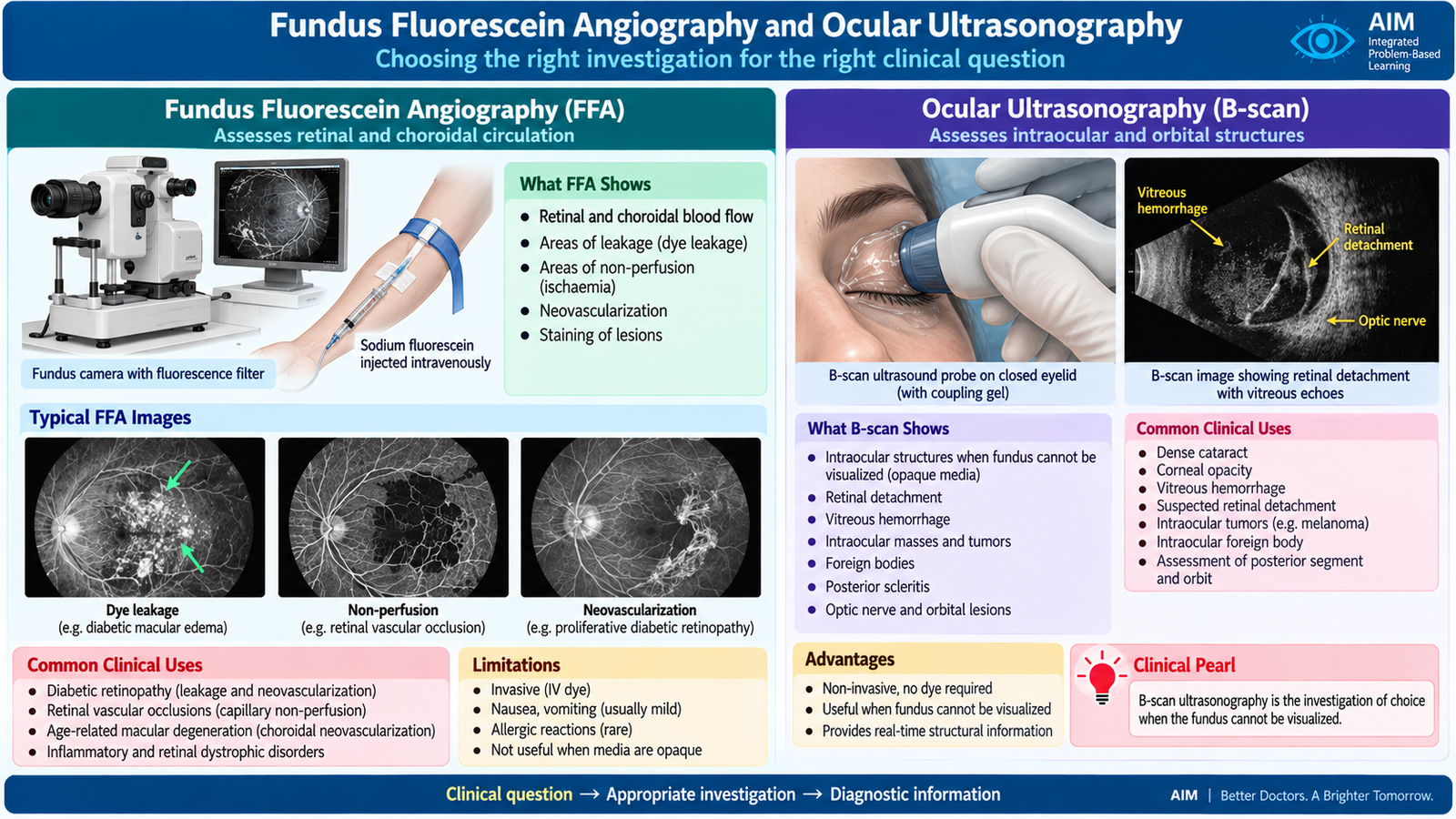
Fundus fluorescein angiography and ocular ultrasonography are selected for different clinical questions. FFA primarily demonstrates the circulation and vascular behavior of the retina and choroid, whereas ultrasonography demonstrates ocular structure using sound waves and is particularly valuable when the posterior segment cannot be seen directly.
Fundus fluorescein angiography
Fundus fluorescein angiography (FFA) involves intravenous administration of fluorescein followed by sequential photography as the dye passes through the ocular circulation. The changing pattern of fluorescence provides information that a routine static fundus photograph cannot provide. Why it is used: FFA is used when the ophthalmologist needs to evaluate retinal vascular perfusion, leakage, abnormal vessels or the vascular behavior of a retinal or choroidal lesion.
How FFA abnormalities are interpreted
- Leakage: fluorescence progressively spreads beyond normal vascular boundaries, indicating breakdown of the blood-retinal barrier or abnormal vessels.
- Non-perfusion: areas fail to fill normally, suggesting inadequate retinal capillary blood flow.
- Blocked fluorescence: blood, pigment or another opacity may prevent normal fluorescence from being seen.
- Abnormal neovascular fluorescence: newly formed vessels often leak because their walls are structurally abnormal.
These patterns make FFA useful in conditions such as diabetic retinopathy, retinal vascular occlusive disease and selected macular disorders in which vascular leakage, ischemia or neovascularization needs to be demonstrated. Fluorescein commonly produces temporary discoloration of the urine and may cause nausea. Allergic reactions can occur and, although uncommon, severe hypersensitivity reactions are possible. The test therefore requires appropriate clinical precautions.
Ocular ultrasonography
Ultrasonography uses reflected high-frequency sound waves to produce information about ocular structures. Unlike optical examination, ultrasound does not require transparent ocular media. This makes it particularly useful when the fundus is hidden by a dense cataract, vitreous hemorrhage or another media opacity.
B-scan ultrasonography
B-scan creates a two-dimensional image of the globe and orbit. It helps identify the shape, position and movement of structures within the posterior segment. Important undergraduate uses include:
- assessment of the retina when direct fundus examination is impossible;
- detection of retinal detachment;
- assessment of vitreous opacities or hemorrhage;
- demonstration of selected intraocular masses;
- assessment of selected intraocular or orbital structural abnormalities.
A detached retina produces a membranous echo separated from the normal posterior contour of the globe. Dynamic scanning can help distinguish retinal tissue from mobile vitreous echoes.
A-scan ultrasonography
A-scan represents returning echoes as spikes rather than a two-dimensional image. An important ophthalmic application is measurement of the axial length of the eye as part of ocular biometry.
Important Comparison — Visual-Field Localization
The pattern of field loss provides a rapid anatomical clue. The most useful examination approach is to move from anterior to posterior along the visual pathway.
| Lesion site | Typical visual-field defect | Why it occurs |
|---|---|---|
| Optic nerve | Ipsilateral monocular loss | All visual fibres from one eye are affected before the chiasm. |
| Central optic chiasm | Bitemporal hemianopia | Crossing nasal retinal fibres from both eyes are damaged. |
| Optic tract | Contralateral homonymous hemianopia | Each tract carries the opposite visual hemifield from both eyes. |
| Temporal optic radiation | Contralateral superior quadrantanopia | Meyer’s loop carries superior visual-field information. |
| Parietal optic radiation | Contralateral inferior quadrantanopia | Parietal radiations carry inferior visual-field information. |
| Occipital cortex | Contralateral homonymous hemianopia, sometimes with macular sparing | Posterior cortical representation of one visual hemifield is lost. |
⭐ AIM High-Yield Review
Focus while watching: Visual pathway anatomy, optic chiasm, visual-field defects, Meyer’s loop, pupillary light reflex, near reflex, RAPD and important pupillary abnormalities.