
This chapter follows the KMU learning outcomes in a logical sequence. First understand where visual loss originates and how intraocular pressure is controlled; then connect these ideas with glaucoma, its investigations and management. Revise the final AIM High-Yield Review only after understanding the explanations.
Topic 8 — Visual Loss, Intraocular Pressure & Open-Angle Glaucoma
Module/Theme: EYE — Ophthalmology 👁️
A structured approach to visual loss, aqueous humor and intraocular pressure, followed by the recognition, investigation and management of primary open-angle glaucoma, normal-tension glaucoma and ocular hypertension.
Topic Introduction
Visual loss is a symptom, not a diagnosis. A student must first decide whether vision is reduced suddenly or gradually, whether the eye is painful or painless, and which ocular structure is responsible. The transparent anterior structures must allow light to reach the retina, while the retina and optic nerve must convert and transmit the visual signal normally. Intraocular pressure depends mainly on the balance between production and drainage of aqueous humor. Disturbance of this system is an important risk factor for glaucoma. This chapter explains the major causes of visual loss, the clinical approach to sudden and gradual loss, aqueous humor physiology, glaucoma classification, and the key differences between primary open-angle glaucoma, normal-tension glaucoma and ocular hypertension.
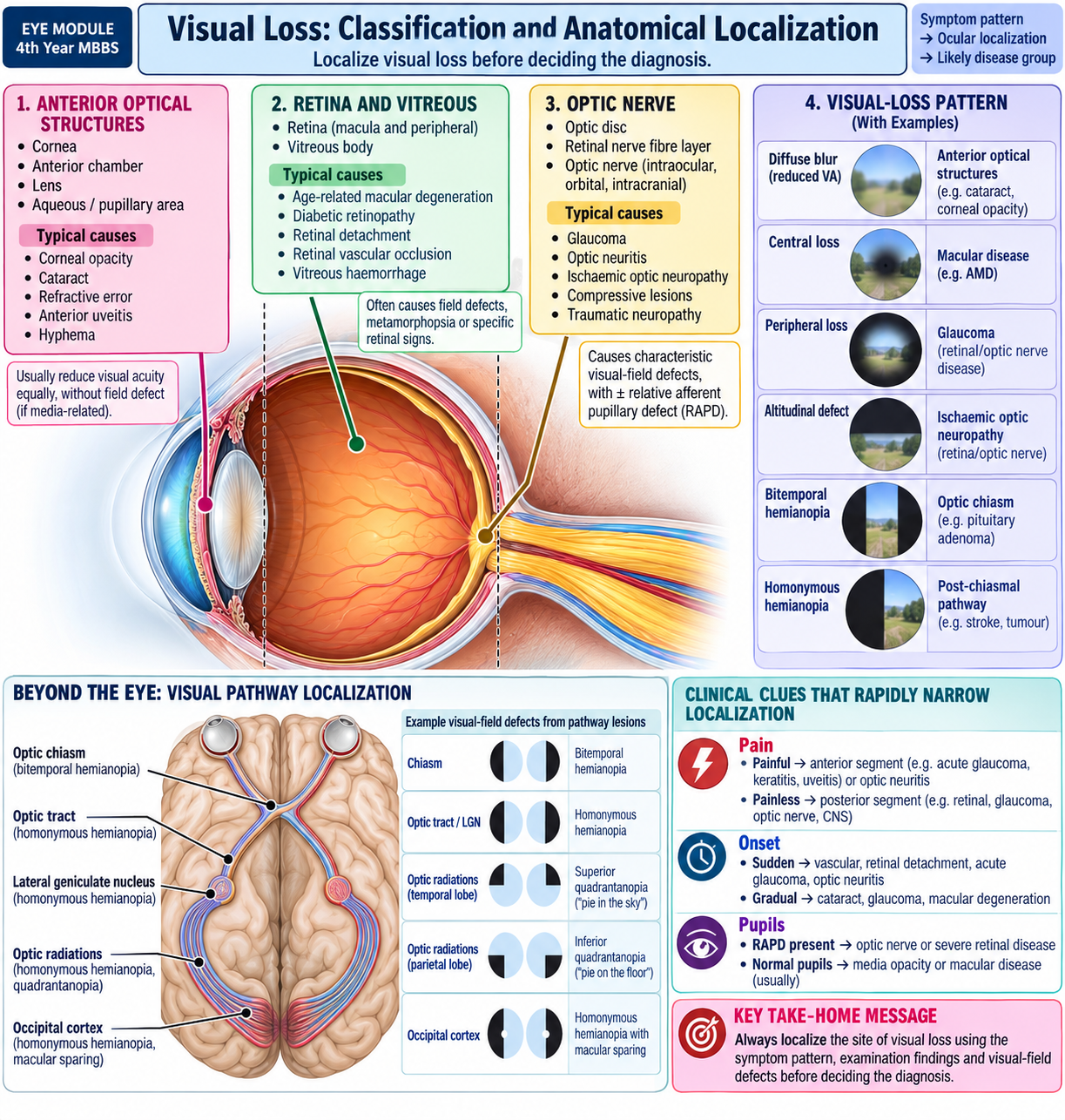
A. Visual Loss: Classification and Anatomical Localization
Visual loss can result from failure of the optical system, retina, optic nerve or visual pathway. The safest clinical approach is therefore to classify the symptom before trying to name a disease. Onset, pain, laterality and ocular examination often localize the problem quickly.
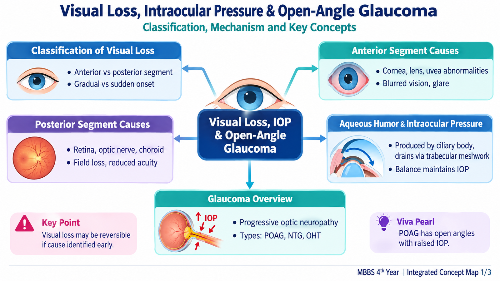
Classification of visual loss
Visual loss can be classified in several complementary ways:
- By onset: sudden or gradual.
- By pain: painful or painless.
- By laterality: unilateral or bilateral.
- By duration: transient or persistent.
- By visual function: loss of central acuity, peripheral field, colour perception or a combination.
- By anatomical site: anterior ocular structures, vitreous/retina/choroid, optic nerve or more posterior visual pathway.
Anterior-segment causes
The cornea, anterior chamber and lens lie in front of the vitreous and retina. Disease in these structures reduces the amount or quality of light reaching the retina. The fundus may therefore be normal even though visual acuity is reduced.
- Cornea: corneal opacity or scar, edema, keratitis and corneal ulceration.
- Anterior chamber: marked inflammation, hypopyon or other severe anterior-segment disease may reduce vision.
- Lens: cataract causes gradual, painless loss by scattering and blocking light.
- Acute angle closure: sudden elevation of intraocular pressure can cause corneal edema and severe painful visual loss.
Posterior ocular causes
Once light reaches the back of the eye, the retina must detect it and the optic nerve must transmit the signal. Posterior disease may therefore produce visual loss despite a relatively clear cornea and lens.
- Vitreous: vitreous hemorrhage blocks light from reaching the retina.
- Retina: retinal detachment, retinal vascular occlusion, macular disease and diabetic retinal disease.
- Optic nerve: glaucoma, optic neuritis, ischemic optic neuropathy and optic atrophy.
How examination helps localization
- Visual acuity: establishes the degree of central visual impairment.
- Pinhole: marked improvement suggests that refractive error contributes importantly to reduced acuity.
- Red reflex and anterior segment: abnormalities may indicate corneal or lenticular obstruction.
- Pupils: a relative afferent pupillary defect suggests significant asymmetric optic-nerve or retinal dysfunction rather than uncomplicated cataract.
- Fundus examination: identifies retinal, macular and optic-disc pathology.
- Visual fields: may reveal characteristic retinal, optic-nerve or visual-pathway defects.
B. Gradual Visual Loss: Major Causes and Management Approach
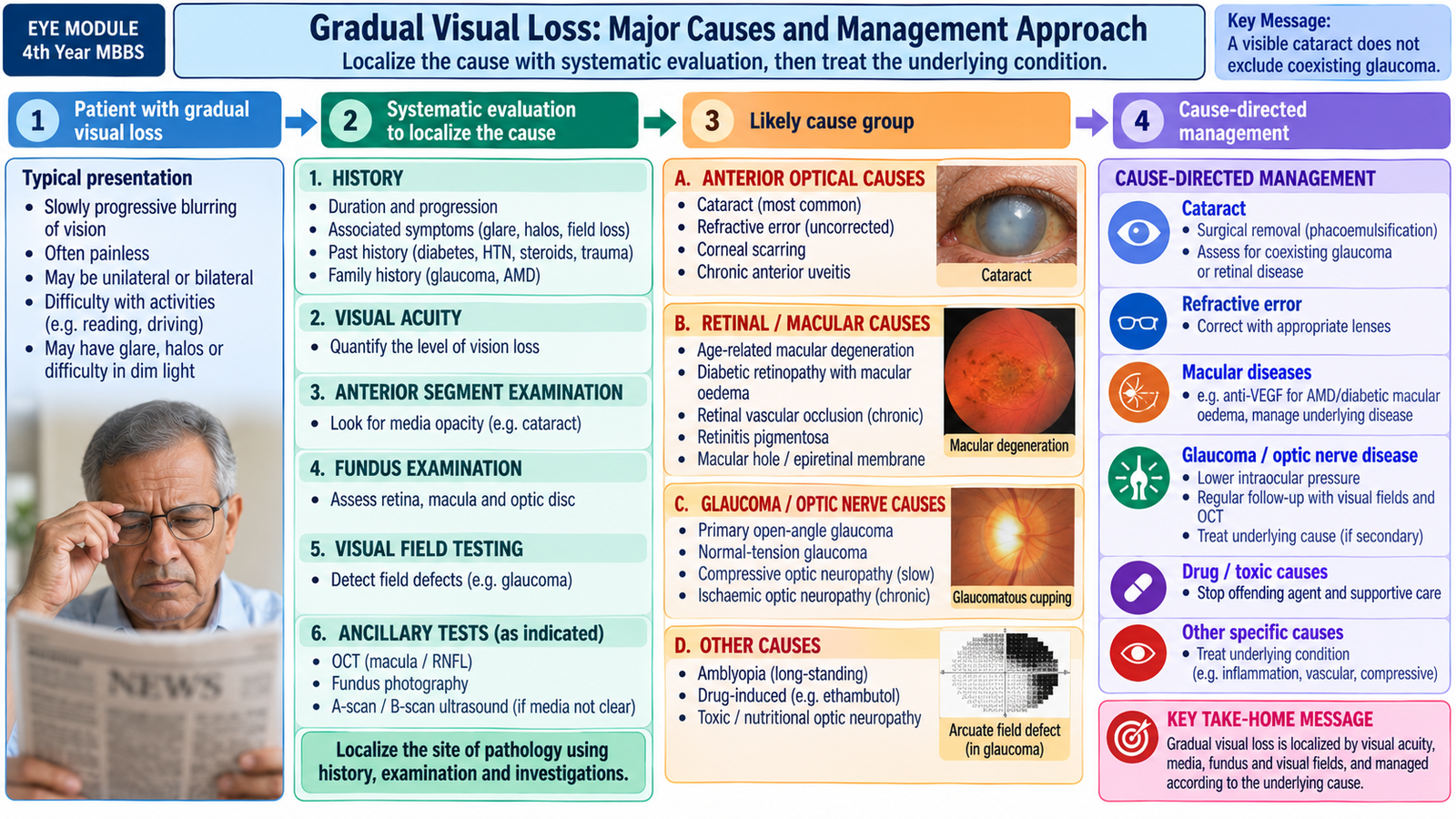
Gradual visual loss develops over weeks, months or years. Because many chronic eye diseases are painless, students should not use absence of pain as reassurance. The important task is to identify whether the problem is optical, retinal, macular or optic-nerve related and then manage the underlying cause before irreversible damage develops.
Common causes
- Refractive error: blurred vision often improves with pinhole and appropriate refractive correction.
- Cataract: progressive painless reduction of vision caused by lens opacity; glare and reduced contrast may accompany the loss.
- Corneal opacity: previous infection, trauma or other corneal disease may interfere with light transmission.
- Primary open-angle glaucoma: typically causes gradual peripheral visual-field loss and may remain unnoticed until disease is advanced.
- Macular disease: commonly affects central vision, reading and recognition of fine detail.
- Diabetic retinal disease: retinal vascular damage or macular involvement may gradually reduce vision.
- Optic-nerve disease: chronic optic neuropathy may impair acuity, colour vision or visual fields depending on the cause.
Stepwise clinical assessment
A gradual complaint still requires a complete eye assessment because the patient may have more than one cause, such as cataract together with glaucoma.
- Measure visual acuity in each eye separately.
- Check pinhole response where reduced acuity may be refractive.
- Examine the cornea and lens for opacity.
- Assess pupils for evidence of asymmetric retinal or optic-nerve dysfunction.
- Measure intraocular pressure when glaucoma is possible.
- Examine the optic disc, macula and retina.
- Assess visual fields when glaucoma or neurological visual loss is suspected.
Management principles
Treatment is directed at the cause rather than the symptom “poor vision.” Correct refractive error when present. Visually significant cataract is managed surgically when it interferes with visual function. Corneal disease requires treatment of the specific cause and visual rehabilitation where needed. Retinal and macular disease require ophthalmic evaluation and disease-specific treatment. Suspected glaucoma requires formal optic-disc, field and intraocular-pressure assessment because lost glaucomatous vision cannot usually be restored; treatment aims to prevent further loss.
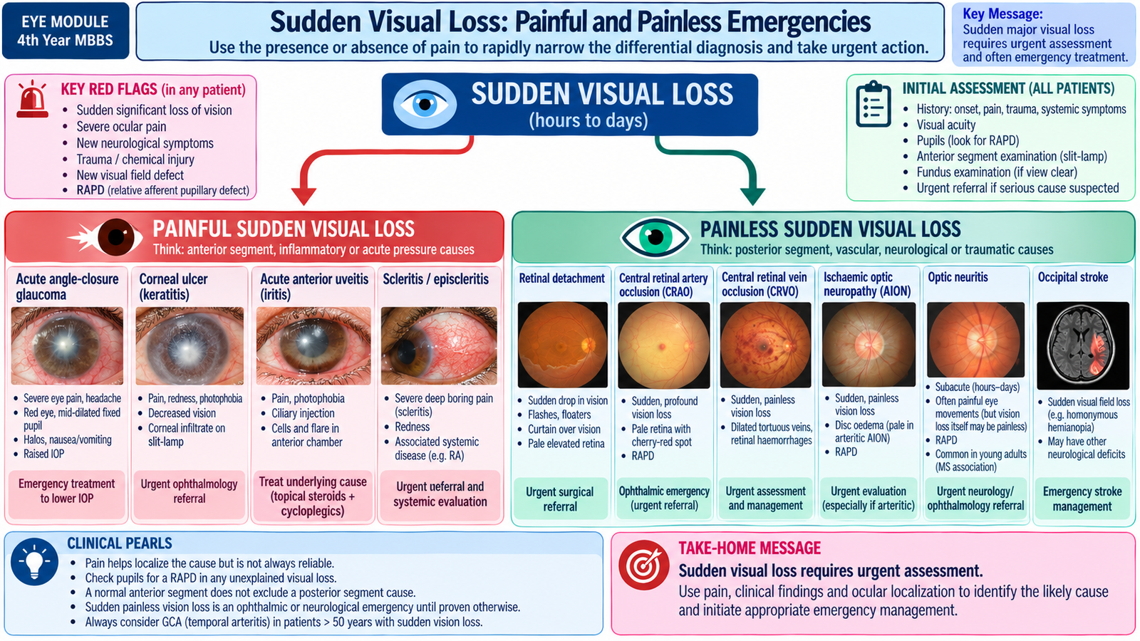
C. Sudden Visual Loss: Painful and Painless Emergencies
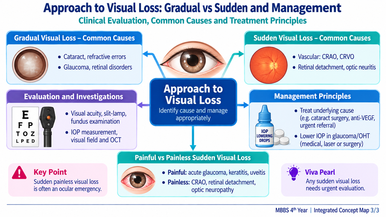
Sudden loss of vision is potentially sight-threatening and should be treated as urgent until a benign explanation is established. Pain is a particularly useful discriminator. Painful visual loss often points toward the cornea, anterior uvea, markedly raised intraocular pressure or an inflamed optic nerve. Painless sudden loss more often suggests retinal vascular disease, retinal detachment, vitreous hemorrhage or ischemic optic-nerve disease.
Painful sudden visual loss
- Acute angle-closure glaucoma: severe ocular pain, headache, blurred vision or halos, red eye, corneal haze and markedly raised IOP.
- Keratitis/corneal ulcer: pain, photophobia, reduced vision and a corneal epithelial or stromal lesion.
- Anterior uveitis: pain, photophobia, circumcorneal redness and anterior-chamber inflammation.
- Optic neuritis: visual reduction commonly associated with pain on eye movement and impaired colour vision; the eye may not be externally red.
- Severe intraocular infection: significant pain and reduced vision after surgery, penetrating injury or ocular infection is an emergency.
Painless sudden visual loss
- Retinal artery occlusion: abrupt profound monocular visual loss caused by sudden retinal ischemia.
- Retinal vein occlusion: sudden or subacute painless visual reduction with widespread retinal hemorrhagic changes.
- Retinal detachment: flashes, floaters and a curtain or shadow progressing across the field.
- Vitreous hemorrhage: sudden haze, floaters or marked visual loss because blood blocks the visual axis.
- Ischemic optic neuropathy: sudden optic-nerve dysfunction, usually painless.
Immediate assessment
The first purpose of examination is to identify a reversible or rapidly progressive sight-threatening process.
- Measure visual acuity in both eyes.
- Ask about exact onset, pain, redness, trauma, surgery, flashes, floaters and field loss.
- Examine the pupils and check for an afferent pupillary defect.
- Inspect the cornea and anterior chamber.
- Measure intraocular pressure when appropriate and when globe integrity is not in doubt.
- Examine the fundus and optic disc where the media are clear.
Management and referral principles
Acute angle closure, severe keratitis, retinal arterial occlusion, retinal detachment, severe intraocular infection and other unexplained sudden major visual loss require urgent ophthalmic assessment. Retinal arterial occlusion also requires urgent systemic vascular evaluation because retinal ischemia may indicate important vascular disease. A suspected retinal detachment needs urgent retinal evaluation before the macula becomes involved or further detachment occurs. Sudden visual loss should never be dismissed as simple conjunctivitis merely because the eye is red.
D. Aqueous Humor Dynamics and Intraocular Pressure
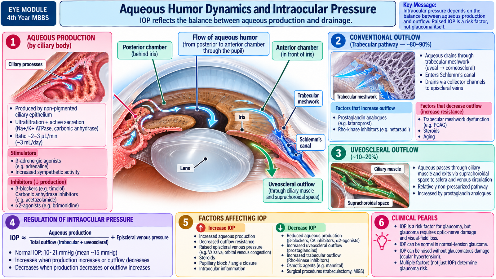
Aqueous humor is the clear fluid that nourishes avascular anterior ocular structures and helps maintain the internal pressure and shape of the eye. Intraocular pressure is not produced simply because aqueous is formed; it reflects a continuous balance between aqueous production, aqueous drainage and resistance to its outflow.
Production and circulation
Aqueous humor is produced mainly by the ciliary processes. It enters the posterior chamber, flows through the pupil into the anterior chamber, and then leaves the eye through two drainage pathways.
Outflow pathways
- Trabecular or conventional pathway: aqueous passes through the trabecular meshwork into Schlemm’s canal, then through collector channels toward the episcleral venous circulation. This is the major drainage pathway.
- Uveoscleral pathway: a smaller proportion passes through tissues around the ciliary muscle into the suprachoroidal region and subsequently leaves the eye.
How intraocular pressure rises
In open-angle glaucoma the drainage angle is physically open, but aqueous encounters increased resistance within the outflow system, particularly the trabecular pathway. Continued aqueous production in the presence of increased resistance can raise intraocular pressure. Raised IOP increases mechanical and physiological stress on the optic-nerve head and is the most important modifiable risk factor for glaucomatous damage.
Factors affecting measured IOP
Interpretation of IOP should consider repeated measurements and the clinical context. Central corneal thickness can influence applanation measurements, while IOP naturally varies over time. Therefore, diagnosis requires correlation with the optic disc, retinal nerve-fibre layer and visual field rather than reliance on one pressure reading.
E. Glaucoma: Definition, Classification and Core Diagnostic Concept
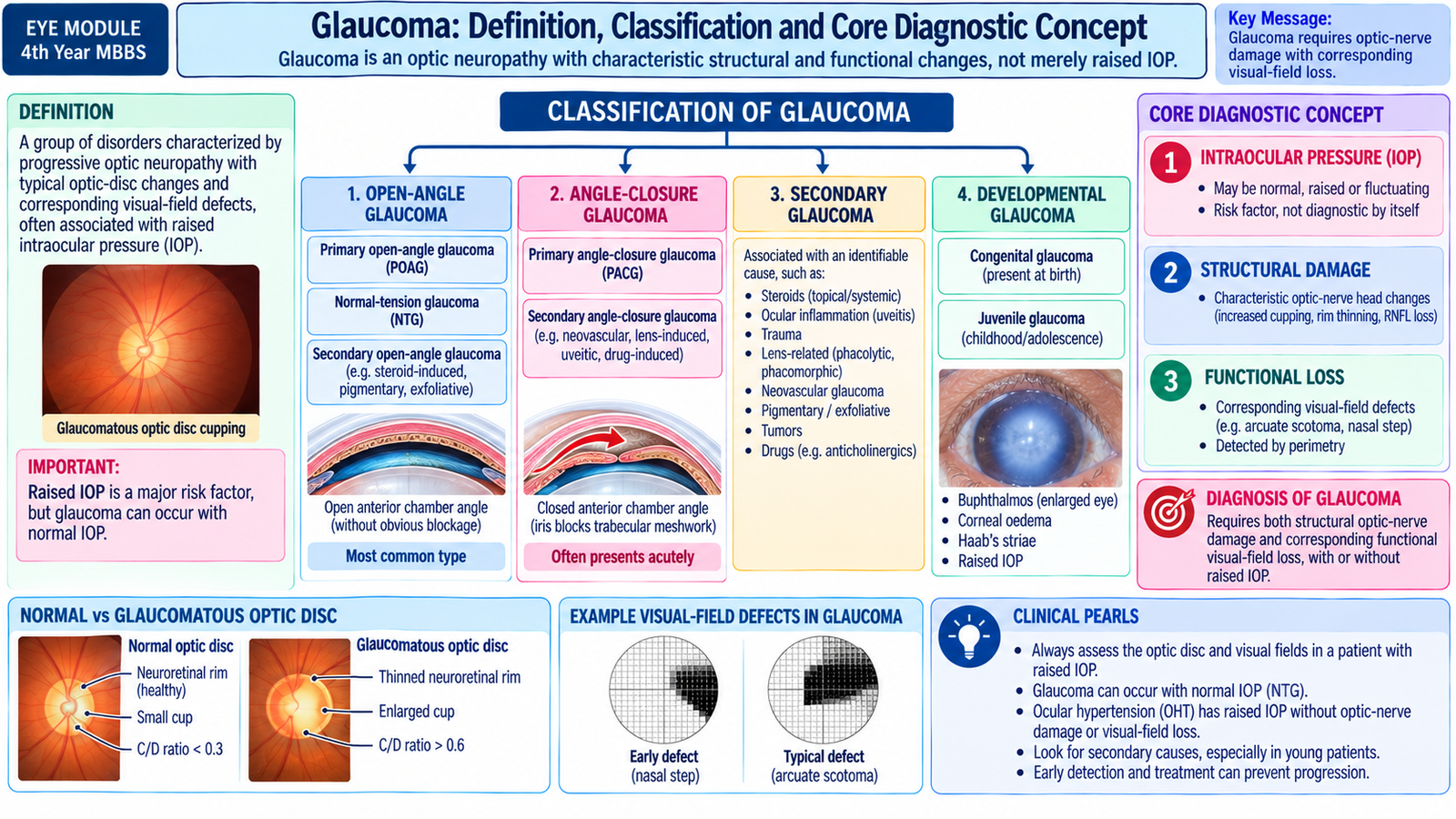
Glaucoma is a group of progressive optic neuropathies characterized by characteristic damage to the optic-nerve head and retinal nerve-fibre layer, usually accompanied by corresponding visual-field loss. Raised intraocular pressure is an important risk factor but is not essential for the diagnosis.
Why vision is lost
Glaucoma damages retinal ganglion-cell axons as they pass through the optic-nerve head. Progressive loss of these fibres produces thinning of the neuroretinal rim and enlargement of the optic cup. Because peripheral retinal ganglion-cell pathways are commonly affected before central fixation, the patient may have useful central acuity while significant visual-field loss is already present.
Classification
Glaucoma is commonly classified according to the configuration of the anterior-chamber angle and whether a primary or secondary cause is present.
- Primary open-angle glaucoma (POAG): open angle with characteristic glaucomatous optic neuropathy and no other ocular disorder sufficient to explain it.
- Normal-tension glaucoma (NTG): an open-angle glaucomatous optic neuropathy in which untreated measured IOP remains within the statistically normal range.
- Primary angle-closure disease/glaucoma: the peripheral iris obstructs the trabecular drainage angle.
- Secondary glaucoma: another ocular or systemic process alters aqueous outflow or otherwise produces glaucoma.
- Developmental/congenital glaucomas: glaucoma associated with developmental abnormalities of the aqueous drainage system.
Three findings that must be separated
- IOP: a risk factor and treatment target.
- Optic-nerve structural damage: cupping, rim loss and retinal nerve-fibre loss.
- Functional damage: corresponding visual-field defects.
This distinction explains why a patient can have glaucoma with apparently normal IOP and why another patient can have ocular hypertension without glaucoma.
F. Primary Open-Angle Glaucoma
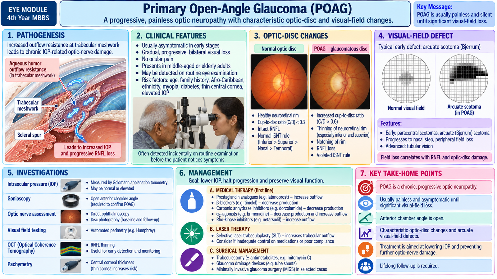
Primary open-angle glaucoma is a chronic progressive glaucomatous optic neuropathy in which the anterior-chamber angle remains open. The key problem is not anatomical closure of the angle but impaired aqueous outflow together with susceptibility of the optic nerve. The disease is especially important because it is usually painless and asymptomatic in its early stages.
Etiology and risk factors
POAG is multifactorial. No single cause explains every case. Increased resistance to aqueous drainage increases IOP in many patients, while age-related and individual susceptibility determine how strongly the optic nerve is affected.
- Increasing age.
- Raised intraocular pressure.
- Family history of glaucoma.
- Myopia.
- Thin central cornea, which is associated with increased risk and also affects interpretation of measured IOP.
- Previous evidence of suspicious optic-disc or retinal nerve-fibre change.
Pathogenesis
Increased resistance to aqueous outflow can raise IOP. Mechanical stress and impaired axonal support at the optic-nerve head contribute to retinal ganglion-cell injury. As axons are lost, the retinal nerve-fibre layer becomes thinner and the neuroretinal rim is reduced. The cup therefore appears progressively enlarged. Structural damage is followed by characteristic visual-field loss.
Clinical features
- Usually bilateral but often asymmetric.
- Usually painless.
- No characteristic red eye in uncomplicated disease.
- Central visual acuity may remain normal until advanced disease.
- Progressive peripheral field loss may go unnoticed by the patient.
- Very advanced disease may produce severe field constriction and major visual disability.
Ophthalmic examination and visual recognition
The optic disc provides structural evidence of glaucomatous damage. The examiner assesses the entire neuroretinal rim rather than using cup size alone.
- Increasing cup-to-disc ratio: reflects loss of neuroretinal tissue, although naturally large physiological cups must be distinguished from pathological cupping.
- Asymmetry of cupping: unexplained asymmetry between the two eyes raises suspicion.
- Neuroretinal-rim thinning or notching: focal loss of rim tissue is particularly suggestive.
- Retinal nerve-fibre-layer defects: represent loss of retinal ganglion-cell axons.
- Optic-disc hemorrhage: may accompany active or progressive glaucomatous damage.
Visual-field changes
Glaucomatous field defects follow the anatomical arrangement of retinal nerve fibres. Early defects may be subtle and localized, while advanced disease causes progressive field constriction.
- Paracentral scotoma.
- Nasal step.
- Arcuate or Bjerrum-type scotoma.
- Expansion and merging of defects with progression.
- Advanced constriction with only small remaining islands of vision in severe disease.
Investigations
Glaucoma diagnosis requires correlation of pressure, anatomy and visual function.
- Tonometry — why: measures IOP. Meaning: raised pressure increases glaucoma risk but does not alone prove glaucoma.
- Gonioscopy — why: directly assesses the anterior-chamber angle. Finding: an open angle supports an open-angle glaucoma diagnosis.
- Optic-disc assessment — why: identifies characteristic structural damage. Finding: rim loss, notching or progressive cupping strengthens the diagnosis.
- Automated perimetry — why: detects functional loss. Finding: reproducible characteristic field defects demonstrate the functional effect of optic-nerve damage.
- Retinal nerve-fibre/optic-nerve imaging such as OCT — why: objectively evaluates structural tissue loss. Finding: focal or diffuse thinning can support diagnosis and help monitor progression.
- Central corneal thickness assessment — why: helps interpret applanation IOP and overall risk.
Management
The purpose of treatment is to preserve the patient’s remaining vision by lowering IOP sufficiently to reduce the risk of further optic-nerve damage. The required pressure reduction is individualized according to disease severity, baseline pressure, progression and other risk factors.
Medical therapy
- Prostaglandin analogues: lower IOP mainly by increasing uveoscleral aqueous outflow.
- Topical beta-blockers: reduce aqueous production by the ciliary body.
- Topical carbonic anhydrase inhibitors: reduce aqueous formation.
- Alpha-2 adrenergic agonists: lower aqueous production and may also increase alternative aqueous outflow.
Laser and surgery
Laser trabeculoplasty can improve aqueous drainage and may be used as initial or additional therapy in suitable open-angle disease. If adequate pressure control cannot be achieved, or glaucoma continues to progress despite medical or laser therapy, glaucoma surgery may be required to create or enhance aqueous drainage. Detailed operative techniques are beyond undergraduate scope.
G. Normal-Tension Glaucoma and Ocular Hypertension
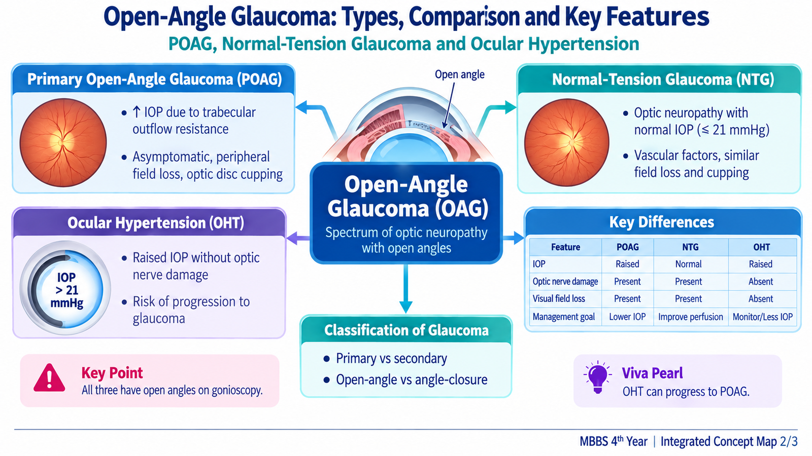
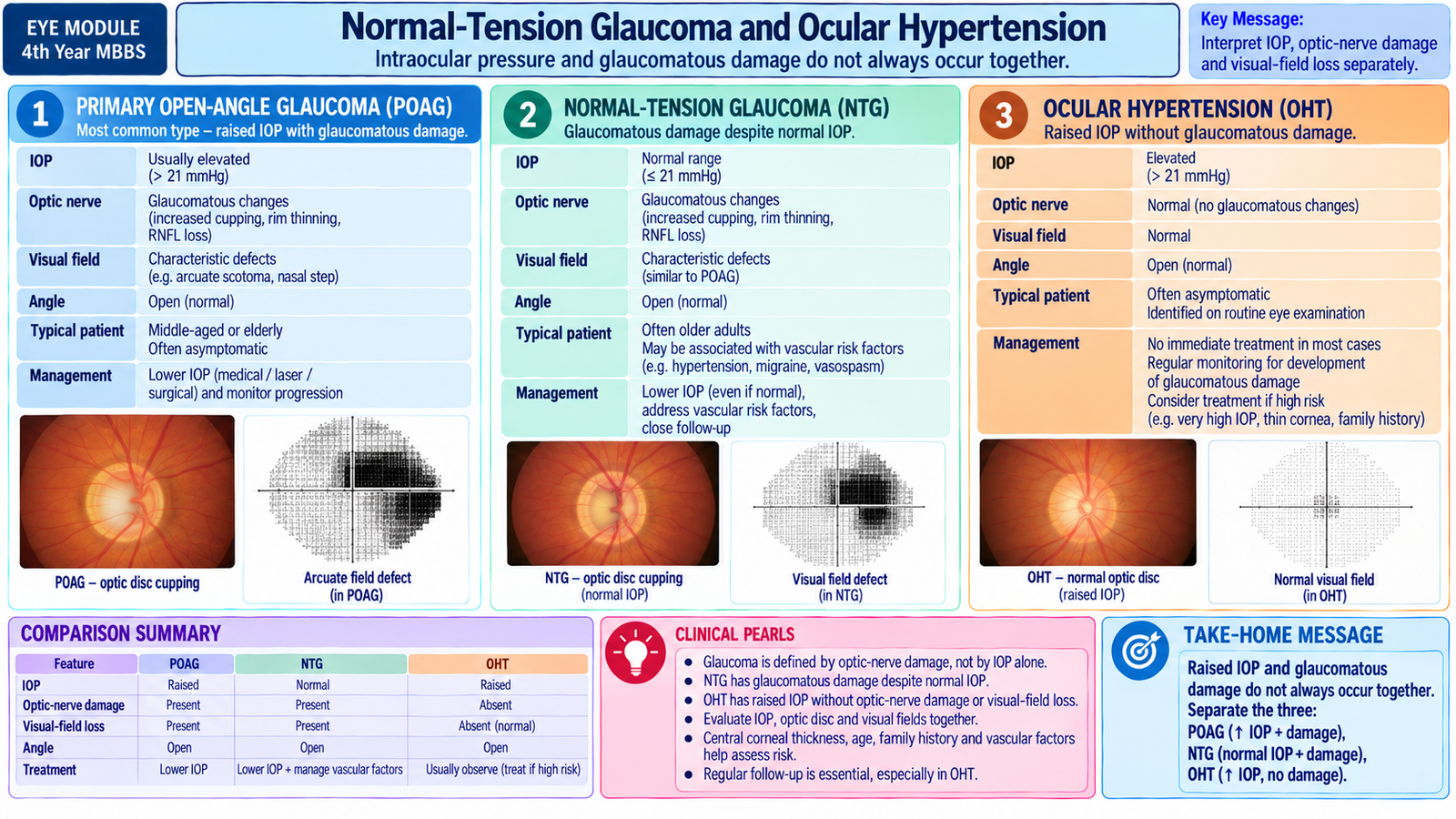
Normal-tension glaucoma and ocular hypertension show why intraocular pressure must never be interpreted in isolation. In NTG the patient already has genuine glaucomatous optic-nerve and visual-field damage even though untreated measured IOP lies within the statistically normal range. In ocular hypertension the pressure is raised, but detectable glaucomatous optic-nerve and visual-field damage is absent.
Normal-tension glaucoma
Etiology and mechanism
The optic nerve in NTG appears vulnerable to damage at pressures tolerated by many other eyes. Mechanical susceptibility and vascular factors may contribute. The important clinical principle is that a pressure described as “normal” for the population is not necessarily safe for every individual optic nerve.
Clinical features
- Painless and usually chronic.
- Open anterior-chamber angle.
- Characteristic glaucomatous optic-disc/RNFL damage.
- Characteristic corresponding visual-field loss.
- Optic-disc hemorrhages and paracentral defects may be prominent in some patients.
Investigations
The same structural and functional glaucoma assessment used for POAG is required: repeated tonometry, gonioscopy, optic-disc assessment, retinal nerve-fibre imaging and perimetry. Repeated IOP measurements help ensure that pressure elevations have not simply been missed. Central corneal thickness should be considered because a thin cornea can affect interpretation of measured IOP. Findings that are atypical for glaucoma should prompt consideration of a non-glaucomatous optic neuropathy.
Management
Despite the apparently normal baseline pressure, treatment is still directed toward further lowering IOP because pressure reduction can reduce continued glaucomatous stress. Medical, laser and surgical approaches are selected according to progression and individual risk. Relevant systemic factors that may impair optic-nerve perfusion should be considered in cooperation with the appropriate physician rather than altering systemic therapy without clinical review.
Ocular hypertension
Definition
Ocular hypertension means that IOP is repeatedly above the statistically normal range while the anterior-chamber angle is open and the optic disc, retinal nerve-fibre layer and visual fields show no definite glaucomatous damage.
Why it matters
OHT is not the same as glaucoma, but it increases the future risk of developing glaucoma. The probability of conversion varies among patients; therefore management is based on overall risk rather than pressure alone.
Investigations
- Repeat tonometry: confirms that the elevation is persistent rather than a single abnormal reading.
- Gonioscopy: confirms that the angle is open and helps exclude other mechanisms.
- Optic-disc/RNFL assessment: establishes that structural glaucomatous damage is absent at baseline.
- Visual-field testing: establishes that functional glaucomatous damage is absent.
- Central corneal thickness: assists interpretation of measured IOP and risk assessment.
Management
Patients at relatively low risk may be observed with regular measurement of IOP, optic-disc/RNFL assessment and visual-field testing. Patients with a greater risk of conversion may be offered pressure-lowering treatment. Development of definite structural or functional glaucomatous damage changes the diagnosis from isolated ocular hypertension to glaucoma.
Integrated Mechanism Flow
The central glaucoma sequence can be understood in six linked steps:
Aqueous production continues
Outflow resistance or susceptible optic nerve
Optic-nerve-head stress
Retinal ganglion-cell axon loss
Rim thinning and optic-disc cupping
Progressive visual-field loss
Intervention point: Lowering IOP reduces an important modifiable source of stress on the optic nerve, including in many patients whose untreated pressure lies within the statistically normal range.
Important Comparison — POAG vs NTG vs Ocular Hypertension
| Feature | POAG | NTG | Ocular Hypertension |
|---|---|---|---|
| Anterior-chamber angle | Open | Open | Open |
| Untreated measured IOP | Often raised | Within statistically normal range | Raised |
| Glaucomatous optic-disc/RNFL damage | Present | Present | Absent |
| Glaucomatous visual-field loss | Present once functional damage develops | Present once functional damage develops | Absent |
| What establishes the diagnosis? | Characteristic structural/functional glaucoma with open angle | Glaucomatous damage despite normal-range measured IOP | Raised IOP without glaucomatous damage |
| Management principle | Lower IOP and monitor progression | Lower IOP despite normal baseline measurement | Risk-based observation or preventive IOP lowering |
⭐ AIM High-Yield Review
🎥 Video Learning — Visual Loss, Intraocular Pressure & Glaucoma
Watch this lecture to reinforce aqueous humor dynamics, intraocular pressure, optic-nerve damage, primary open-angle glaucoma, visual-field changes, investigations and management principles.
Topic 8 — Visual Loss, Intraocular Pressure & Open-Angle Glaucoma