
Topic 2 — Visual Optics, Refraction & Refractive Errors
Topic Introduction
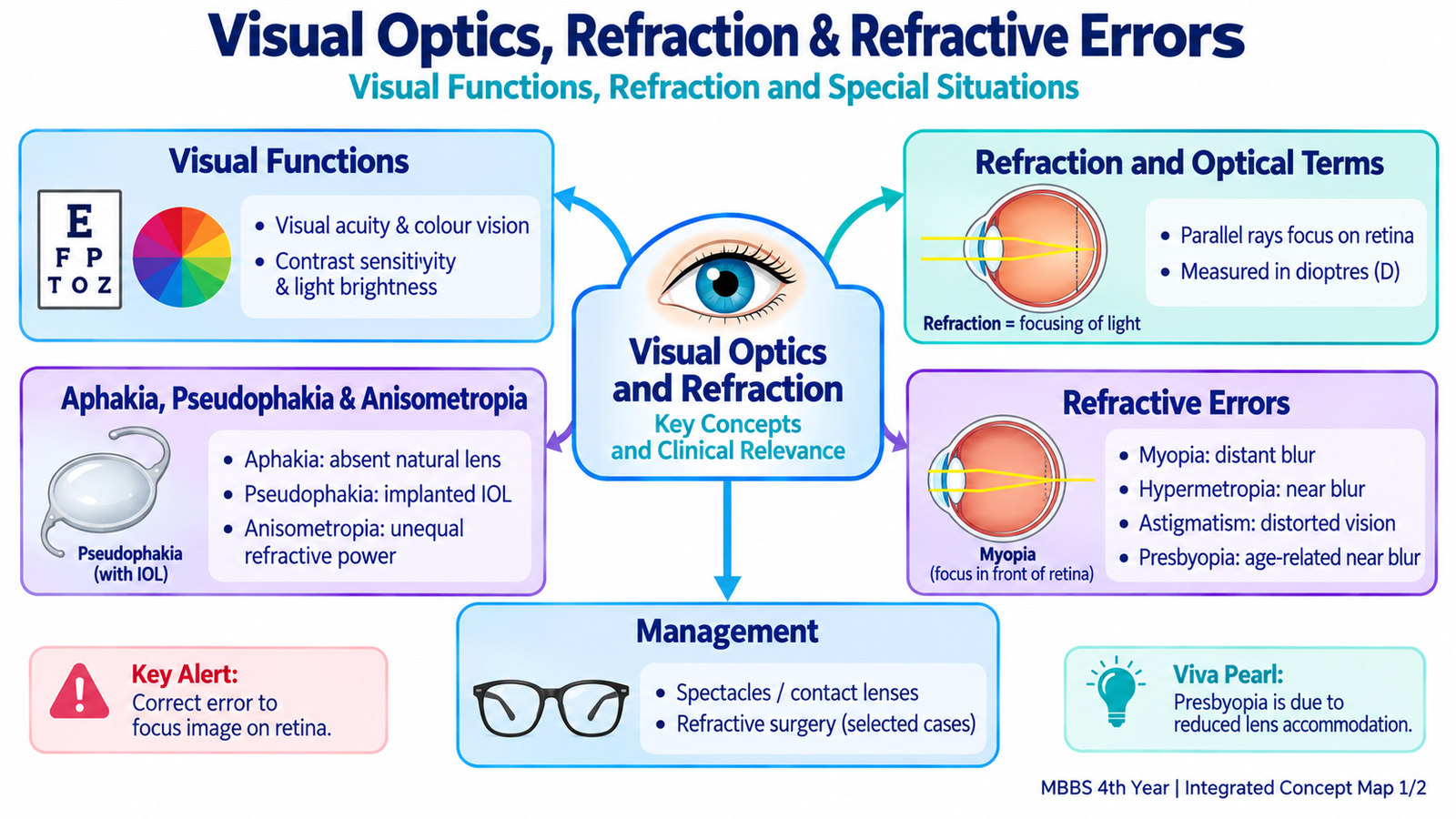
Clear vision depends on more than simply seeing a letter on a chart. The eye must focus light accurately on the retina, resolve fine detail, distinguish colours and contrast, and function across different levels of illumination. When the optical power of the eye does not match its length, the image becomes defocused and a refractive error results. In this chapter, you will first understand the main visual functions and the principles of refraction. You will then learn why myopia, hypermetropia, astigmatism and presbyopia occur, how they present clinically, and how they are corrected. Anisometropia, aphakia and pseudophakia are also explained as important related optical states.
A. Visual Functions: How Useful Vision Is Assessed
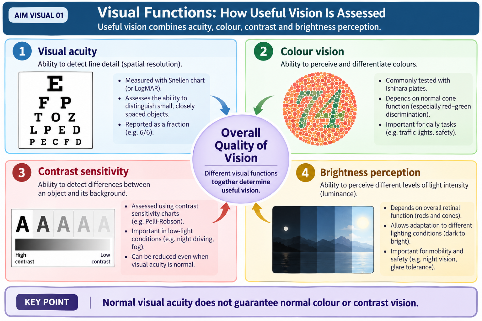
Vision is a group of related functions rather than a single ability. A person may have good visual acuity but still have abnormal colour vision or reduced contrast sensitivity. Understanding these components helps explain why routine visual acuity alone does not describe the complete quality of vision.
Visual acuity
Visual acuity is the ability of the eye to resolve fine spatial detail and distinguish two closely separated points as separate. It depends on a sharply focused retinal image, particularly at the fovea, and an intact visual pathway.
Distance visual acuity is commonly measured with a Snellen-type chart. A notation of 6/6 means that the person can read at 6 metres the detail that a person with standard vision should read at 6 metres. Reduced acuity may result from refractive error or from ocular disease.
A pinhole allows mainly central rays of light to enter the eye and reduces the blur produced by many refractive errors. Therefore, improvement of vision through a pinhole suggests that optical defocus is contributing to the reduced acuity. Failure to improve makes a purely refractive cause less likely.
Colour vision
Colour vision is the ability to distinguish light of different wavelengths as different colours. It depends mainly on the cone photoreceptors of the retina and on normal transmission through the visual pathway. Different cone populations have different spectral sensitivities, and the brain compares their activity to generate colour perception.
Colour vision can be assessed clinically with colour plates such as Ishihara plates, particularly for red-green discrimination. Abnormal colour perception can be congenital or acquired, but detailed classification is outside the supplied outcomes.
Contrast sensitivity
Contrast sensitivity is the ability to detect an object when it differs only slightly in brightness from its background. It is particularly important in real-life situations such as recognizing a face in dim light or seeing an object in fog. A patient may therefore have reasonably good high-contrast visual acuity but still report poor quality of vision when contrast is low.
Light and brightness perception
Brightness is the visual perception produced by the intensity of light reaching and stimulating the retina. Cones function best in brighter conditions, while rods are especially important in dim illumination. Retinal adaptation allows vision to continue over a wide range of environmental light levels. Thus, brightness perception reflects both the amount of light entering the eye and the retina’s response to that light.
B. Refraction: How the Eye Focuses Light
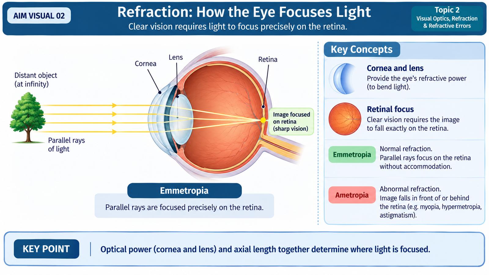
Refraction is the bending of light as it passes between media with different refractive properties. In the eye, the cornea and crystalline lens together bend incoming light so that a clear image is formed on the retina. The cornea provides most of the fixed refractive power, while the crystalline lens can alter its power during accommodation.
Emmetropia
An emmetropic eye has the correct relationship between its optical power and axial length. With accommodation relaxed, parallel rays from a distant object are focused sharply on the retina. No correcting lens is required for clear distance vision.
Ametropia
Ametropia means that parallel rays are not focused precisely on the retina when accommodation is relaxed. This occurs because the length of the eye and its refractive power do not match appropriately. Myopia, hypermetropia and astigmatism are major examples.
Accommodation
Accommodation allows the eye to increase its refractive power for near objects. When the ciliary muscle contracts, tension on the zonular fibres decreases and the elastic lens becomes more convex. The increased lens curvature gives it greater converging power, allowing near objects to be focused on the retina.
The refractive power of a lens is expressed in diopters (D). A converging lens has positive power, whereas a diverging lens has negative power.
Clinical assessment of refraction
Refraction determines the lens power needed to provide the clearest retinal image. It is approached using objective and subjective methods.
- Objective refraction: estimates the refractive state without relying on the patient’s judgement. Retinoscopy and automated refraction are common examples.
- Subjective refraction: refines the prescription by asking the patient which lens provides clearer vision.
- Cycloplegic refraction: temporary paralysis of accommodation may be needed when active accommodation would otherwise hide or alter the refractive error, particularly in younger patients.
C. Myopia
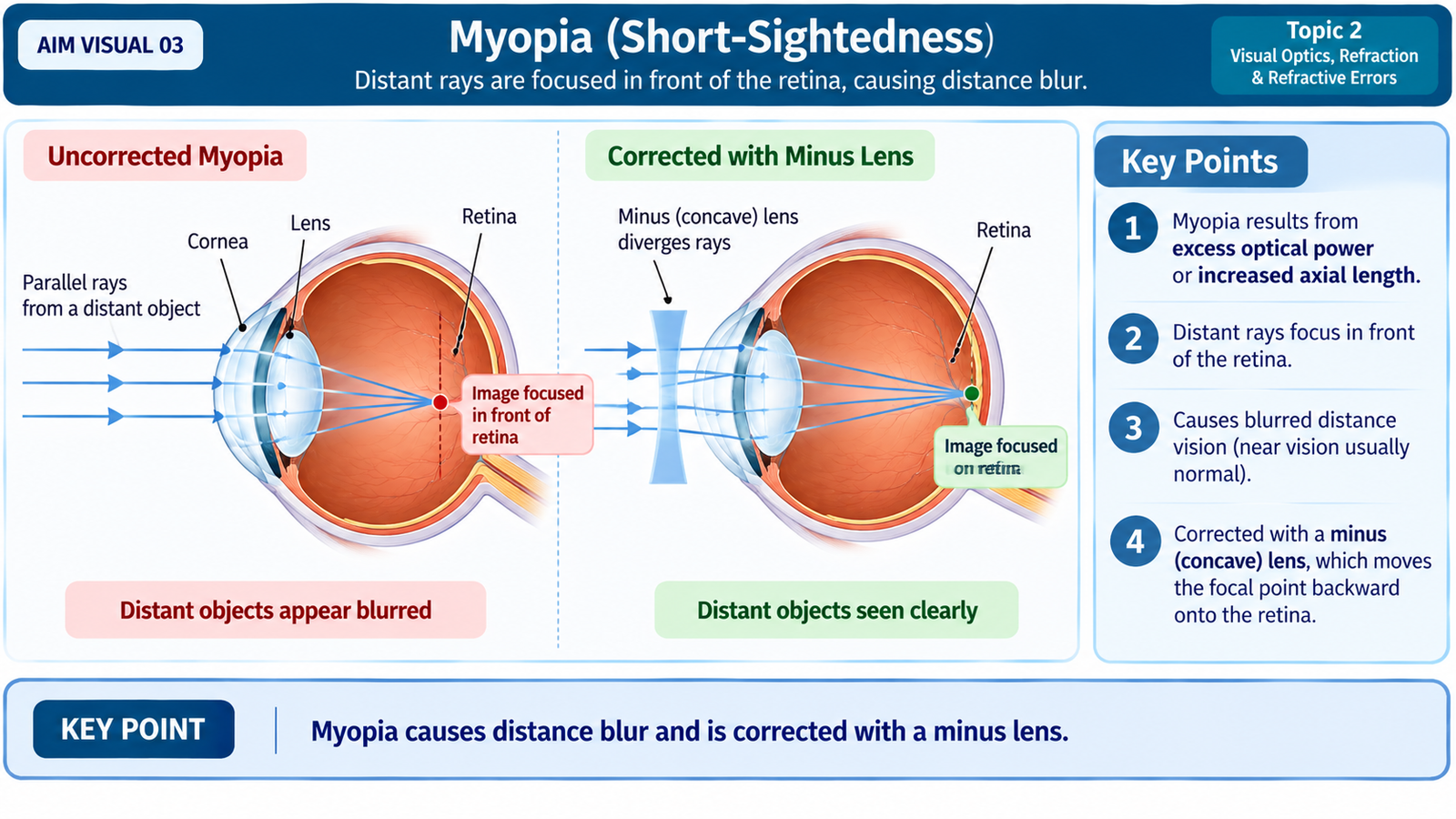
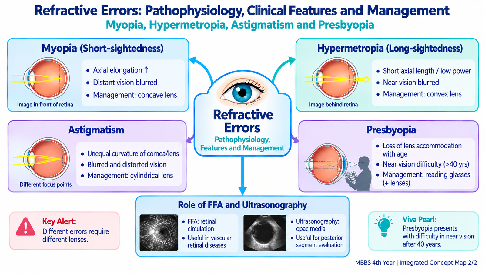
Myopia, or short-sightedness, is a refractive state in which parallel rays from a distant object are focused in front of the retina when accommodation is relaxed. As a result, distant objects appear blurred.
Pathophysiology
Myopia develops when the eye is optically too strong for its axial length or, more commonly, when the axial length is too great for its refractive power. In either situation, incoming parallel rays converge before reaching the retina. By the time they reach the retinal surface they have begun to diverge again, producing a blurred rather than sharply focused image.
Clinical presentation
The characteristic complaint is poor distance vision with relatively better near vision. The patient may narrow the palpebral aperture or squint because this reduces the effective blur circle and can temporarily improve image clarity.
- Blurred vision for distant objects.
- Near objects are seen more clearly than distant ones.
- Squinting may temporarily improve distance clarity.
- Uncorrected refractive effort may be associated with visual discomfort or headache.
Correction
Concave, diverging or minus lenses reduce the converging power of the optical system. They cause incoming rays to diverge slightly before entering the eye, allowing the eye’s optics to focus them farther backward, directly on the retina. Contact lenses and appropriately selected refractive procedures are additional options where suitable.
D. Hypermetropia
Hypermetropia, or hyperopia, is a refractive state in which parallel rays would come to focus behind the retina when accommodation is relaxed. The eye therefore has insufficient refractive power in relation to its axial length.
Pathophysiology
A common optical basis is an eye that is relatively too short, although reduced refractive power can produce the same result. Because the retina lies in front of the point at which the rays would otherwise meet, the image reaching the retina is defocused.
A young person can compensate for some hypermetropia by accommodation. Increasing lens curvature adds converging power and moves the focal point forward onto the retina. This explains why a young hypermetropic patient may initially maintain clear distance vision but develop symptoms of accommodative effort.
Clinical presentation
- Visual fatigue or eyestrain, especially during sustained near work.
- Headache may occur after prolonged accommodative effort.
- Near vision may become blurred when compensation is inadequate.
- With greater refractive error or reduced accommodative ability, distance vision may also be blurred.
Correction
Convex, converging or plus lenses add refractive power before light enters the eye. They move the focal point forward so that it lies on the retina and also reduce the amount of accommodation required for clear vision.

E. Astigmatism
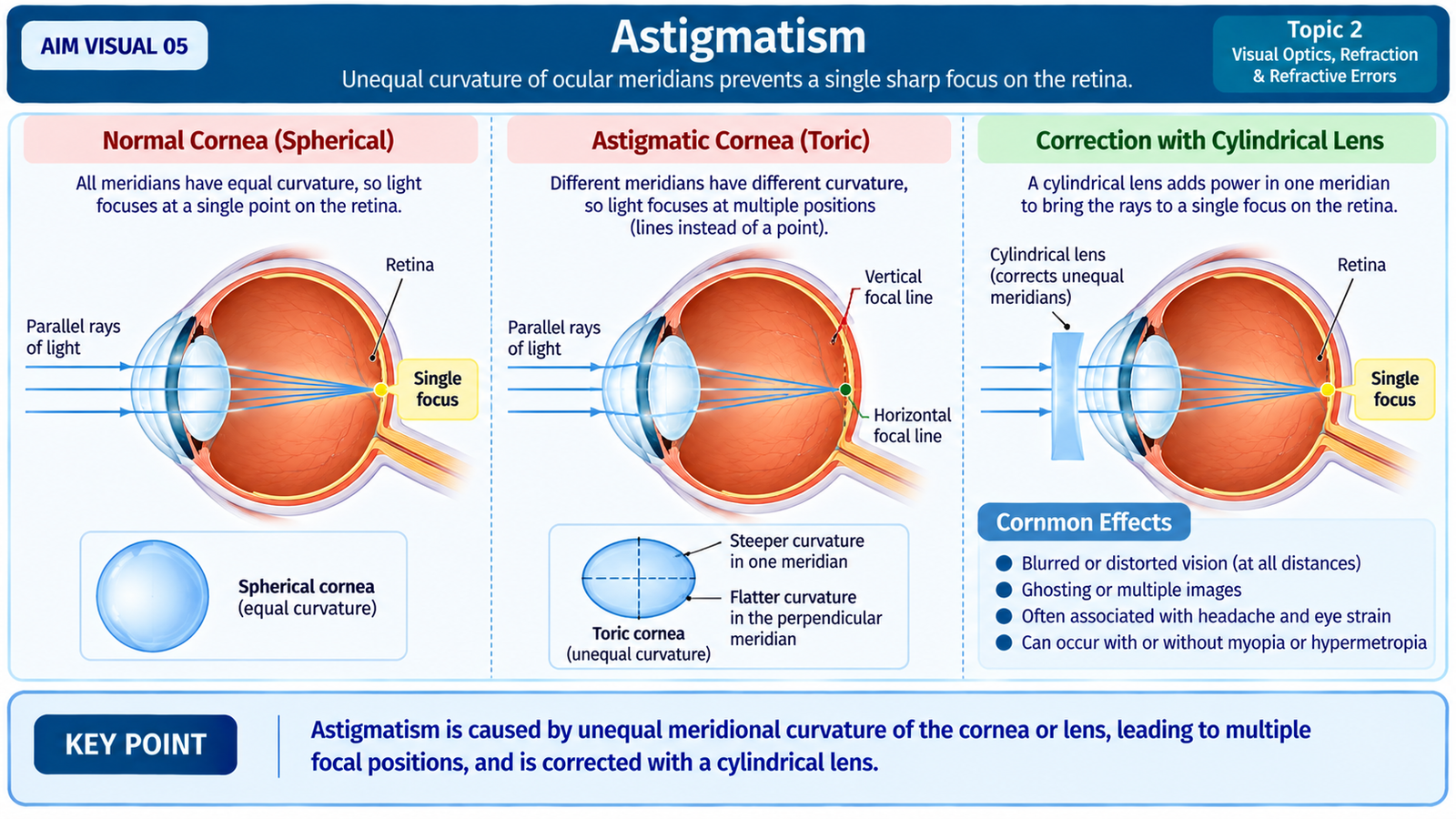
Astigmatism is a refractive error in which the optical system does not have the same refractive power in all meridians. Instead of light from a point object forming a single point focus, different meridians focus light differently. The retinal image therefore becomes blurred or distorted.
Pathophysiology
The usual optical basis is unequal curvature of the cornea in different meridians, although the lens can also contribute. A surface that is steeper in one meridian bends light more strongly than a flatter meridian. Consequently, rays travelling through different planes do not meet at one common retinal point.
Regular and irregular astigmatism
In regular astigmatism, the principal meridians are orderly and usually perpendicular to each other, so the error can generally be corrected with cylindrical lens power. In irregular astigmatism, refractive power varies irregularly across the optical surface, making simple spectacle correction less effective.
Clinical presentation
- Blurred or distorted vision.
- Difficulty obtaining a uniformly sharp image at one distance.
- Eyestrain during visual tasks.
- Headache may accompany sustained visual effort.
Correction
Cylindrical or sphero-cylindrical lenses provide different refractive power in selected meridians and compensate for the meridional difference in the eye. Contact lenses may be useful, especially when the corneal surface contributes significantly to the optical irregularity.
F. Presbyopia
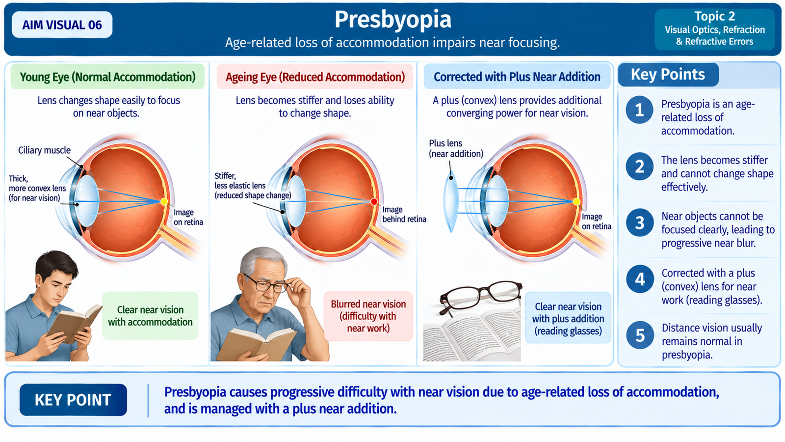
Presbyopia is the age-related reduction in the eye’s ability to accommodate for near vision. It is therefore different from myopia, hypermetropia and astigmatism: the central problem is a loss of accommodative reserve rather than simply a fixed mismatch between axial length and refractive power.
Pathophysiology
With increasing age, the crystalline lens becomes progressively less able to change shape effectively during accommodation. When the patient looks at a near object, the optical system can no longer increase its power sufficiently to maintain a sharp retinal image.
Clinical presentation
The typical complaint is increasing difficulty with near work. Because a more distant object requires less accommodation, patients often move reading material farther away to obtain a clearer image.
- Difficulty reading small print at the usual working distance.
- Tendency to hold reading material farther away.
- Eyestrain during prolonged near work.
- Near tasks may feel easier under good illumination because the visual target becomes easier to discriminate.
Correction
Plus-powered near lenses provide the converging power that the patient’s accommodation can no longer supply. Depending on the person’s distance refractive status and visual needs, near correction may be provided through reading spectacles, bifocal lenses or progressive-addition lenses.
G. Anisometropia, Aphakia and Pseudophakia
These conditions are important because they alter the optical relationship between the two eyes or change the lens component of the eye. Understanding them becomes easier once normal refraction and the major refractive errors are clear.
Anisometropia
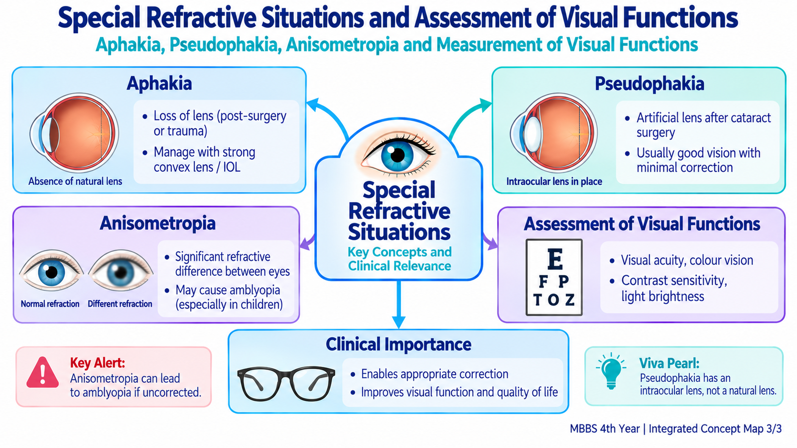
Anisometropia means that the two eyes have significantly different refractive states. For example, one eye may be more myopic or hypermetropic than the other. The two eyes can therefore form retinal images of different clarity and, with spectacle correction, sometimes different apparent sizes.
A difference in perceived image size between the eyes is called aniseikonia. Marked unequal image quality can interfere with binocular vision. During visual development, persistent unequal refractive error may also contribute to amblyopia because the brain preferentially uses the clearer image.
Aphakia
Aphakia means absence of the natural crystalline lens. Because the lens normally contributes important positive refractive power, its absence produces a major loss of converging power and therefore a marked hypermetropic optical state. Physiological accommodation is also lost.
On examination, the natural lens is absent. Depending on the situation, the anterior chamber may appear relatively deep and movement of the unsupported iris may be visible. Optical rehabilitation can be achieved with a strong plus spectacle lens, a contact lens, or more commonly by replacement of lens power with an intraocular lens where appropriate.
Pseudophakia
Pseudophakia means that the natural crystalline lens has been replaced by an artificial intraocular lens (IOL), most commonly in association with cataract surgery. The IOL restores much of the refractive power lost when the natural lens is removed.
A conventional monofocal IOL provides fixed optical power and does not reproduce the normal dynamic accommodation of a young natural lens. The patient may therefore require additional near correction even when distance vision is well corrected.
| State | Central optical issue | Important consequence |
|---|---|---|
| Anisometropia | Different refractive states in the two eyes | Unequal image clarity or size; binocular difficulty |
| Aphakia | Natural lens absent | Marked loss of refractive power and accommodation |
| Pseudophakia | Artificial IOL replaces natural lens | Optical power restored, but normal accommodation usually absent |

H. Management of Refractive Errors
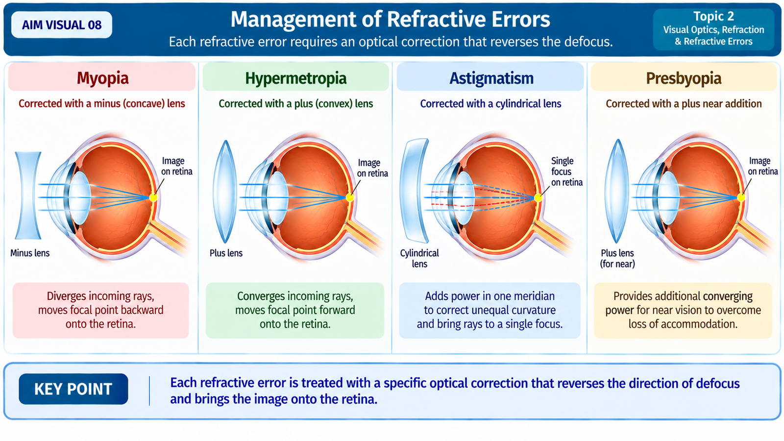
Management aims to place a sharply focused image on the retina while meeting the patient’s distance, near and everyday visual requirements. The correcting optical system should oppose the direction of the refractive error: excessive convergence is reduced, insufficient convergence is increased, and unequal meridional power is corrected selectively.
Spectacle correction
- Myopia: corrected with concave, diverging, minus lenses.
- Hypermetropia: corrected with convex, converging, plus lenses.
- Astigmatism: corrected with cylindrical or sphero-cylindrical power directed in the appropriate axis.
- Presbyopia: corrected by additional plus power for near work.
Contact lenses
Contact lenses provide correction at the corneal surface and can be used for several refractive errors. They can be particularly useful when spectacle-induced image-size differences are troublesome, as may occur in significant anisometropia, and in selected forms of astigmatism.
Refractive procedures
Selected patients may undergo procedures that alter the optical power of the eye, particularly by changing corneal curvature. At undergraduate level, the important principle is that refractive surgery attempts to reduce dependence on external optical correction. Suitability requires proper ophthalmic assessment; operative details are not required here.
Why proper refraction matters
The prescription should follow assessment of visual acuity and refractive status rather than simply matching a patient’s symptom to a lens type. Reduced vision that does not behave like an uncomplicated refractive error requires assessment for an ocular cause rather than repeated changes in spectacle power.
Integrated Mechanism Flow
Important Comparison — Common Refractive Errors
| Feature | Myopia | Hypermetropia | Astigmatism | Presbyopia |
|---|---|---|---|---|
| Main problem | Eye optically too strong / relatively long | Eye optically too weak / relatively short | Unequal power in different meridians | Reduced accommodation |
| Optical result | Focus in front of retina | Focus tends behind retina | Different focal positions | Insufficient near focusing power |
| Typical complaint | Distance blur | Eyestrain; near or distance blur depending on compensation | Blurred or distorted vision | Progressive near difficulty |
| Correction | Minus lens | Plus lens | Cylindrical / sphero-cylindrical lens | Plus near addition |
⭐ AIM High-Yield Review
Refractive Errors of the Eye
Reinforce the concepts of refraction, myopia, hypermetropia, astigmatism and presbyopia after completing the AIM learning material.