
This chapter follows the supplied KMU learning outcomes and explains the corneal and retinal causes of visual loss in a logical clinical sequence. First understand how each disorder changes ocular structure or retinal function, then use the final AIM High-Yield Review for rapid revision.
Topic 12 — Corneal & Retinal Causes of Visual Loss
A structured approach to important corneal disorders, systemic retinal disease, retinal vascular occlusion, retinal detachment, choroidal melanoma, inherited retinal degeneration and vitamin A deficiency.
1. Topic Introduction
Visual loss may result from failure of the transparent cornea to focus light correctly, damage to the retina and its blood supply, separation of the retina from its supporting tissues, or progressive degeneration of photoreceptors. In this topic, the important corneal causes are keratoconus and an overview of corneal dystrophies and degenerations. The posterior-segment disorders include diabetic and hypertensive retinal disease, central retinal vein and artery occlusion, retinal detachment, choroidal melanoma, retinitis pigmentosa and vitamin A deficiency. The central learning approach is to connect the disease process with the characteristic symptoms, ophthalmic examination findings, investigations and appropriate management.
A. Keratoconus: Progressive Corneal Ectasia
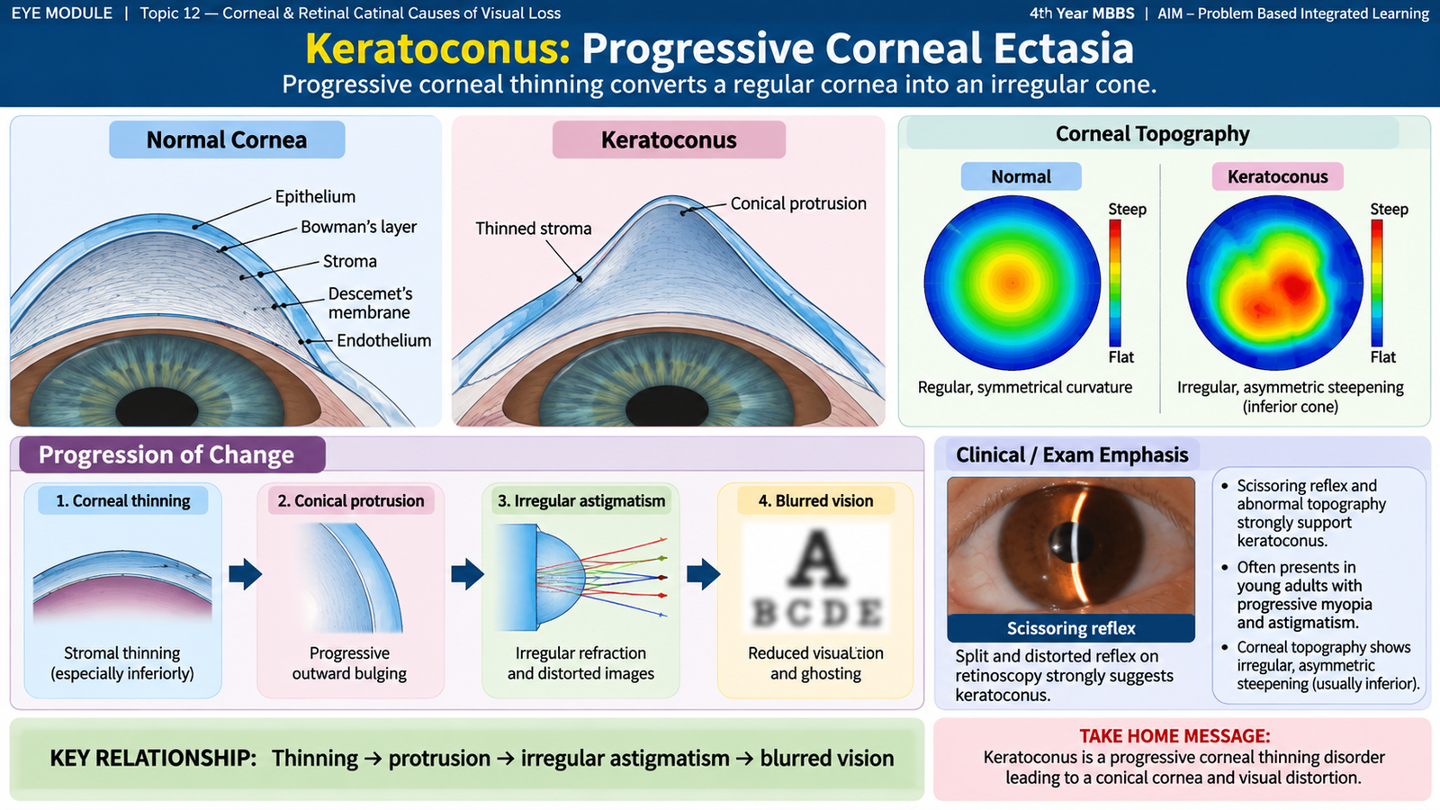
Keratoconus is a usually bilateral but often asymmetrical disorder in which the central or paracentral cornea becomes progressively thin and protrudes forward into a cone-like shape. Because the cornea provides most of the eye’s refractive power, distortion of its normally regular curvature produces increasing irregular astigmatism and reduced vision.
Etiology and Risk Factors
The exact cause is multifactorial. Structural weakness of the corneal stroma allows the cornea to become progressively ectatic. Genetic susceptibility is important in some patients, while mechanical factors can accelerate progression.
- Family history or genetic predisposition.
- Repeated or vigorous eye rubbing.
- Atopic disease and ocular allergy, particularly when associated with chronic rubbing.
- Some connective-tissue and systemic disorders may be associated.
How the Disease Produces Visual Loss
Unlike ordinary regular astigmatism, the distorted corneal surface in established keratoconus cannot always be fully corrected by ordinary spectacles.
Clinical Features
The disease commonly begins in adolescence or early adult life and may progress over several years. Early disease may resemble ordinary refractive error, so repeated changes in spectacle prescription can be an important clue.
- Progressive blurring of vision.
- Increasing myopia and irregular astigmatism.
- Frequent change in spectacle prescription.
- Glare, halos or distorted images.
- Monocular ghost images or multiple images may occur.
On examination, important findings may include:
- Scissoring reflex on retinoscopy because different parts of the irregular cornea refract light differently.
- Corneal thinning and conical protrusion in more advanced disease.
- Fleischer ring, an iron-deposition ring near the base of the cone.
- Vogt striae, fine vertical stress lines in the deep cornea.
- Munson sign in advanced disease, where the lower eyelid develops a V-shaped indentation on downgaze because of the protruding cone.
Investigations
Corneal topography or tomography is the key investigation because it maps the corneal curvature and demonstrates abnormal steepening and asymmetry. It can detect disease before obvious clinical signs develop.
- Keratometry: demonstrates increased and irregular corneal curvature.
- Corneal topography/tomography: defines the cone pattern, asymmetry and progression.
- Pachymetry: measures corneal thickness and identifies thinning.
- Slit-lamp examination: identifies stromal thinning, Fleischer ring, Vogt striae and scarring.
Management
Management depends mainly on visual function and whether the ectasia is progressing. The aim is both to improve vision and, when appropriate, to prevent further structural progression.
- Spectacles may correct vision in early disease when astigmatism is still reasonably regular.
- Rigid gas-permeable or scleral contact lenses provide a more regular refracting surface and can improve vision when spectacles become inadequate.
- Corneal collagen cross-linking strengthens the corneal stroma and is used to reduce further progression in appropriate progressive disease.
- Intracorneal ring segments may improve corneal shape in selected patients.
- Corneal transplantation may be required for advanced disease with severe irregularity, contact-lens intolerance or significant corneal scarring.
- Patients should be advised to avoid vigorous eye rubbing and to treat associated ocular allergy.
B. Corneal Dystrophies and Degenerations
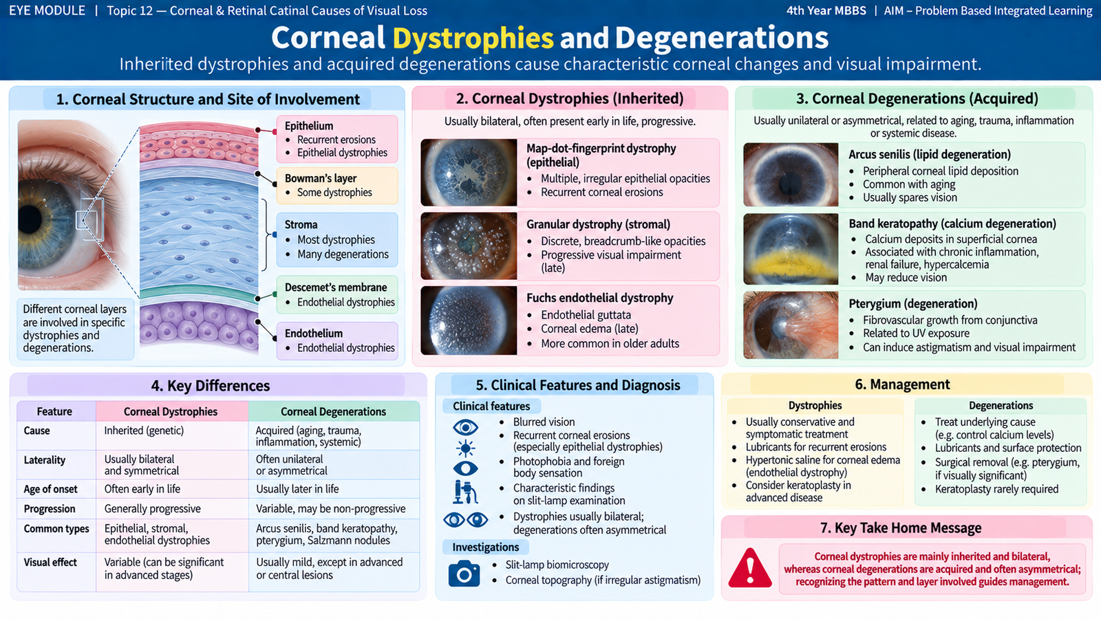
Corneal dystrophies and degenerations both produce abnormal material or structural change within the cornea, but their basic patterns differ. Corneal dystrophies are usually inherited disorders affecting one or more corneal layers and are commonly bilateral and relatively symmetrical. Corneal degenerations are generally acquired changes related to aging, local disease, metabolic disturbance or environmental exposure.
Corneal Dystrophies
The clinical effect depends on which corneal layer is involved. A lesion affecting the central visual axis may reduce transparency and visual acuity, while epithelial abnormalities may cause recurrent painful erosions.
- Epithelial/anterior dystrophies: may produce recurrent epithelial breakdown, discomfort and irregular vision.
- Stromal dystrophies: produce deposits within the corneal stroma and may progressively interfere with transparency.
- Endothelial dystrophies: impair endothelial function. Because the endothelium normally keeps the corneal stroma relatively dehydrated, endothelial failure leads to corneal edema and reduced transparency.
Examples important for recognition include granular, lattice and macular stromal dystrophies and Fuchs endothelial corneal dystrophy. The purpose at undergraduate level is to recognize the general pattern rather than memorize every rare subtype.
Corneal Degenerations
Degenerations are usually acquired and may be related to age, chronic inflammation, systemic metabolic disturbance or environmental exposure. They may be unilateral or asymmetrical and often begin peripherally.
- Arcus: peripheral lipid deposition producing a grey-white ring near the limbus.
- Band keratopathy: calcium deposition in the superficial cornea, usually in the exposed interpalpebral area.
- Other degenerative changes may develop after chronic ocular disease or repeated inflammation.
Clinical Assessment and Management Principles
Assessment includes visual acuity and careful slit-lamp examination to determine the affected corneal layer, distribution of opacity and effect on the visual axis. Management is directed at symptoms, optical impairment and the underlying cause where one exists. Lubrication, treatment of epithelial disease, optical correction or corneal procedures may be required depending on the specific disorder.
| Feature | Corneal Dystrophy | Corneal Degeneration |
|---|---|---|
| General nature | Usually inherited | Usually acquired |
| Laterality | Often bilateral and symmetrical | May be asymmetrical |
| Typical influence | Genetic corneal disorder | Age, local disease, environment or metabolism |

C. Diabetes and Diabetic Eye Disease
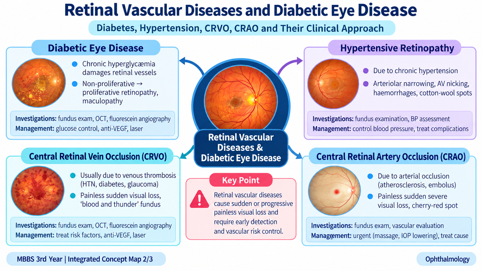
Diabetes mellitus can affect several ocular structures, but its most important sight-threatening posterior-segment complication is diabetic retinopathy. Chronic hyperglycemia damages the retinal microcirculation, producing vascular leakage, retinal ischemia and, in advanced disease, abnormal new blood vessels. Diabetes may therefore reduce vision through both macular edema and proliferative retinal disease.
Effects of Diabetes on the Eye
- Temporary refractive changes associated with changing blood glucose.
- Earlier development or progression of cataract.
- Diabetic retinopathy and diabetic macular edema.
- Greater susceptibility to some ocular surface and infectious problems.
- Ocular motor nerve palsies may occur because of diabetic microvascular disease.
- Severe retinal ischemia may contribute to anterior-segment neovascularization and secondary glaucoma.
Pathogenesis of Diabetic Retinopathy
The same microvascular injury therefore produces two major problems. Leakage causes retinal edema and hard exudates, while capillary non-perfusion causes ischemia. When ischemia becomes sufficiently extensive, the retina produces signals that stimulate fragile new vessels.
Non-Proliferative Diabetic Retinopathy
Non-proliferative diabetic retinopathy (NPDR) describes retinal microvascular abnormalities without retinal or disc neovascularization.
- Microaneurysms: tiny focal capillary dilatations and often the earliest visible retinal lesions.
- Dot and blot hemorrhages: intraretinal hemorrhage within deeper retinal layers.
- Hard exudates: lipid-rich residues left after chronic vascular leakage.
- Cotton-wool spots: superficial retinal ischemic lesions caused by interrupted axoplasmic flow.
- Venous abnormalities and intraretinal microvascular abnormalities: suggest increasing retinal ischemia in more severe disease.
Proliferative Diabetic Retinopathy
Proliferative diabetic retinopathy (PDR) is defined by retinal or optic-disc neovascularization. These new vessels develop in response to retinal ischemia but are structurally fragile.
Because the vessels are fragile, they may bleed into the vitreous. Associated fibrovascular tissue can later contract and pull on the retina, producing tractional retinal detachment. These mechanisms explain why advanced diabetic retinopathy can cause severe visual loss even when the anterior segment appears relatively quiet.
Diabetic Maculopathy
The macula is responsible for detailed central vision. Leakage from damaged retinal capillaries near the macula produces retinal thickening and edema, while macular ischemia reduces the blood supply to this highly specialized region. Therefore diabetic macular disease commonly causes blurred or distorted central vision.
Diabetic macular edema can occur with different stages of diabetic retinopathy and is an important cause of visual impairment.
Investigations
- Visual acuity: establishes the functional effect of the disease.
- Dilated fundus examination: identifies microaneurysms, hemorrhages, exudates, ischemic signs and neovascularization.
- Fundus photography: documents retinal changes and allows comparison over time.
- Optical coherence tomography (OCT): provides cross-sectional retinal imaging and is especially useful for identifying and monitoring macular edema.
- Fluorescein angiography: demonstrates vascular leakage and areas of capillary non-perfusion when this information is required for management.
Management
Management combines systemic risk-factor control with treatment of sight-threatening retinal disease. Good control of diabetes and associated hypertension reduces continuing vascular injury but does not replace ophthalmic treatment when significant retinal disease has already developed.
- Optimize blood glucose, blood pressure and other relevant systemic risk factors.
- Regular retinal assessment allows disease to be identified before irreversible visual loss occurs.
- Intravitreal anti-VEGF therapy is important for diabetic macular edema and is also used in selected proliferative disease.
- Panretinal laser photocoagulation reduces the drive for neovascularization in proliferative diabetic retinopathy.
- Vitrectomy may be required for selected advanced complications such as non-clearing vitreous hemorrhage or tractional retinal detachment threatening vision.

D. Hypertension and Hypertensive Retinopathy
Systemic hypertension affects the eye mainly by damaging small retinal vessels. The retinal circulation is directly visible during ophthalmoscopy, so the fundus can provide evidence of chronic vascular damage as well as severe acute hypertension. The pattern ranges from arteriolar narrowing and arteriovenous crossing changes to hemorrhages, retinal ischemia, exudation and optic-disc edema.
Effects of Hypertension on the Eye
- Retinal arteriolar narrowing and sclerosis.
- Hypertensive retinopathy with hemorrhages, exudates and ischemic lesions.
- Choroidal vascular injury may occur in severe hypertension.
- Optic-disc edema may occur in severe hypertensive disease and indicates serious systemic vascular injury.
- Hypertension also increases the risk of retinal vascular occlusive disease.
Pathogenesis
Clinical and Fundus Features
Most patients do not develop a specific ocular symptom during early chronic hypertensive retinopathy. The abnormalities are therefore often detected on fundus examination. Severe retinal involvement may be accompanied by visual disturbance.
- Generalized or focal arteriolar narrowing reflects vasoconstriction and chronic vascular change.
- Arteriovenous nicking occurs where a thickened arteriole affects the underlying vein at a crossing point.
- Flame-shaped hemorrhages occur within the superficial nerve-fiber layer.
- Cotton-wool spots indicate focal retinal ischemia.
- Hard exudates result from vascular leakage.
- A macular star may develop when lipid exudates are arranged around the macula.
- Optic-disc edema in a patient with severe hypertension is a major warning finding.
Investigations
Diagnosis is based mainly on retinal examination together with assessment of systemic blood pressure and associated organ damage.
- Measure blood pressure accurately.
- Perform visual acuity and dilated fundus examination.
- Fundus photography can document the vascular and retinal abnormalities.
- OCT may be used if macular or optic-disc swelling requires further anatomical assessment.
- Systemic evaluation is guided by the severity of hypertension and evidence of target-organ involvement.
Management
The essential treatment is appropriate control of the underlying systemic hypertension. Retinal signs generally represent the effect of systemic vascular disease rather than an isolated eye disorder.
- Arrange medical assessment and control of blood pressure.
- Identify associated systemic vascular risk factors.
- Severe hypertension with retinal hemorrhages, exudates or disc edema requires urgent systemic evaluation.
- Specific ophthalmic treatment is directed at associated complications rather than uncomplicated arteriolar changes alone.

E. Retinal Vascular Occlusion: CRVO and CRAO
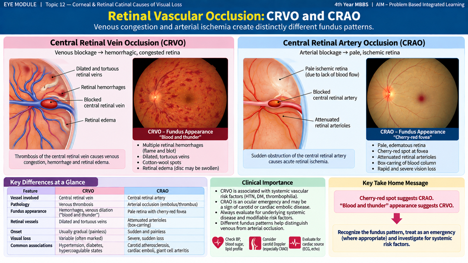
Occlusion of the central retinal vein and central retinal artery both cause retinal dysfunction, but they do so through different mechanisms. Central retinal vein occlusion (CRVO) obstructs retinal venous drainage, producing venous congestion, hemorrhage and edema. Central retinal artery occlusion (CRAO) stops arterial inflow, causing sudden inner-retinal ischemia. Recognizing the different fundus appearances is essential.
Central Retinal Vein Occlusion — Etiology and Risk Factors
CRVO results from obstruction or thrombosis of the central retinal vein. Impaired venous outflow raises pressure within the retinal venous circulation and causes widespread vascular leakage.
- Increasing age.
- Systemic hypertension.
- Diabetes mellitus.
- Glaucoma and raised intraocular pressure.
- Other vascular or hyperviscosity states in appropriate clinical settings.
CRVO Clinical Features
The usual presentation is sudden or subacute painless monocular visual loss. The severity varies according to the amount of retinal ischemia and whether the macula is edematous.
The classic fundus appearance is sometimes described as a “blood-and-thunder” retina because hemorrhages are widespread.
- Dilated and tortuous retinal veins.
- Retinal hemorrhages in multiple quadrants.
- Optic-disc swelling and hyperemia may occur.
- Cotton-wool spots may indicate ischemia.
- Macular edema is an important cause of reduced central vision.
Ischemic CRVO causes more severe retinal non-perfusion and has a greater risk of neovascular complications than non-ischemic CRVO.
CRVO Investigations
- Dilated fundus examination: establishes the characteristic venous and hemorrhagic pattern.
- OCT: identifies and measures macular edema.
- Fluorescein angiography: may demonstrate retinal non-perfusion and vascular leakage.
- Assess blood pressure, diabetes, glaucoma and other relevant systemic vascular risk factors.
CRVO Management
- Manage associated systemic vascular risk factors.
- Intravitreal anti-VEGF therapy is an important treatment for macular edema due to CRVO.
- Monitor for retinal, disc or anterior-segment neovascularization.
- Laser treatment is used for appropriate neovascular complications rather than simply because a vein occlusion is present.
Central Retinal Artery Occlusion — Etiology
CRAO occurs when blood flow through the central retinal artery is suddenly interrupted. The usual mechanism is embolic or thrombotic arterial occlusion. Because the inner retina has a very high metabolic requirement, prolonged loss of arterial perfusion rapidly threatens irreversible vision.
- Embolic disease associated with carotid or cardiac sources.
- Atherosclerotic vascular disease.
- Thrombotic arterial disease.
- Giant cell arteritis is an important arteritic cause in an appropriate older patient and must not be missed.
CRAO Clinical Features
- Sudden, profound, painless monocular visual loss.
- A relative afferent pupillary defect is expected when the unilateral retinal ischemia is severe.
- The retina becomes pale and opaque because of ischemic intracellular edema.
- A cherry-red spot appears at the fovea because the surrounding ischemic retina becomes pale while the thin foveal region retains the underlying reddish choroidal colour.
- Retinal arteries appear narrowed and blood flow may appear segmented.
CRAO Investigations
- Immediate fundus examination: looks for retinal whitening and a cherry-red spot.
- OCT: may show acute swelling and increased reflectivity of the inner retinal layers.
- Fluorescein angiography: can demonstrate delayed or absent retinal arterial filling when required.
- Urgent systemic vascular assessment is required to identify an embolic source and associated cerebrovascular risk.
- If giant cell arteritis is clinically suspected, urgent inflammatory and medical assessment is required because the fellow eye and systemic circulation may also be at risk.
CRAO Management
CRAO is an ocular and systemic vascular emergency. The key undergraduate principle is immediate recognition and urgent referral for ophthalmic and stroke-oriented systemic assessment. Traditional ocular measures have uncertain benefit and must not delay urgent evaluation of the vascular cause.
- Arrange immediate ophthalmic assessment.
- Arrange urgent evaluation for cerebral and cardiovascular vascular disease.
- Identify and treat the underlying embolic or arteritic cause.
- Suspected giant cell arteritis requires urgent medical treatment to protect the fellow eye and reduce systemic complications.
Important Comparison — CRVO versus CRAO
| Feature | CRVO | CRAO |
|---|---|---|
| Vessel affected | Central retinal vein | Central retinal artery |
| Mechanism | Venous congestion and leakage | Acute retinal ischemia |
| Fundus | Dilated tortuous veins with widespread hemorrhages | Pale retina with cherry-red spot |
| Important complication | Macular edema and neovascularization | Severe irreversible retinal ischemic damage |
| Urgency | Prompt ophthalmic assessment | Immediate vascular emergency assessment |
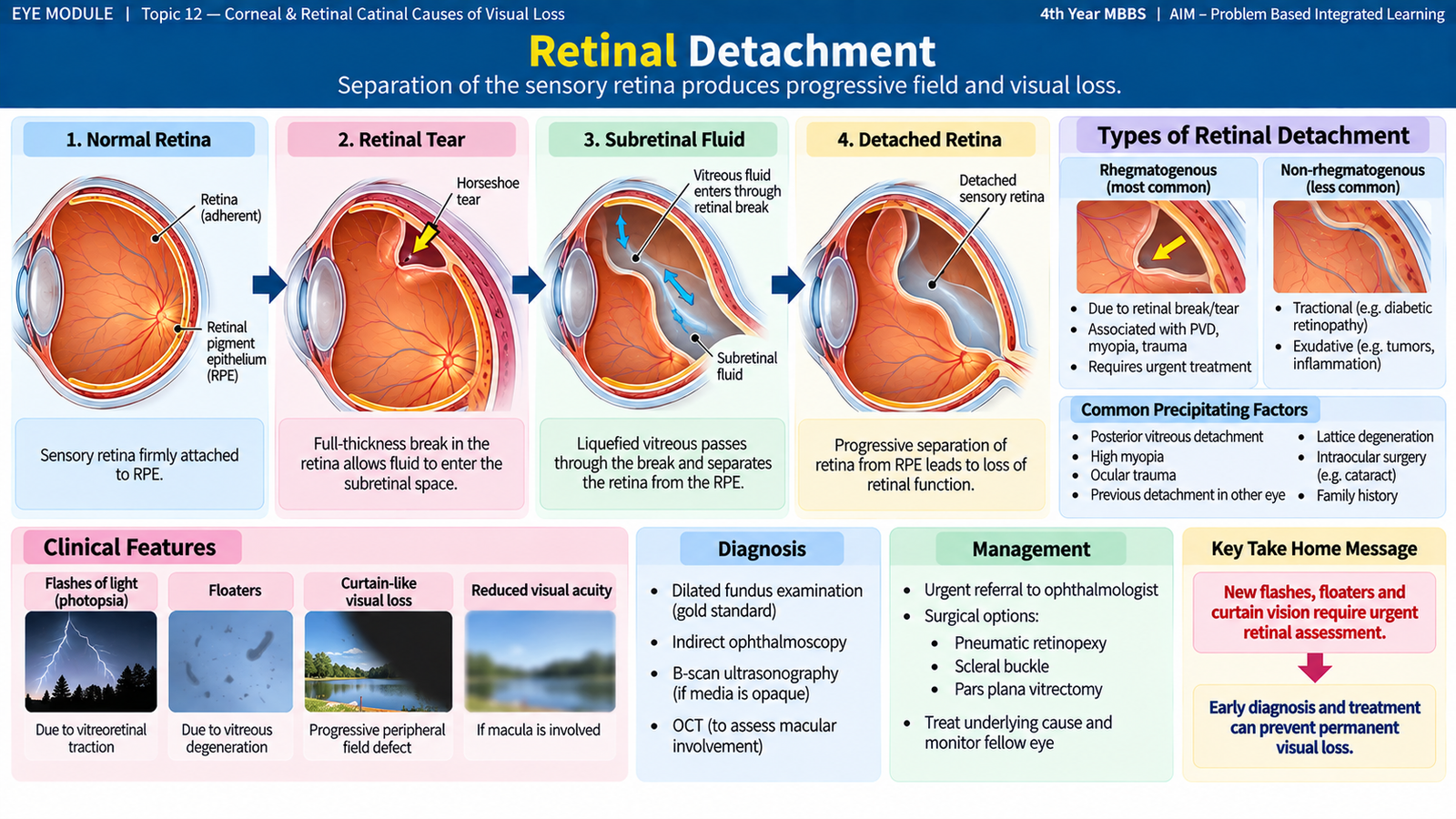
F. Retinal Detachment
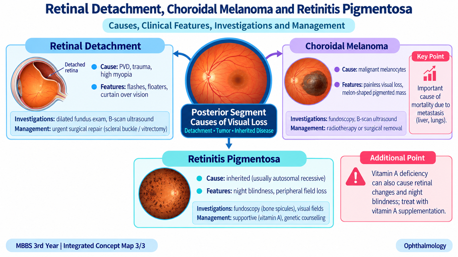
Retinal detachment is separation of the neurosensory retina from the underlying retinal pigment epithelium. This separation interferes with normal support of the photoreceptors and can lead to permanent visual loss, particularly when the macula becomes detached. Recognition of warning symptoms is therefore extremely important.
Types and Etiology
The three major types are defined by the mechanism that separates the retina.
1. Rhegmatogenous retinal detachment
A full-thickness retinal break allows liquefied vitreous to pass through the tear and collect beneath the sensory retina.
- Posterior vitreous separation with a retinal tear.
- Peripheral retinal degeneration.
- High myopia.
- Previous intraocular surgery or ocular trauma may increase risk.
2. Tractional retinal detachment
Fibrovascular or fibrous tissue contracts and physically pulls the retina away from the retinal pigment epithelium. Proliferative diabetic retinopathy is an important cause.
3. Exudative retinal detachment
Fluid accumulates beneath the retina without a retinal tear because of inflammation, vascular leakage or an underlying lesion.
Clinical Features
Rhegmatogenous detachment classically begins with symptoms caused by vitreoretinal traction and then progresses as the detached retina no longer functions normally.
- Photopsia: flashes of light produced by mechanical stimulation of the retina during vitreoretinal traction.
- Sudden increase in floaters: may result from vitreous change, pigment cells or small hemorrhage associated with a retinal tear.
- Dark curtain or shadow: represents loss of the visual field corresponding to the detached retina.
- Central visual acuity may initially remain good if the macula is still attached.
- Marked central visual loss develops when the detachment involves the macula.
Ophthalmic Examination
Dilated retinal examination may show an elevated, grey and mobile retina. Rhegmatogenous detachment is often corrugated, and careful examination seeks the causative retinal tear. The associated visual-field defect corresponds anatomically to the detached retinal region.
Investigations
- Dilated indirect ophthalmoscopy: identifies the detachment and searches for retinal breaks.
- Slit-lamp biomicroscopy with an appropriate retinal lens: provides detailed assessment of the posterior segment.
- B-scan ultrasonography: is particularly useful when the fundus cannot be visualized because of media opacity such as dense vitreous hemorrhage.
- OCT: can help define macular involvement in suitable cases.
Management
Retinal detachment requires prompt ophthalmic assessment. The treatment principle is to remove or counteract the cause of separation and restore retinal contact where possible.
- Symptomatic retinal tears may be sealed with laser photocoagulation or cryotherapy before a major detachment develops.
- Rhegmatogenous retinal detachment may require procedures such as pneumatic retinopexy, scleral buckling or pars plana vitrectomy, depending on the retinal break and clinical situation.
- Tractional detachment may require vitreoretinal surgery to release traction.
- Exudative detachment is managed primarily by treating the underlying inflammatory, vascular or neoplastic cause.
G. Choroidal Melanoma and Its Mortality Importance
Choroidal melanoma is a malignant tumor arising from melanocytes within the choroid and is an important primary intraocular malignancy in adults. It may threaten vision locally, but its greatest clinical importance is its ability to spread through the bloodstream and cause fatal metastatic disease.
Etiology and Predisposition
Most cases do not have a single identifiable cause. The tumor develops through malignant transformation of uveal melanocytes. Important associations include:
- Light skin and lighter ocular pigmentation.
- Pre-existing choroidal nevus in a small proportion of cases.
- Ocular or oculodermal melanocytosis.
- Increasing age is associated with greater occurrence.
Clinical Features
A small choroidal melanoma may be discovered incidentally. Symptoms depend on its position and whether it affects the macula, causes subretinal fluid or produces retinal detachment.
- Blurred or reduced vision.
- Photopsia or visual-field disturbance.
- Metamorphopsia if the macular region is affected.
- Secondary exudative retinal detachment may cause further visual loss.
Ophthalmic Appearance
Fundus examination may show a raised choroidal mass that is pigmented, variably pigmented or occasionally relatively non-pigmented. A melanoma may have a dome-shaped appearance, while breakthrough through Bruch membrane can produce a characteristic mushroom or collar-button configuration. Orange pigment and associated subretinal fluid increase suspicion in a melanocytic choroidal lesion.
Investigations
- Dilated fundus examination and fundus photography: document the lesion’s colour, shape and dimensions.
- Ocular ultrasonography: defines the intraocular mass and its thickness and internal characteristics.
- OCT: can demonstrate overlying retinal changes and subretinal fluid.
- Additional ocular imaging is selected when differentiation from other choroidal lesions is required.
- Systemic assessment is important after diagnosis because metastatic disease determines overall survival.
Management
Treatment depends on tumor size, location, visual potential, extension and the patient’s general condition. The objective is control of the primary tumor while preserving the eye and useful vision when safely possible.
- Selected small suspicious lesions may be closely monitored when diagnostic uncertainty exists.
- Plaque brachytherapy is an important eye-preserving treatment for many suitable tumors.
- Other forms of focused radiation may be used in selected patients.
- Enucleation may be required for large tumors, painful eyes or disease not suitable for conservative ocular treatment.
- Patients require systemic assessment and follow-up for metastatic disease.
Why Choroidal Melanoma Is Important in Relation to Mortality
The mortality importance of choroidal melanoma comes from hematogenous metastasis. Because the choroid is highly vascular and lacks lymphatic drainage, metastatic spread occurs primarily through the bloodstream. The liver is the most important site of distant metastasis. Metastatic disease may become clinically apparent even after successful control of the ocular tumor.
Therefore, successful local eye treatment does not completely eliminate long-term systemic risk. Larger tumor size, extraocular extension and unfavorable biological features are associated with a worse prognosis. Long-term systemic surveillance is therefore important after treatment.

H. Retinitis Pigmentosa
Retinitis pigmentosa (RP) is a group of inherited retinal dystrophies characterized by progressive degeneration of photoreceptors. In the common rod-cone pattern, rod photoreceptors are affected first, so difficulty seeing in dim light and progressive peripheral field loss usually precede major central visual loss.
Etiology
RP is genetically heterogeneous and can follow different patterns of inheritance. The underlying genetic abnormality disrupts normal photoreceptor structure or function. Rod photoreceptors usually degenerate first, followed later by cone involvement.
Clinical Features
- Night blindness is an early and characteristic symptom because rods are responsible for vision in dim illumination.
- Progressive loss of peripheral vision produces narrowing of the visual field.
- Patients may describe difficulty navigating in darkness or bumping into objects outside their central field.
- Advanced disease may produce tunnel vision.
- Central visual acuity can remain relatively preserved until later stages when cone function or the macula becomes significantly affected.
Fundus Findings
The classic fundus triad reflects progressive retinal degeneration:
- Bone-spicule pigmentation, especially in the mid-peripheral retina.
- Attenuated retinal arterioles.
- Waxy pallor of the optic disc.
Bone-spicule pigmentation develops because retinal pigment epithelial cells migrate into the retina around vessels as photoreceptor degeneration progresses.
Investigations
- Visual-field testing: demonstrates progressive peripheral field loss and may show a ring scotoma before severe concentric constriction develops.
- Electroretinography (ERG): assesses global retinal electrical responses and shows reduced photoreceptor function.
- OCT: demonstrates structural loss of the outer retinal layers and identifies associated macular complications.
- Genetic assessment: may help identify the inherited retinal disorder and guide family counseling.
Management
There is no single conventional treatment that restores all lost photoreceptor function in the broad group of RP disorders. Management therefore focuses on preserving useful function, treating associated ocular problems and supporting the patient.
- Correct refractive error and optimize remaining vision.
- Provide low-vision rehabilitation when visual-field or acuity loss becomes functionally important.
- Treat associated cataract when appropriate.
- Assess and manage associated macular edema when present.
- Offer genetic counseling and specialist inherited-retinal-disease assessment where appropriate.
I. Vitamin A Deficiency and Ocular Disease
Vitamin A is essential for normal visual function and maintenance of healthy conjunctival and corneal epithelium. Deficiency therefore affects both dark adaptation and the integrity of the ocular surface. Early disease may produce night blindness, whereas severe deficiency can progress to destructive corneal disease and irreversible blindness.
Etiology
Vitamin A deficiency develops when body stores become inadequate because intake is insufficient or absorption and handling of the vitamin are impaired.
- Severe nutritional deficiency.
- Malabsorption disorders.
- Chronic conditions interfering with fat absorption.
- Liver disease may impair storage and metabolism of vitamin A.
- Children with severe malnutrition are particularly vulnerable to serious ocular consequences.
Why Night Blindness Occurs
Vitamin A contributes to the visual cycle within rod photoreceptors. Rods are essential for vision under low-light conditions. Deficiency interferes with regeneration of visual pigment and therefore impairs dark adaptation.
Clinical Features
The ocular manifestations of vitamin A deficiency are collectively termed xerophthalmia. They represent progressive failure of normal conjunctival and corneal epithelial health.
- Night blindness: an early functional manifestation.
- Conjunctival xerosis: the conjunctiva becomes dry and loses its normal moist appearance.
- Bitot spots: foamy, whitish areas of abnormal keratinized conjunctival epithelium.
- Corneal xerosis: drying and loss of normal corneal surface quality.
- Corneal ulceration or keratomalacia: severe deficiency can cause rapid corneal softening and destruction.
- Corneal scarring may produce permanent visual loss after healing.
Investigations
The diagnosis is often primarily clinical when characteristic ocular findings occur in a patient with nutritional or malabsorptive risk. Investigations are directed at confirming deficiency and identifying its cause.
- History of nutrition and systemic disease.
- Slit-lamp examination of conjunctiva and cornea.
- Assessment of visual function, including history of night blindness.
- Serum vitamin A assessment may support the diagnosis when available and clinically appropriate.
- Evaluate for malnutrition, malabsorption or hepatic disease when suggested by the history.
Management
Established ocular vitamin A deficiency requires replacement of vitamin A according to an accepted age-appropriate clinical regimen and correction of the underlying nutritional or absorptive problem. Severe corneal disease requires urgent ophthalmic care because corneal destruction can progress rapidly.
- Replace vitamin A using an established clinical protocol appropriate to the patient.
- Correct dietary deficiency and general malnutrition.
- Investigate and treat malabsorption or other underlying systemic causes.
- Protect and manage the ocular surface.
- Urgently refer severe corneal xerosis, ulceration or keratomalacia for ophthalmic treatment.

⭐ AIM High-Yield Review
Topic 12 — Corneal & Retinal Causes of Visual Loss
Use these selected videos after reading the AIM Learning Material to reinforce important corneal, retinal and fundus-recognition concepts.
Keratoconus
Reinforces etiology, clinical features, characteristic corneal findings, investigations and management principles of keratoconus.
Corneal Dystrophies
Reviews the major epithelial, stromal and endothelial corneal dystrophies and helps relate the affected corneal layer to the clinical appearance.
Retinal Disorders — Integrated Pathology Review
High-yield integrated review of diabetic retinopathy, hypertensive retinal disease, retinal vascular occlusions, retinal detachment and retinitis pigmentosa.
Uveal / Choroidal Melanoma
Explains uveal melanoma, its clinical significance and metastatic behavior. Relate this to the AIM learning material on choroidal melanoma and its mortality importance.
Xerophthalmia — Vitamin A Deficiency
Reviews night blindness, conjunctival xerosis, Bitot spots, corneal xerosis, keratomalacia and the major management principles of vitamin A deficiency.