
Topic 5 — Red Eye, Conjunctival, Corneal & Ocular Surface Disorders
1. Topic Introduction
A red eye is produced when vessels of the conjunctiva, episclera, sclera or deeper anterior segment become dilated because of irritation, inflammation, infection or raised intraocular pressure. Many causes are minor, but corneal infection, scleritis and other deep ocular disease can threaten vision. The safest approach is therefore not to treat every red eye as conjunctivitis. Instead, look at the pattern: pain, discharge, photophobia, visual acuity, corneal clarity and the depth of redness. This chapter explains conjunctival inflammation, infectious and non-infectious corneal disease, lacrimal drainage obstruction and dacryocystitis, dry eye, blepharitis, and the important differences between pterygium, pseudo-pterygium, episcleritis and scleritis.
A. Red Eye — Causes, Localization and Recognition of Danger
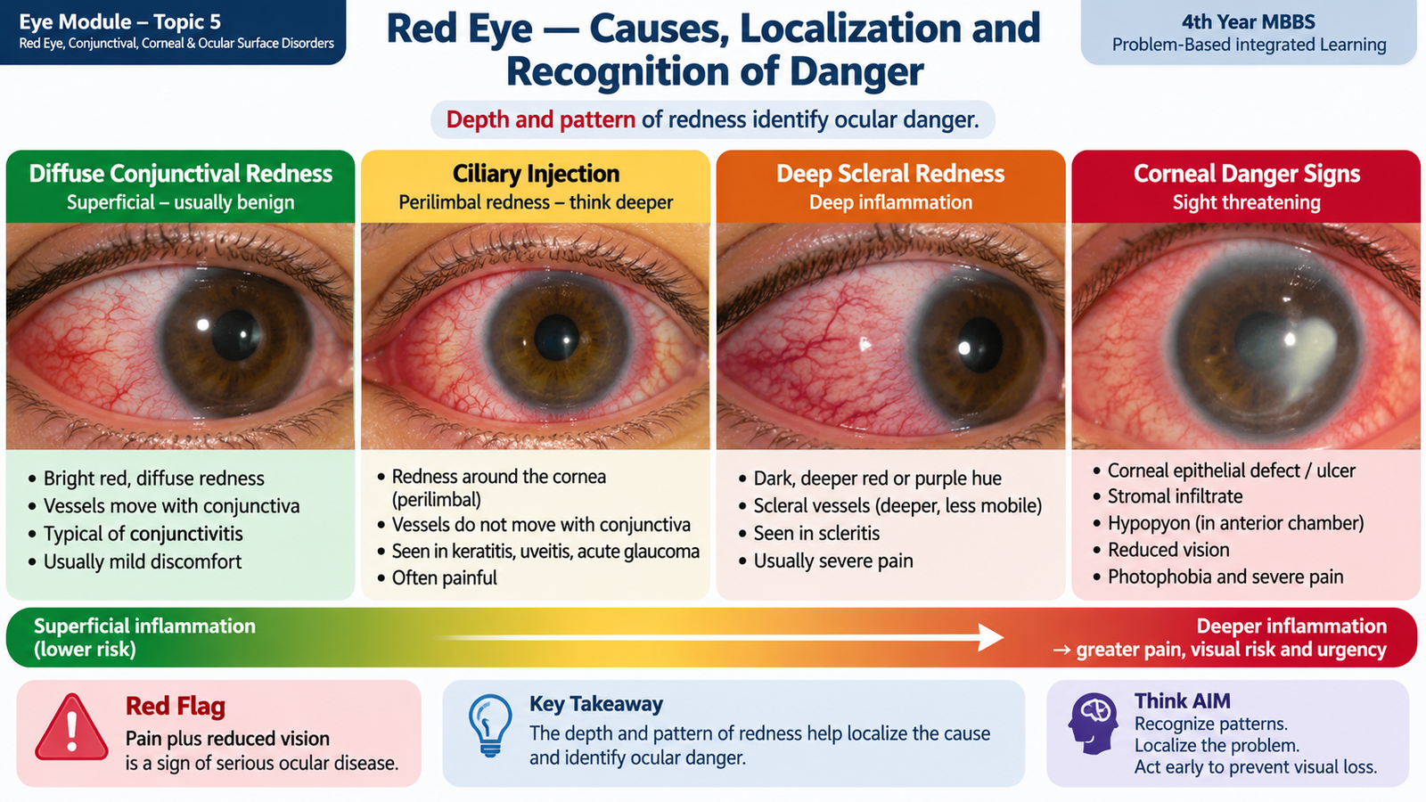
Redness alone does not identify the disease. The first task is to decide whether inflammation is mainly superficial, as in conjunctivitis and episcleritis, or involves the cornea or deeper ocular structures. Superficial disease usually produces irritation with relatively preserved vision. Severe pain, photophobia, reduced vision or an abnormal cornea suggests deeper disease and requires much greater urgency.
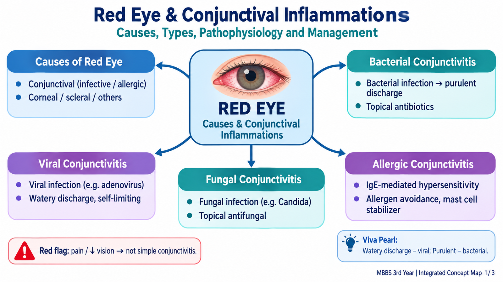
Major causes of red eye
- Conjunctivitis: bacterial, viral, fungal and allergic inflammation.
- Keratitis and corneal ulceration: bacterial, fungal, viral, Acanthamoeba or non-infectious inflammation.
- Dry-eye disease and ocular surface irritation.
- Blepharitis and meibomian gland dysfunction.
- Episcleritis and scleritis.
- Anterior uveitis, which often causes pain, photophobia and circumcorneal redness.
- Acute angle-closure glaucoma, an important painful red-eye emergency with reduced vision.
- Corneal abrasion, foreign body, contact-lens related injury and chemical or other surface trauma.
- Inflamed pterygium or other focal conjunctival lesions.
How to assess a red eye
Begin with visual acuity. Significant new reduction in vision is unusual in uncomplicated conjunctivitis and immediately raises concern for corneal or deeper ocular disease. Ask about pain, photophobia, contact-lens use, trauma and the nature of discharge. Examine the distribution of redness and the cornea.
Common in conjunctivitis. Conjunctival vessels are bright red and usually most obvious away from the limbus.
Redness is concentrated around the corneal limbus and suggests corneal or anterior uveal involvement.
Together with severe boring pain, this strongly suggests scleritis.
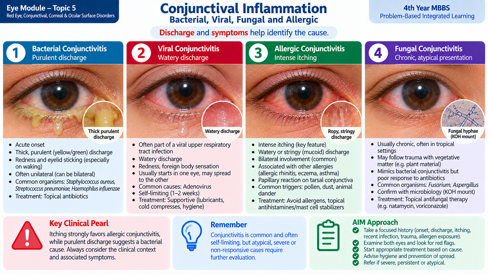
B. Conjunctival Inflammation — Bacterial, Viral, Fungal and Allergic
The conjunctiva is a vascular mucous membrane covering the inner eyelids and anterior sclera. Inflammation causes vasodilatation, producing redness, while increased mucus and inflammatory exudate produce discharge. Most uncomplicated conjunctivitis does not cause severe pain or major visual loss because the corneal optical surface remains relatively intact.
Bacterial conjunctivitis
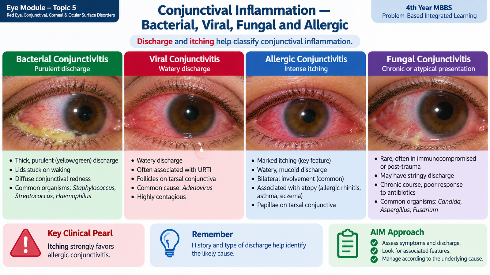
Bacteria infect the conjunctival epithelium and produce an acute inflammatory response. Common organisms include staphylococci, streptococci and Haemophilus; the exact organism varies with age and setting.
- Red eye with mucopurulent or purulent discharge.
- Lids may be stuck together on waking.
- Usually begins in one eye and may spread to the other.
- Mild irritation or gritty discomfort is more typical than severe pain.
- Vision is generally preserved except for temporary blur from discharge.
Diagnosis is usually clinical. Conjunctival smear and culture are reserved for severe, recurrent, neonatal, hyperacute or treatment-resistant disease. Management includes cleaning discharge, hand hygiene and an appropriate topical broad-spectrum antibiotic when bacterial infection is suspected. Contact lenses should be stopped during active infection.
Viral conjunctivitis
Adenoviruses are a common cause. Viral replication damages conjunctival epithelial cells and activates inflammation, producing watery rather than purulent discharge.
- Watery discharge and prominent redness.
- Foreign-body or gritty sensation.
- Follicles may be visible on the palpebral conjunctiva.
- Preauricular lymph-node enlargement supports a viral cause.
- May accompany fever, sore throat or upper respiratory symptoms.
- Highly contagious forms can spread rapidly between family members or students.
Management is mainly supportive with cool compresses, lubricants and strict hygiene. Sharing towels and touching the eyes should be avoided. Antibiotics do not treat viral infection. Herpes simplex should be considered when disease is markedly unilateral, recurrent, associated with lid vesicles or accompanied by corneal disease.
Fungal conjunctival inflammation
Fungal conjunctivitis is uncommon. It is more likely when conjunctival inflammation is atypical or chronic, particularly after exposure to contaminated organic material or in a susceptible host. Because fungi can also invade the cornea, persistent focal disease should not be treated repeatedly without examination.
Suspicious lesions require conjunctival sampling for direct microscopy and culture. Treatment is with an appropriate topical antifungal selected according to the suspected or identified organism under ophthalmic supervision. Corneal involvement changes the problem into potentially sight-threatening fungal keratitis.
Allergic conjunctivitis
Allergic conjunctivitis is usually an IgE-mediated hypersensitivity reaction. Exposure to an allergen activates conjunctival mast cells, which release histamine and other mediators. Histamine causes itching, vasodilatation and increased vascular permeability.
- Itching is the hallmark symptom.
- Usually bilateral.
- Watery or stringy mucoid discharge.
- Conjunctival chemosis and papillary reaction may occur.
- Frequently associated with other allergic disorders.
Management begins with allergen avoidance, cool compresses and lubricants. Topical antihistamine or combined antihistamine/mast-cell stabilizing therapy is useful when symptoms persist. Topical corticosteroids should not be used casually because of important ocular adverse effects and should be reserved for specialist-directed treatment.
C. Non-Infectious Corneal Inflammation
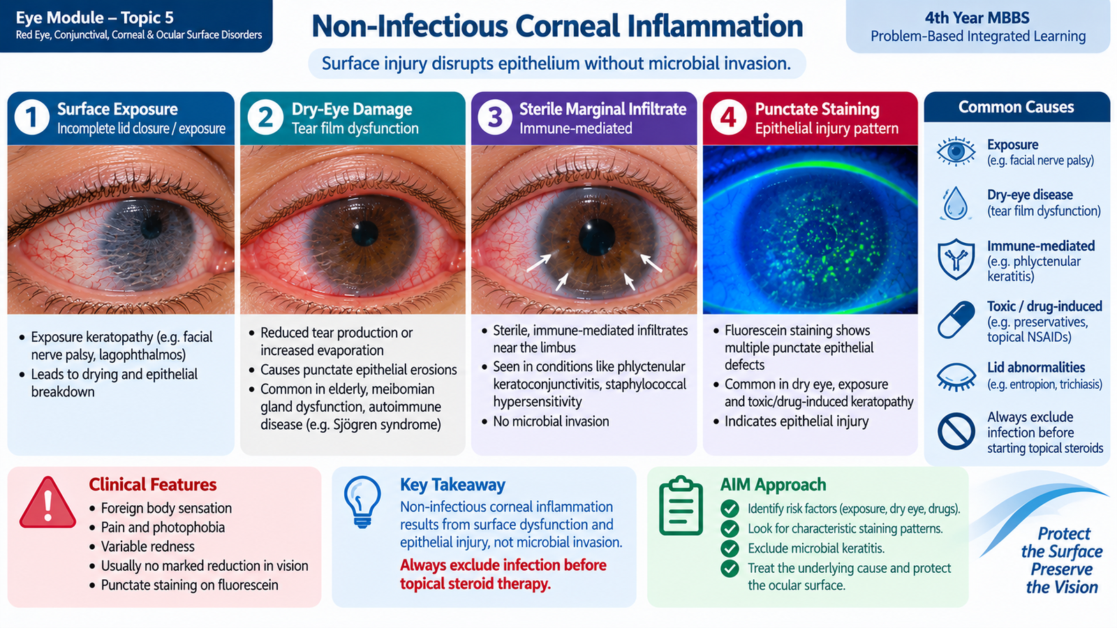
The cornea is densely supplied with sensory nerves and must remain transparent for clear vision. Inflammation therefore produces a different clinical pattern from uncomplicated conjunctivitis: pain, photophobia, tearing and visual disturbance become much more important. Non-infectious keratitis means corneal inflammation that is not primarily caused by active microbial invasion, although infection must always be excluded when an epithelial defect and infiltrate are present.
Important causes
- Exposure keratopathy: inadequate lid closure exposes the corneal epithelium and allows excessive evaporation.
- Severe dry-eye disease: an unstable tear film produces epithelial stress and punctate damage.
- Marginal sterile keratitis: often an immune response to bacterial antigens associated with chronic blepharitis.
- Ultraviolet injury or photokeratitis: UV exposure damages corneal epithelial cells.
- Neurotrophic corneal disease: loss of normal corneal sensory innervation reduces epithelial healing and protective reflexes.
- Immune-mediated peripheral corneal inflammation may occur with systemic inflammatory disease and can become sight-threatening if thinning develops.
Clinical features
The common symptoms are foreign-body sensation, pain, watering, photophobia and blurred vision. Fluorescein staining may show punctate epithelial erosions or a larger epithelial defect. A sterile marginal infiltrate is usually peripheral, whereas central stromal inflammation is more threatening to vision because subsequent scarring lies within the visual axis.
Neurotrophic disease deserves special attention because a damaged cornea may be surprisingly painless. Reduced corneal sensation explains this apparent contradiction and is itself an important diagnostic clue.
Investigations and interpretation
- Visual acuity: determines whether the optical function of the cornea is affected.
- Slit-lamp examination: localizes epithelial defects, infiltrates, edema and anterior chamber reaction.
- Fluorescein staining: identifies areas where epithelium is absent or damaged.
- Corneal sensation: reduced sensation supports neurotrophic disease and can occur in herpetic disease.
- Microbiological investigation: becomes necessary when infection cannot confidently be excluded.
Management principles
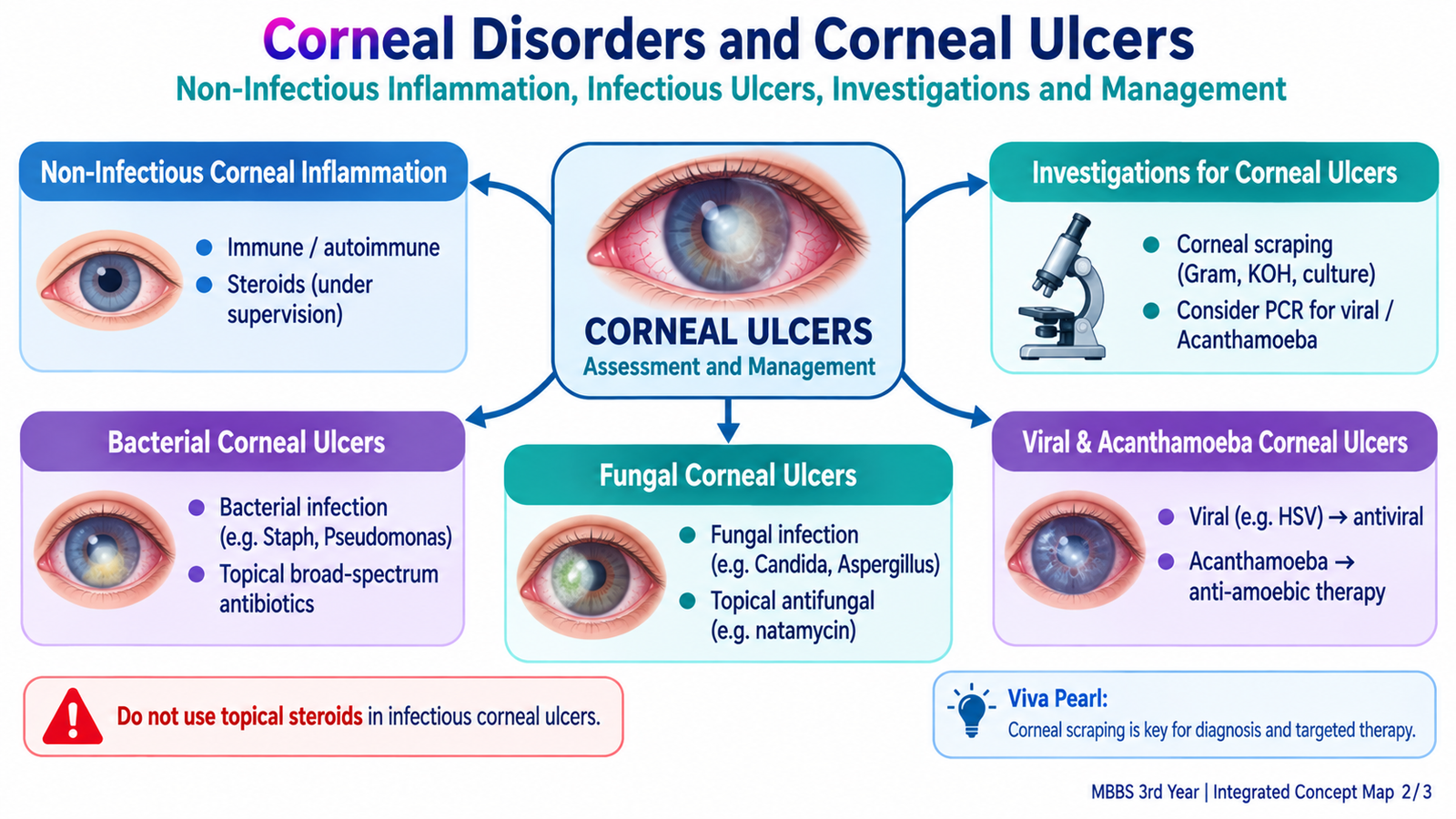
Treatment is directed at the cause. Lubrication and protection of the ocular surface are central in exposure and dry-eye related disease. Lid closure problems must be corrected or protected. Blepharitis should be treated when it is driving marginal inflammation. Anti-inflammatory therapy may be useful in selected sterile inflammatory conditions, but topical corticosteroids should not be started until infectious keratitis has been excluded.
D. Corneal Ulcer Investigation and Bacterial Keratitis
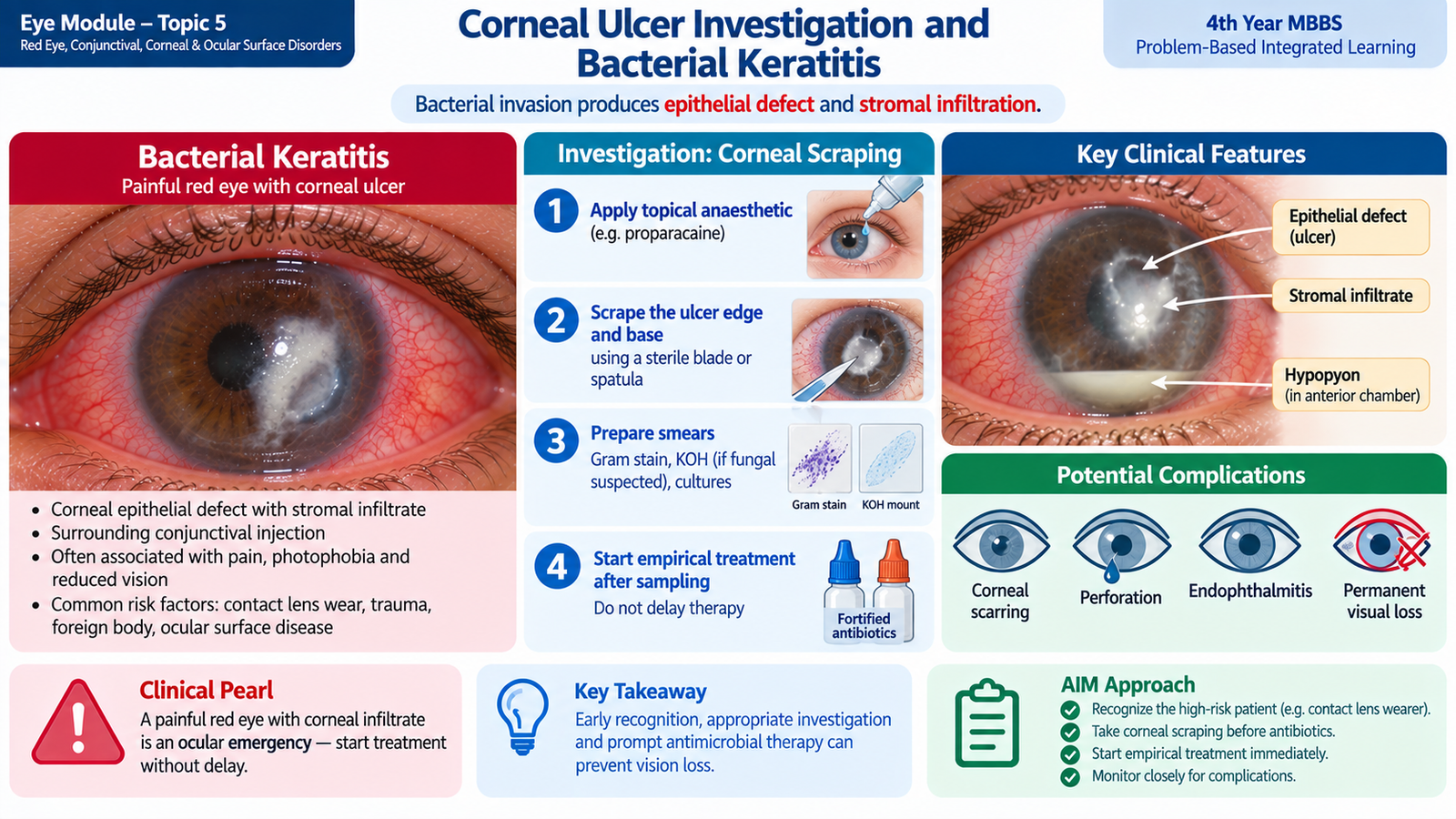
A corneal ulcer is an epithelial defect associated with underlying stromal inflammation or infiltration. When caused by infection, microorganisms penetrate a damaged epithelial barrier, multiply within the cornea and trigger intense inflammation. Stromal destruction can progress rapidly, so suspected microbial keratitis should be treated as a potential threat to vision.
Why bacterial keratitis occurs
An intact corneal epithelium is an effective barrier. Trauma, contact-lens wear, severe ocular-surface disease, previous surgery or inappropriate topical corticosteroid use can weaken this barrier and permit bacterial invasion.
Clinical features
- Acute painful red eye.
- Marked photophobia and tearing.
- Reduced vision, particularly with a central lesion.
- Discharge may be present.
- A focal grey-white or yellow-white stromal infiltrate.
- Overlying epithelial defect that stains with fluorescein.
- Corneal edema and anterior chamber inflammation.
- Hypopyon may occur in severe disease.
Investigations for a corneal ulcer
The purpose of investigation is not merely to name the organism. It also determines how much vision is threatened, how deep the infection has extended and whether the cornea is becoming dangerously thin.
- Visual acuity: provides a baseline and shows the functional effect of the lesion.
- Slit-lamp examination: defines the location, size and depth of the stromal infiltrate, surrounding edema and anterior chamber reaction.
- Fluorescein staining: demonstrates the epithelial defect overlying the ulcer.
- Corneal scraping: material is taken from the active edge and base of a clinically significant ulcer for microscopy and culture, ideally before antimicrobial treatment when this can be done without delaying urgent therapy.
- Direct microscopy: Gram staining assists bacterial identification, while fungal stains or preparations are used when fungus is suspected.
- Culture: identifies the organism and allows antimicrobial susceptibility testing where appropriate.
- Seidel testing: is used when perforation is suspected; aqueous leaking through a corneal wound dilutes the fluorescein and indicates an ocular emergency.
Management of bacterial corneal ulcer
Treatment should begin promptly because continued bacterial replication and host inflammation can destroy stromal tissue. The main treatment is intensive topical antibacterial therapy. Initial treatment is broad enough to cover likely pathogens and is modified according to clinical response and microbiological results. Severe, large, central or rapidly progressive ulcers require urgent specialist-directed therapy.
- Stop contact-lens wear.
- Take appropriate corneal specimens when indicated.
- Begin intensive topical antimicrobial treatment promptly.
- Cycloplegic medication may be used to reduce painful ciliary spasm and associated anterior uveal inflammation.
- Monitor epithelial defect, infiltrate, stromal thinning and visual acuity.
- Do not use unsupervised topical corticosteroids during active microbial infection.
- Impending or actual perforation may require surgical corneal intervention.
E. Fungal, Viral and Acanthamoeba Corneal Ulcers
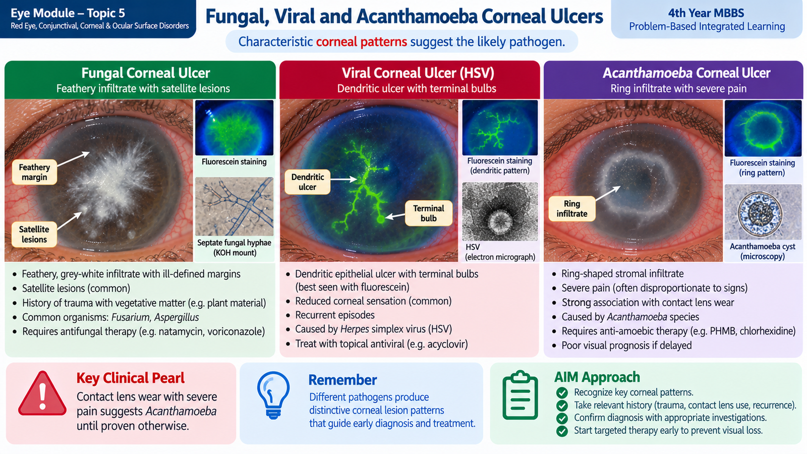
Different microorganisms can produce overlapping corneal ulcers, but several clinical patterns help guide the initial diagnosis. These clues are particularly important because bacterial, fungal, viral and Acanthamoeba keratitis require different antimicrobial strategies.
Fungal keratitis
Fungal infection commonly follows disruption of the corneal epithelium, especially after trauma involving plant or soil material. Corticosteroid exposure and chronic ocular-surface disease can also increase susceptibility.
- Pain, redness, photophobia and reduced vision.
- A relatively dry-looking stromal infiltrate may be present.
- Feathery or irregular infiltrate margins are an important clue.
- Small satellite lesions may appear around the main infiltrate.
- Hypopyon can occur in severe disease.
Corneal scraping is examined for fungal elements and cultured on suitable fungal media. Treatment requires prolonged topical antifungal therapy selected according to the likely organism and microbiological findings. Natamycin is commonly used for filamentous fungal keratitis, while other antifungal agents may be required for different organisms. Corticosteroids should be avoided during active fungal infection. Progressive thinning or perforation may require therapeutic corneal surgery.
Viral keratitis
Herpes simplex virus is a major viral cause of recurrent corneal disease. After primary infection, the virus remains latent in sensory ganglia and may reactivate. Repeated episodes can affect the epithelium, stroma or both.
The classic epithelial lesion is a dendritic ulcer: a branching epithelial defect with terminal bulbs that becomes visible with corneal staining. Corneal sensation may be reduced. Disease is frequently unilateral and recurrent.
Epithelial herpes simplex keratitis is treated with antiviral therapy. Topical corticosteroid used alone is dangerous in active epithelial herpetic disease because it can promote viral replication and enlarge the epithelial ulcer. Stromal herpetic inflammation is managed differently and requires ophthalmic supervision, often with antiviral cover when anti-inflammatory therapy is required.
Acanthamoeba keratitis
Acanthamoeba is a free-living protozoan associated particularly with contact-lens exposure to contaminated water. Infection can be difficult to recognize early because initial signs may appear less dramatic than the patient’s symptoms.
- Severe pain that may appear out of proportion to early examination findings.
- History of contact-lens wear, especially with water exposure or poor lens hygiene.
- Photophobia and reduced vision.
- Perineural corneal infiltrates may occur.
- A ring-shaped stromal infiltrate is a classic later clue but is not required for diagnosis.
Corneal scraping for microscopy and specialized culture is important. Specialist investigations such as in-vivo confocal microscopy may support diagnosis when available. Treatment uses prolonged topical anti-Acanthamoeba agents, commonly from the biguanide group, under close ophthalmic supervision. Delay in diagnosis can lead to severe stromal damage.
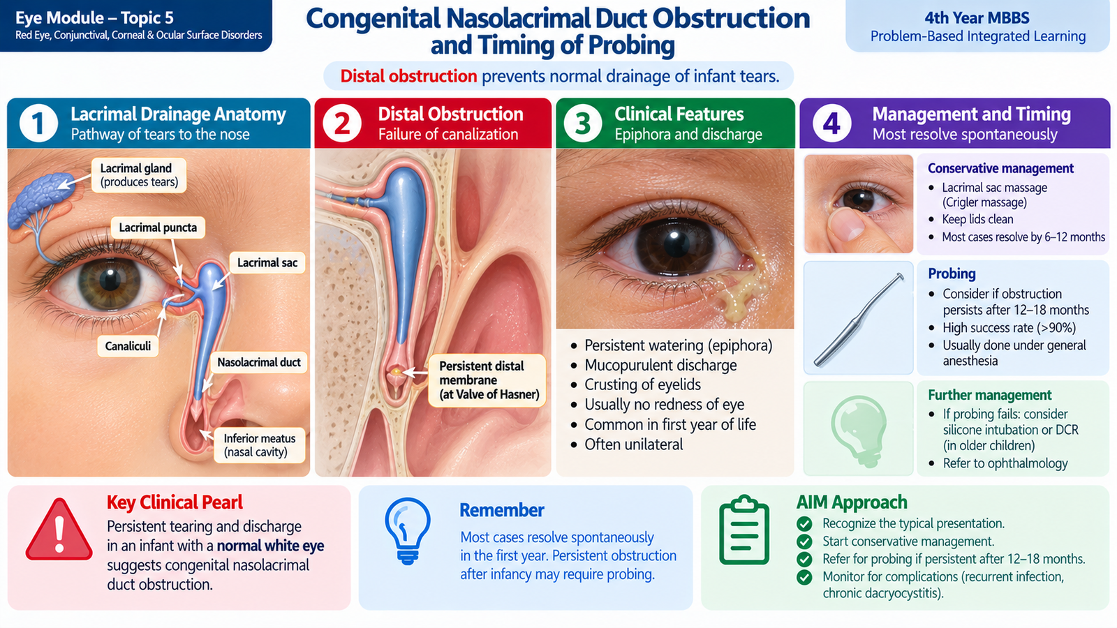
F. Congenital Nasolacrimal Duct Obstruction and Timing of Probing
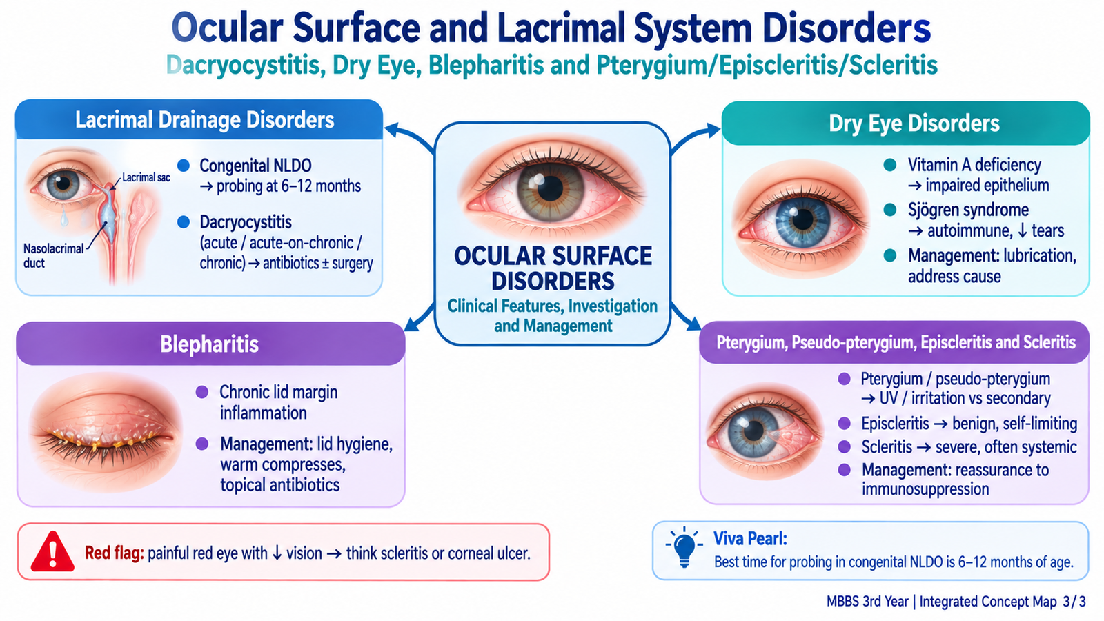
Tears normally pass from the puncta through the canaliculi into the lacrimal sac and then through the nasolacrimal duct into the nose. In congenital nasolacrimal duct obstruction, drainage is usually blocked because the distal end of the duct has not opened completely, commonly near the valve of Hasner.
Clinical features
- Persistent watering or epiphora beginning in infancy.
- Mucous or mucopurulent material around the eyelashes.
- Reflux of mucus or fluid from the punctum when the lacrimal sac is gently compressed.
- The eye itself is usually white unless secondary conjunctival irritation or infection develops.
The diagnosis is mainly clinical. An infant with tearing must still be examined carefully because other causes, especially congenital glaucoma or corneal disease, are more serious. Corneal enlargement, clouding or marked photophobia is not explained by simple nasolacrimal duct obstruction and requires urgent assessment.
Conservative management
Many congenital obstructions open spontaneously during infancy. Initial treatment therefore consists of cleaning accumulated discharge and regular lacrimal sac massage. Proper massage raises hydrostatic pressure within the sac and may help open the persistent distal membrane.
Topical antibiotics may be used when significant mucopurulent discharge indicates secondary bacterial infection, but antibiotics do not remove the underlying mechanical obstruction.
When should probing be considered?
Probing mechanically opens the persistent obstruction by passing a fine probe through the lacrimal drainage pathway. Because many infants improve spontaneously, immediate routine probing is usually unnecessary.
Persistent symptomatic obstruction after approximately the first year of life is a common indication to consider probing, often during the following months depending on the child and local ophthalmic practice. Earlier intervention may be appropriate when there are recurrent significant infections or other complications. The decision therefore combines the child’s age, persistence of symptoms, response to massage and infection history rather than relying on age alone.
G. Dacryocystitis — Acute, Acute-on-Chronic and Chronic Disease
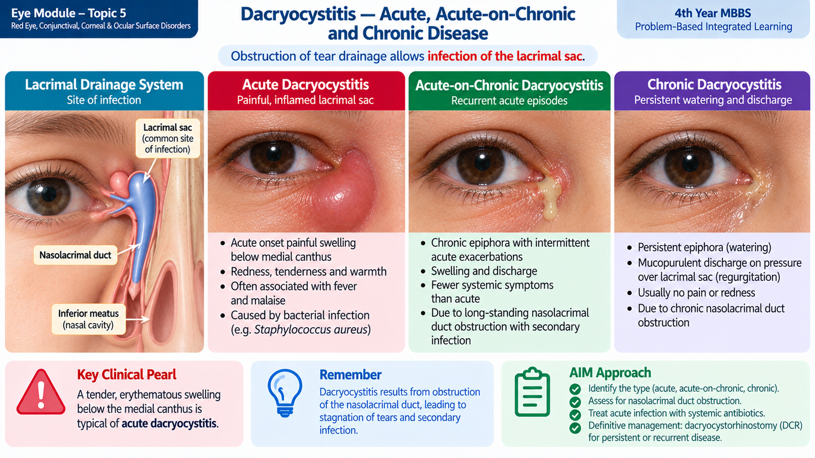
Dacryocystitis is inflammation and usually infection of the lacrimal sac. The usual underlying problem is impaired drainage through the nasolacrimal duct. Tear stagnation allows organisms to multiply inside the sac, explaining why obstruction and infection frequently occur together.
Acute dacryocystitis
Acute infection produces sudden inflammatory swelling of the lacrimal sac. The patient develops a painful, red and tender swelling below the medial canthus. Surrounding edema may be marked, and systemic fever or malaise can occur. An abscess may form if infection becomes localized.
Treatment during the acute phase is directed at infection and inflammation. Systemic antimicrobial therapy, analgesia and warm compresses are commonly required. A localized abscess may require drainage. Manipulation or routine probing of an acutely inflamed lacrimal system is avoided because it is painful and may spread infection. Once acute infection settles, the underlying drainage obstruction is assessed for definitive treatment.
Chronic dacryocystitis
Chronic obstruction produces persistent tear stagnation and low-grade infection. The patient commonly complains of epiphora and recurrent discharge rather than severe pain. Pressure over the lacrimal sac may cause mucus or pus to reflux through the puncta; this is referred to clinically as regurgitation on pressure over the lacrimal sac.
Lacrimal syringing can help demonstrate the level or presence of drainage obstruction in chronic disease. Nasal assessment may also be relevant when planning definitive treatment. The main definitive procedure in an adult with persistent symptomatic nasolacrimal duct obstruction is usually dacryocystorhinostomy (DCR), which creates a new drainage pathway between the lacrimal sac and the nose.
Acute-on-chronic dacryocystitis
Acute-on-chronic disease occurs when a previously obstructed and chronically infected lacrimal sac develops an acute inflammatory flare. The patient therefore has the background history of watering or recurrent discharge plus the sudden pain, redness and tenderness of acute infection.
Sudden pain, erythema, tenderness and swelling over lacrimal sac.
Long-standing epiphora, discharge and regurgitation with little acute pain.
Chronic watering/discharge followed by an acute painful inflammatory episode.
 .
.H. Dry-Eye Disease — Including Vitamin A Deficiency and Sjögren Syndrome
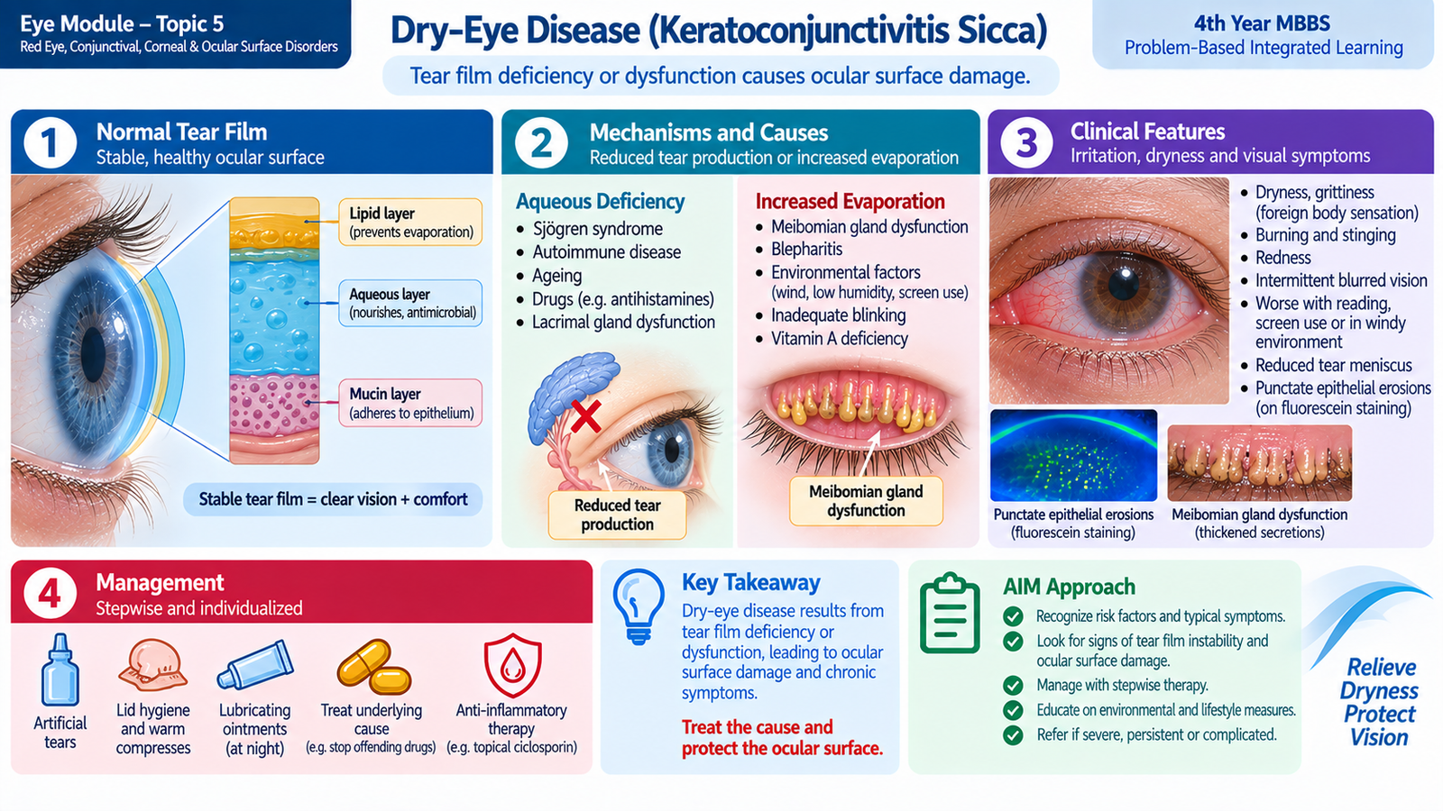
The tear film lubricates the ocular surface, provides a smooth optical interface and protects the corneal and conjunctival epithelium. Dry-eye disease develops when tear-film homeostasis is lost. The film becomes unstable and often hyperosmolar, which promotes epithelial stress and inflammation. This creates a cycle in which surface damage further destabilizes the tear film.
Main mechanisms
Lacrimal secretion is reduced, so there is insufficient aqueous tear volume.
Tears evaporate too rapidly, commonly because of meibomian gland dysfunction or excessive ocular exposure.
These mechanisms often overlap. Reduced blinking, prolonged visual tasks, lid disease and systemic disorders can further disturb the tear film.
Clinical features
- Burning, stinging or gritty sensation.
- Foreign-body sensation.
- Intermittent blurred or fluctuating vision.
- Redness and ocular fatigue.
- Paradoxical watering may occur because surface irritation triggers reflex tear production.
- More severe disease can produce photophobia and epithelial injury.
Investigations
- Slit-lamp examination: assesses tear meniscus, lid margin and ocular surface.
- Fluorescein tear-film break-up assessment: demonstrates premature disruption of an unstable tear film.
- Corneal and conjunctival staining: demonstrates epithelial surface damage.
- Schirmer testing: helps assess aqueous tear production when aqueous deficiency is suspected.
- Meibomian gland examination: identifies obstructed glands and abnormal lipid secretion in evaporative disease.
Sjögren syndrome
Sjögren syndrome is an autoimmune disorder in which lymphocytic inflammation damages exocrine glands. Damage to the lacrimal glands decreases aqueous tear secretion, producing significant aqueous-deficient dry eye. Patients commonly also report dry mouth, and other systemic autoimmune features may be present.
Management includes regular ocular lubrication and treatment of significant ocular-surface inflammation under ophthalmic supervision. Because the condition is systemic, patients with suggestive features require appropriate medical evaluation rather than treatment of the eye alone.
Vitamin A deficiency
Vitamin A is necessary for normal differentiation of conjunctival and corneal epithelium and for maintenance of mucin-producing goblet cells. Deficiency therefore causes ocular surface dryness and keratinization. Early manifestations may include conjunctival xerosis and Bitot spots. Severe deficiency can progress to corneal xerosis, ulceration and keratomalacia.
Night blindness may coexist because vitamin A is also required for normal visual pigment function. Management requires vitamin A replacement and correction of the nutritional or systemic cause. Corneal ulceration or softening represents an ophthalmic emergency because irreversible blindness can develop.
General management of dry eye
- Regular preservative-minimized lubricating drops or gels when frequent use is needed.
- Reduce environmental evaporation and address prolonged incomplete blinking where relevant.
- Treat associated blepharitis and meibomian gland dysfunction.
- Manage the underlying systemic or nutritional disorder.
- More severe inflammatory dry eye may require specialist anti-inflammatory therapy or other ocular-surface measures.
I. Blepharitis
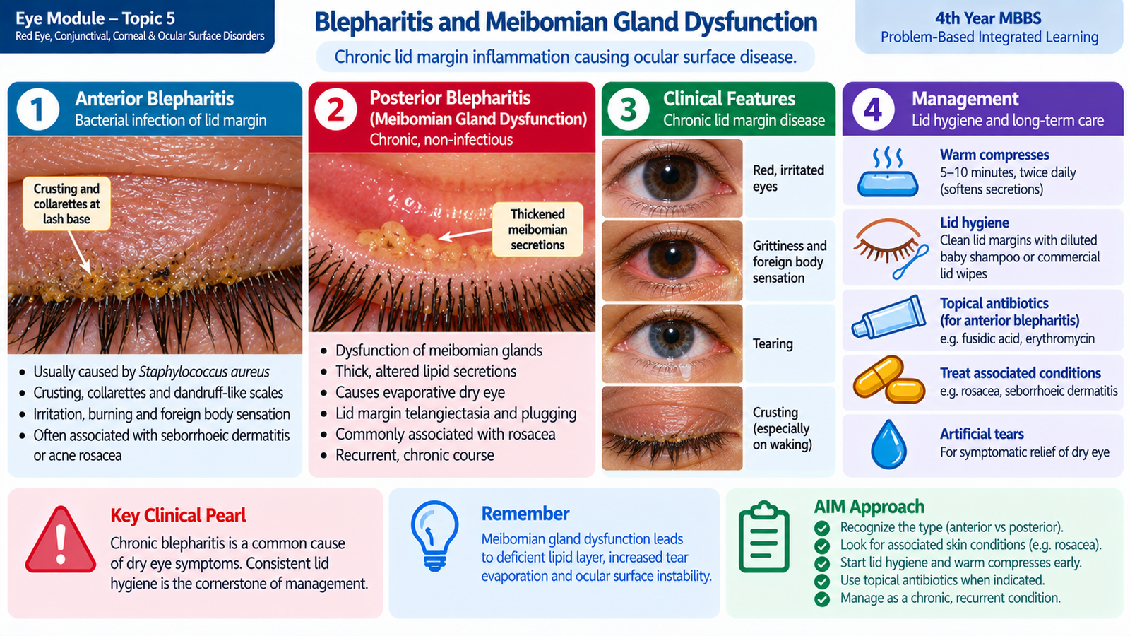
Blepharitis is chronic inflammation of the eyelid margins. It is important because the lid margin and meibomian glands contribute directly to tear-film stability. Chronic lid inflammation can therefore produce irritation, recurrent conjunctival inflammation, dry eye and secondary corneal disease.
Types and pathogenesis
Anterior blepharitis mainly affects the skin and lash follicles at the front of the lid margin and is commonly associated with staphylococcal colonization or seborrheic disease. Posterior blepharitis is closely related to meibomian gland dysfunction. Thick or obstructed meibomian secretions reduce the normal lipid layer of the tear film, increasing evaporation.
Clinical features
- Burning, irritation and foreign-body sensation.
- Itching and intermittent redness.
- Crusting around the eyelashes, especially on waking.
- Red, thickened or irregular lid margins.
- Scales or collarettes around lashes in anterior disease.
- Capped or poorly functioning meibomian gland openings in posterior disease.
- Foamy or unstable tear film.
- Recurrent chalazia may accompany meibomian gland dysfunction.
Investigations
Diagnosis is mainly clinical. Slit-lamp examination demonstrates lid-margin abnormalities, lash debris, meibomian gland plugging and associated corneal or conjunctival staining. Routine laboratory investigation is usually unnecessary unless the presentation is atypical or resistant to treatment.
Management
Blepharitis is often chronic, so treatment aims to control rather than instantly eliminate the condition. Regular lid hygiene is the foundation of management.
- Warm compresses soften crusts and meibomian secretions.
- Gentle lid-margin cleaning removes debris and reduces inflammatory load.
- Lid massage can help express obstructed meibomian glands.
- Lubricants reduce secondary dry-eye symptoms.
- Topical antibiotic ointment may be useful in selected anterior bacterial blepharitis.
- Persistent posterior disease or associated rosacea may require specialist-directed systemic therapy.
J. Pterygium, Pseudo-Pterygium, Episcleritis and Scleritis
These disorders may all produce visible redness near the cornea, but they arise from different tissues. Pterygium and pseudo-pterygium are conjunctival lesions extending toward or onto the cornea, whereas episcleritis and scleritis are inflammatory disorders involving increasingly deeper coats of the eye. Depth of involvement explains the major differences in pain and risk to vision.
Pterygium
A pterygium is a triangular fibrovascular growth of conjunctival tissue onto the cornea, usually from the nasal side. Chronic ultraviolet exposure, dust and surface irritation are important associations. Progressive growth can distort the cornea and induce astigmatism; advanced lesions may approach the visual axis.
Small asymptomatic lesions can be managed with UV protection, avoidance of surface irritants and lubricants. Surgical excision is considered when the lesion is progressive, interferes with vision, produces significant astigmatism or causes persistent troublesome inflammation.
Pseudo-pterygium
A pseudo-pterygium is an adhesion of conjunctiva to the peripheral cornea following previous inflammation, trauma or chemical injury. Unlike a typical pterygium, it may occur in any quadrant. The conjunctival fold is generally attached only at its apex, so a probe can often be passed beneath its body near the limbus.
Management is directed at the underlying cause and the functional significance of the adhesion. Surgery may be required if it causes important symptoms or interferes with ocular function.
Episcleritis
Episcleritis is inflammation of the superficial episcleral vascular tissue. It usually produces a localized or sectoral area of bright redness with mild tenderness or discomfort rather than severe pain. Vision is generally normal. Many cases are self-limiting.
Examination shows superficial mobile vessels. These vessels tend to blanch with a vasoconstrictor such as phenylephrine, helping distinguish episcleritis from deeper scleral inflammation. Lubrication and simple anti-inflammatory treatment may be sufficient, while recurrent or atypical disease may prompt evaluation for systemic association.
Scleritis
Scleritis is a much deeper and potentially destructive inflammation of the sclera. Because the sclera and adjacent tissues are richly innervated, inflammation commonly produces severe deep or boring pain, sometimes radiating to the forehead, brow or jaw. The pain may disturb sleep and is often aggravated by eye movement.
The eye may have a deep bluish-red or violaceous appearance. Deep vessels do not blanch in the same way as superficial episcleral vessels. Scleritis is important because it may be associated with systemic autoimmune disease and can cause scleral thinning, keratitis, uveitis and visual loss.
Management requires urgent ophthalmic assessment. Treatment is usually systemic rather than simple topical therapy and depends on severity and any associated systemic disease. Infectious scleritis must be excluded before immunosuppressive treatment is used.
Important Comparison — High-Yield Red-Eye Patterns
| Condition | Pain / discomfort | Discharge | Vision | Key clue |
|---|---|---|---|---|
| Conjunctivitis | Mild irritation / gritty feeling | Watery or mucopurulent | Usually preserved | Diffuse superficial redness |
| Corneal ulcer | Significant pain and photophobia | Variable | Often reduced | Corneal infiltrate + fluorescein defect |
| Anterior uveitis | Pain and photophobia | Usually absent | May be reduced | Ciliary injection; intraocular inflammation |
| Acute angle closure | Severe pain | Absent | Markedly reduced | Corneal haze, abnormal pupil, raised IOP |
| Episcleritis | Mild discomfort | Absent | Normal | Sectoral superficial redness |
| Scleritis | Severe deep boring pain | Absent | May be reduced | Deep violaceous redness |
Important Comparison — Pterygium, Pseudo-Pterygium, Episcleritis and Scleritis
| Feature | Pterygium | Pseudo-pterygium | Episcleritis | Scleritis |
|---|---|---|---|---|
| Main process | Fibrovascular growth | Post-inflammatory adhesion | Superficial inflammation | Deep scleral inflammation |
| Typical location | Usually nasal | Any quadrant | Sectoral or diffuse episclera | Sclera |
| Pain | Usually little | Depends on cause | Mild | Severe, deep, boring |
| Key distinction | Triangular lesion crossing limbus | Probe may pass beneath body | Superficial vessels blanch | Deep vessels do not readily blanch |
| Threat to vision | If progressive or inducing astigmatism | Usually related to underlying injury | Usually low | Potentially sight-threatening |