
Topic 7 — Uveitis: Classification, Evaluation & Management
Topic Introduction
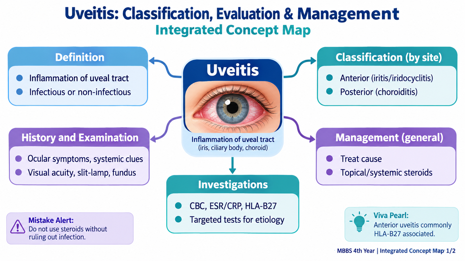
Uveitis means inflammation involving the uveal tract, which consists of the iris, ciliary body and choroid. In clinical practice, inflammation may also involve nearby structures such as the vitreous, retina and optic nerve. The most useful first step is to identify where the inflammation is mainly located, because anterior and posterior disease produce different symptoms and examination findings. This chapter explains the definition and classification of uveitis, the important history and ophthalmic workup, and the causes, clinical features, investigations and management of anterior and posterior uveitis. The aim is to develop a simple clinical sequence: recognize the pattern, localize the inflammation, investigate intelligently and protect vision.
A. Definition, Uveal Anatomy & Classification of Uveitis
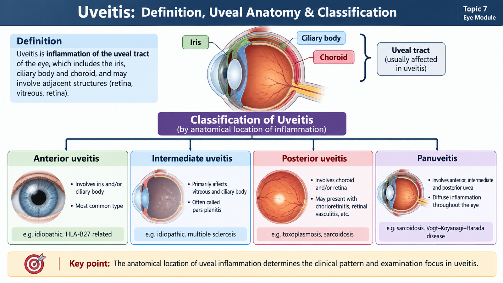
The uvea is the vascular middle coat of the eye. Anteriorly it forms the iris and ciliary body, while posteriorly it forms the choroid. Inflammation can be concentrated in one part or extend through several parts of the eye. Therefore, classification by the main anatomical site of inflammation is clinically important because it predicts the patient’s symptoms, guides examination and helps organize the investigation.
Definition
Uveitis is an inflammatory disorder primarily affecting the uveal tract. The term is commonly used more broadly when inflammation extends to adjacent ocular tissues, particularly the vitreous, retina and optic nerve.
Anatomical classification
- Anterior uveitis: inflammation is mainly in the anterior chamber, usually involving the iris with or without the anterior ciliary body. Terms such as iritis and iridocyclitis may be used.
- Intermediate uveitis: inflammation is mainly centered in the vitreous and peripheral retina/ciliary body region.
- Posterior uveitis: inflammation primarily involves the choroid, retina or both. It may appear as choroiditis, retinitis or chorioretinitis.
- Panuveitis: significant inflammation involves the anterior chamber, vitreous and posterior segment without one site clearly dominating.
Other useful ways to describe uveitis
After identifying the anatomical site, the ophthalmologist also describes the disease according to its clinical behavior. It may be acute, recurrent or chronic, and it may affect one eye or both eyes. Etiologically, uveitis may be broadly considered infectious or non-infectious. These descriptions help narrow the possible cause.
B. Clinical Evaluation: History & Ophthalmic Examination
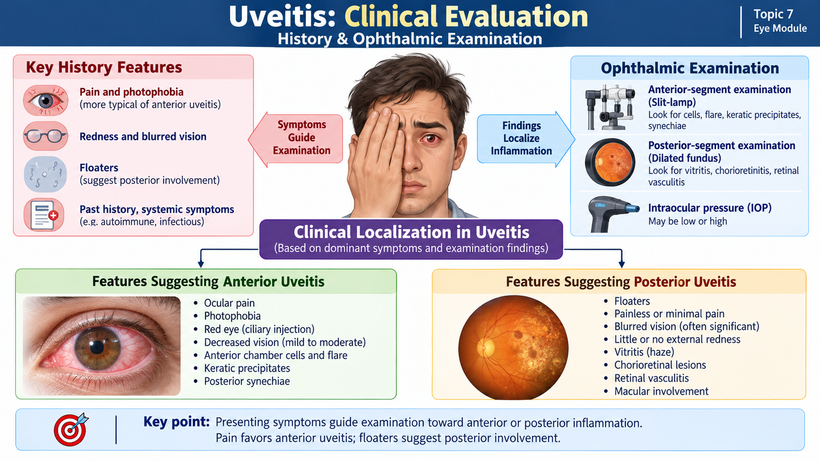
Evaluation of uveitis begins by deciding where the inflammation is located and how severe it is. A good history often gives the first clue. Pain, photophobia and circumcorneal redness suggest anterior inflammation, whereas floaters and painless reduction of vision are more suggestive of posterior involvement. The examination then confirms the anatomical localization and identifies complications that may threaten sight.
History
The ocular history should establish the pattern of the present episode before searching for systemic associations.
- Onset and duration: sudden or gradual; first episode or recurrence.
- Laterality: unilateral, alternating or bilateral.
- Pain and photophobia: particularly important in anterior uveitis because inflammation and ciliary muscle spasm stimulate sensory nerves.
- Redness: especially redness concentrated around the corneal limbus.
- Visual disturbance: blurred vision may occur in either anterior or posterior disease.
- Floaters: suggest inflammatory cells or debris within the vitreous and therefore favor intermediate or posterior involvement.
- Previous episodes: recurrent attacks may suggest a systemic inflammatory association.
- Previous ocular trauma or surgery: may be relevant to the cause of intraocular inflammation.
Relevant systemic history
Uveitis can be an ocular manifestation of systemic disease. The history should therefore be directed rather than excessively broad.
- Inflammatory back pain, arthritis or joint swelling.
- Oral or genital ulceration.
- Skin lesions or rashes.
- Respiratory or constitutional symptoms suggesting systemic inflammatory or infectious disease.
- Neurological symptoms when clinically relevant.
- Exposure or risk factors for infections such as tuberculosis or sexually transmitted infection when suggested by the presentation.
- Immune suppression, which increases the possibility of opportunistic ocular infection.
Ophthalmic examination
Visual acuity should be documented in each eye. Reduced acuity may result from anterior chamber inflammation, corneal changes, cataract, vitritis, macular involvement or posterior-segment disease. Marked reduction of vision should make the examiner actively look for posterior involvement or a complication.
Pupillary examination may reveal a small or irregular pupil in anterior uveitis. An irregular pupil often results from posterior synechiae, in which the inflamed posterior surface of the iris adheres to the anterior lens capsule.
Slit-lamp examination is central to anterior uveitis. Inflammatory leukocytes circulating in the aqueous are seen as anterior chamber cells. Increased protein leaking through inflamed iris and ciliary-body vessels produces flare, which appears as a smoky beam of light in the anterior chamber.
Keratic precipitates are collections of inflammatory cells deposited on the posterior corneal surface. Their size and appearance can provide information about the inflammatory pattern.
Intraocular pressure (IOP) should be assessed because uveitis may be associated with either low or raised pressure. Inflammation can initially reduce aqueous production, while inflammatory debris, trabecular involvement, synechiae or treatment-related effects can later raise IOP.
Dilated fundus examination is necessary when posterior involvement is suspected. The examiner looks for vitreous cells or haze, retinal or choroidal inflammatory lesions, retinal vasculitis, macular involvement and optic-disc changes.
C. Diagnostic Workup of Uveitis
The purpose of the uveitis workup is not to order every available laboratory test. The first task is to define the anatomical type, clinical appearance, laterality and course. Investigations are then selected according to the suspected cause. This targeted approach reduces unnecessary testing and makes each result easier to interpret.
Step 1 — Confirm and localize ocular inflammation
Slit-lamp examination confirms anterior chamber cells, flare, keratic precipitates and synechiae. Dilated fundus examination identifies vitritis, retinal or choroidal lesions, vasculitis and macular or optic-disc involvement. These findings determine whether the disease is mainly anterior or posterior.
Step 2 — Assess structural effects and complications
Optical coherence tomography (OCT) is particularly useful when central vision is reduced or macular involvement is suspected. It can demonstrate retinal thickening and macular edema, an important cause of visual impairment in uveitis.
Fundus fluorescein angiography may be used when retinal vascular leakage, retinal vasculitis or inflammatory activity in the posterior segment needs clarification. Leakage of dye indicates breakdown of the normal vascular barrier.
When the fundus cannot be seen adequately because of severe media opacity, ocular ultrasonography can help assess the posterior segment.
Step 3 — Search for the cause when indicated
Laboratory and systemic investigations should follow clues from the history and ocular phenotype. Examples include:
- Tuberculosis assessment: considered when ocular appearance, exposure history or systemic features raise suspicion; appropriate testing and chest assessment may support the diagnosis.
- Syphilis serology: important when syphilitic uveitis is clinically possible because the disease can produce varied ocular appearances.
- Toxoplasma assessment: considered particularly in compatible focal posterior retinochoroidal inflammation.
- HLA-B27 testing: may support evaluation of recurrent acute anterior uveitis when the clinical history suggests an associated spondyloarthropathy.
- Tests for systemic inflammatory disease: selected according to symptoms and clinical suspicion rather than requested routinely in every patient.
In selected severe or atypical cases in which infection is strongly suspected, ophthalmologists may obtain ocular fluid for specialized microbiological or molecular testing. This is not part of routine investigation for every patient.

D. Anterior Uveitis: Etiology, Features, Investigations & Management
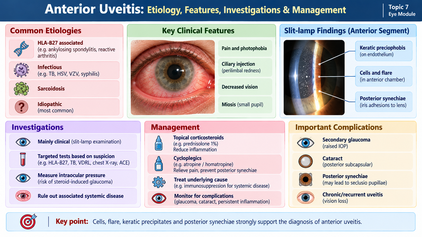
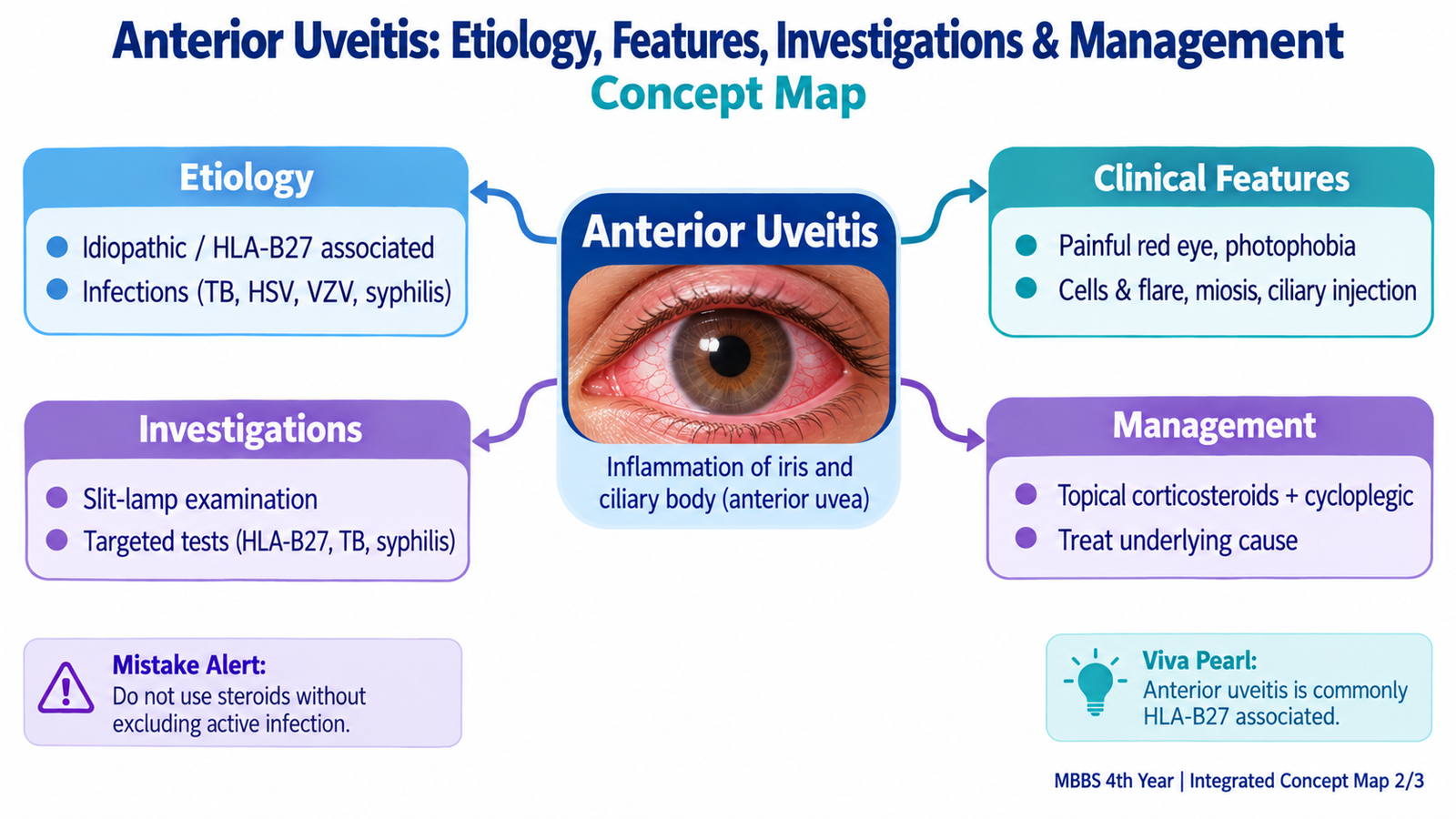
Anterior uveitis is inflammation predominantly affecting the iris and anterior ciliary body. It is the form of uveitis most likely to present as a painful, photophobic red eye. The inflammation disrupts the blood-aqueous barrier, allowing inflammatory cells and plasma proteins to enter the anterior chamber. This explains the characteristic slit-lamp findings of cells and flare.
Etiology
Anterior uveitis may occur without an identified systemic cause, or it may be associated with inflammatory or infectious disease. The clinical pattern helps determine which causes need investigation.
- Immune-mediated or systemic inflammatory disease: especially HLA-B27-associated disorders such as ankylosing spondylitis and related spondyloarthropathies.
- Juvenile idiopathic arthritis: an important association in children.
- Sarcoidosis: may produce granulomatous ocular inflammation.
- Infectious causes: herpes simplex and varicella-zoster viruses can produce anterior uveitis; tuberculosis and syphilis may also involve the uveal tract.
- Trauma or ocular surgery: can produce intraocular inflammation.
- Idiopathic: in some patients no definite cause is identified despite appropriate evaluation.
How the inflammation produces symptoms
Pain results partly from inflammation and spasm of the ciliary muscle. Photophobia occurs because light-induced pupillary movement stimulates the inflamed iris and ciliary region. Redness is usually most marked around the corneal limbus because the deeper anterior ciliary circulation becomes congested.
Clinical features and examination findings
- Painful red eye.
- Photophobia.
- Blurred or reduced vision.
- Watering may occur, but a purulent discharge is not a typical feature.
- Ciliary or circumcorneal injection: a violaceous-red flush around the limbus.
- Anterior chamber cells: inflammatory cells visible in the slit-lamp beam.
- Flare: a hazy appearance of the slit-lamp beam caused by increased protein in the aqueous.
- Keratic precipitates: inflammatory deposits on the corneal endothelium.
- Miotic pupil: the pupil may be relatively small because of iris inflammation and spasm.
- Posterior synechiae: adhesion of the posterior iris to the lens may make the pupil irregular.
Granulomatous and non-granulomatous patterns
This clinical distinction can help guide etiological thinking. Granulomatous uveitis tends to produce larger, greasy-looking “mutton-fat” keratic precipitates and may be associated with iris nodules. Non-granulomatous uveitis more often produces smaller keratic precipitates and may present more acutely. These appearances suggest a pattern; they do not establish the cause by themselves.
Investigations
The most important initial investigation is a careful slit-lamp examination, because it confirms anterior chamber inflammation and detects synechiae and keratic precipitates. IOP measurement identifies pressure-related complications. A dilated fundus examination is required to make sure significant posterior involvement has not been missed.
Additional laboratory investigation depends on the clinical pattern. For example, recurrent acute anterior uveitis with inflammatory back symptoms may prompt assessment for an HLA-B27-associated disease, whereas a granulomatous or atypical presentation may require investigation for infectious or systemic inflammatory causes.
Management
Management has three major aims: suppress harmful inflammation, relieve pain and prevent structural complications. The underlying cause must also be identified and treated where possible.
- Topical corticosteroids: are a main treatment for non-infectious anterior uveitis. They reduce inflammatory-cell activity and vascular leakage within the anterior chamber.
- Cycloplegic/mydriatic drugs: relax the ciliary muscle, reducing painful spasm. Dilating the pupil also helps prevent or limit posterior synechiae.
- Treatment of infection: when an infectious cause is suspected or confirmed, specific antimicrobial or antiviral treatment is required. Corticosteroid treatment must be planned with the infectious process in mind.
- Control of raised IOP: pressure should be monitored and treated when necessary because inflammation may produce secondary ocular hypertension or glaucoma.
- Systemic or other specialist anti-inflammatory therapy: may be required in severe, recurrent or chronic non-infectious disease under ophthalmology supervision.
- Management of associated systemic disease: may require coordinated care with the relevant specialty.
Complications and red flags
Persistent or inadequately controlled inflammation may lead to posterior synechiae, secondary glaucoma, cataract and macular edema. Severe reduction of vision, marked IOP elevation, dense inflammation, hypopyon or suspicion of ocular infection requires urgent ophthalmic assessment.
E. Posterior Uveitis: Etiology, Features, Investigations & Management
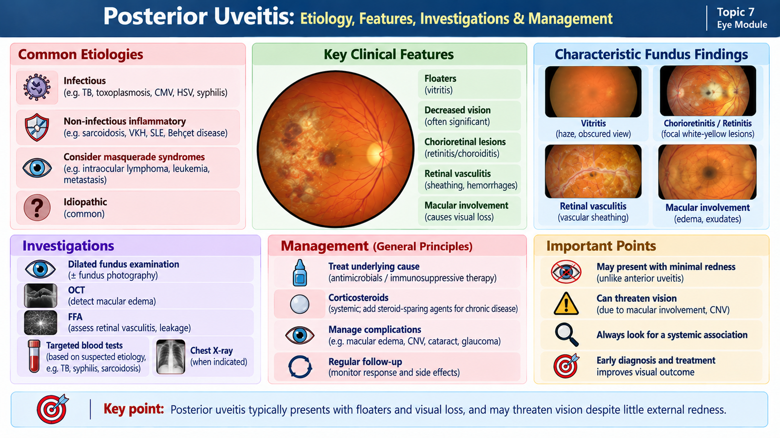
Posterior uveitis is inflammation primarily affecting structures behind the vitreous, particularly the choroid and retina. Because the inflammation lies away from the highly sensitive anterior uveal structures, the eye may show little external redness and pain may be absent. The major complaints are therefore often floaters and reduced vision.
Etiology
Posterior uveitis can result from infection or from immune-mediated inflammation. Distinguishing these groups is particularly important because corticosteroids can worsen uncontrolled infection if given without appropriate antimicrobial treatment.
- Toxoplasmosis: an important infectious cause of focal retinochoroiditis.
- Tuberculosis: may produce choroidal or retinal inflammatory disease.
- Syphilis: can involve several ocular structures and may present with posterior uveitis.
- Viral retinitis: becomes particularly important in susceptible or immunocompromised patients.
- Systemic inflammatory disorders: conditions such as sarcoidosis and Behçet disease may involve the posterior segment.
- Other immune-mediated uveitic disorders: may produce choroidal, retinal or combined inflammation.
Pathological basis of the visual symptoms
Floaters occur because inflammatory cells and debris within the vitreous cast moving shadows on the retina. Visual acuity falls particularly when inflammation involves the macula, produces macular edema, causes marked vitreous haze or damages retinal tissue.
Clinical features
- Blurred or reduced vision.
- Floaters.
- Scotoma or distortion of vision when specific retinal areas are affected.
- External redness may be mild or absent.
- Pain and photophobia are usually less prominent than in isolated anterior uveitis, unless anterior structures are also inflamed.
Fundus and posterior-segment findings
Vitritis appears as inflammatory cells and haze in the vitreous. This can partly obscure the retinal view. Retinitis appears as areas of active retinal inflammation, while choroiditis produces inflammatory lesions originating in the choroid. When both tissues are affected, the term chorioretinitis is used.
Retinal vasculitis may produce perivascular inflammatory changes and vascular leakage. Involvement of the macula may cause macular edema, while optic-disc inflammation or edema may accompany some posterior inflammatory disorders.
Investigations
Dilated fundus examination is essential because it directly demonstrates the site and pattern of posterior inflammation. The distribution and appearance of lesions help narrow the differential diagnosis.
OCT is especially useful when macular disease is suspected. Demonstration of retinal thickening or cystic fluid confirms macular edema and explains central visual loss.
Fundus fluorescein angiography may demonstrate retinal vascular leakage, vasculitis or areas of active inflammatory involvement. Ocular ultrasonography is useful when dense vitreous opacity prevents adequate visualization of the fundus.
Cause-directed laboratory investigations are selected according to the fundus appearance and systemic history. Testing may be directed toward toxoplasmosis, tuberculosis, syphilis or systemic inflammatory disease when those diagnoses are clinically suspected.
Management
The most important management decision is whether the posterior inflammation is infectious or non-infectious. The wrong assumption can be harmful; therefore suspected infectious posterior uveitis requires prompt specialist evaluation.
- Infectious posterior uveitis: requires treatment directed against the responsible organism. Examples include appropriate antiparasitic, antibacterial, antituberculous or antiviral therapy according to the diagnosis.
- Non-infectious posterior uveitis: corticosteroids may be used to suppress damaging inflammation. The route depends on severity and anatomical involvement and is determined by the ophthalmologist.
- Recurrent or chronic immune-mediated disease: may require steroid-sparing immunomodulatory therapy under specialist supervision.
- Systemic disease: should be treated in collaboration with the relevant medical specialty when present.
- Complications: macular edema, raised IOP, cataract and other structural complications require appropriate ophthalmic management.
Integrated Mechanism Flow
Infectious or immune-mediated stimulus
Leukocyte activation and vascular permeability
Cells and protein enter ocular compartments
Anterior cells/flare or posterior lesions/vitritis
Synechiae, glaucoma, cataract, macular or retinal damage
Control inflammation and treat the cause
Important Comparison — Anterior versus Posterior Uveitis
| Feature | Anterior Uveitis | Posterior Uveitis |
|---|---|---|
| Main site | Iris / anterior ciliary body | Choroid / retina |
| Pain | Common | Often absent or mild |
| Photophobia | Prominent | Usually less prominent |
| External redness | Ciliary injection common | May be minimal or absent |
| Floaters | Less prominent | Common with vitritis |
| Key examination | Slit lamp: cells, flare, KPs, synechiae | Fundus: vitritis, retinitis, choroiditis, vasculitis |
| Major visual threat | Synechiae, glaucoma, cataract, macular edema | Macular, retinal or optic-nerve damage |
| Management emphasis | Topical anti-inflammatory treatment plus cycloplegia when appropriate | Establish infectious versus non-infectious cause before definitive therapy |
⭐ AIM High-Yield Review
- Uveitis is inflammation of the uveal tract and may involve adjacent retina, vitreous and optic nerve.
- ⭐ Anatomical classification includes anterior, intermediate, posterior and panuveitis.
- Pain + photophobia + ciliary redness strongly suggest anterior uveitis.
- Floaters + reduced vision with little redness should raise suspicion of posterior involvement.
- Anterior chamber cells represent inflammatory leukocytes; flare represents increased aqueous protein.
- An irregular pupil in anterior uveitis may result from posterior synechiae.
- Slit-lamp examination is fundamental for anterior uveitis; dilated fundus examination is essential for posterior uveitis.
- Uveitis investigations should be targeted to the clinical pattern, not ordered as an indiscriminate laboratory panel.
- OCT is particularly useful for detecting uveitic macular edema.
- Topical corticosteroids control non-infectious anterior inflammation, while cycloplegics reduce ciliary spasm and help prevent synechiae.
- ⭐ In posterior uveitis, determining whether inflammation is infectious or non-infectious is a critical management decision.
- Important sight-threatening complications include secondary glaucoma, cataract, macular edema and retinal damage.
- Marked visual loss, severe intraocular inflammation or suspected infection requires urgent ophthalmology assessment.