
Topic 15 — Squint, Diplopia & Ocular Motor Nerve Palsies
Topic Introduction
Normal binocular vision requires the two eyes to remain accurately aligned so that both foveae are directed toward the same object. A squint (strabismus) occurs when this alignment is lost. The deviation may be inward, outward, vertical or torsional, and it may remain similar in different directions of gaze or vary according to the affected muscle or nerve. In children, the brain can suppress the image from the deviating eye, which may contribute to amblyopia and loss of binocular vision. In adults, new misalignment commonly produces diplopia. This chapter develops a practical approach to defining and examining squint, understanding esotropia and exotropia, identifying causes of diplopia, and localizing third, fourth and sixth cranial nerve palsies.
A. Understanding Squint: Definitions and Basic Classification
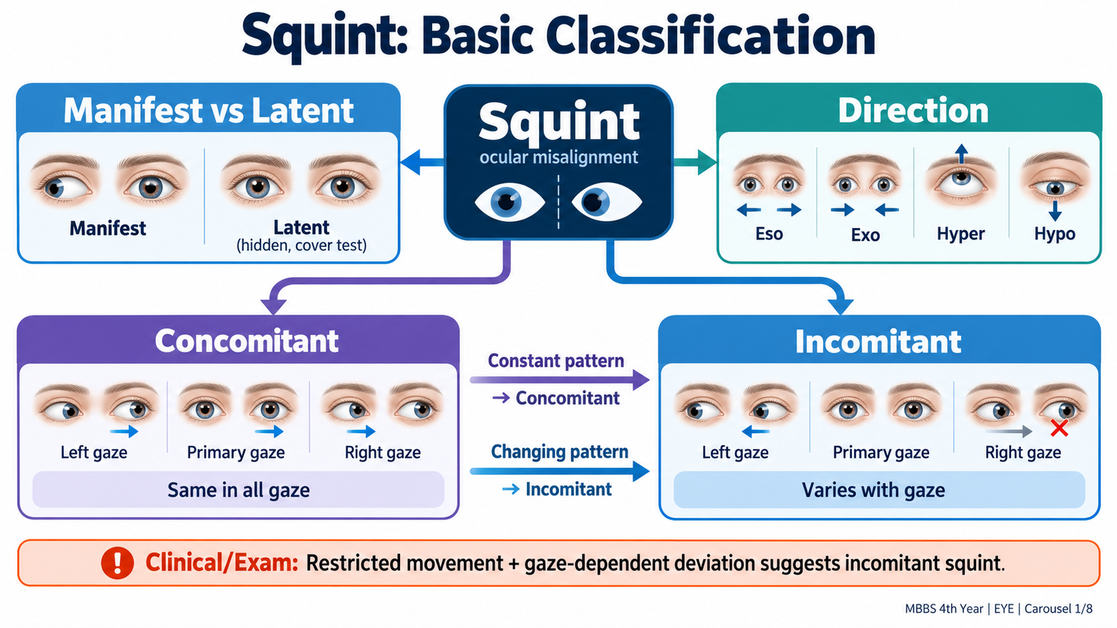
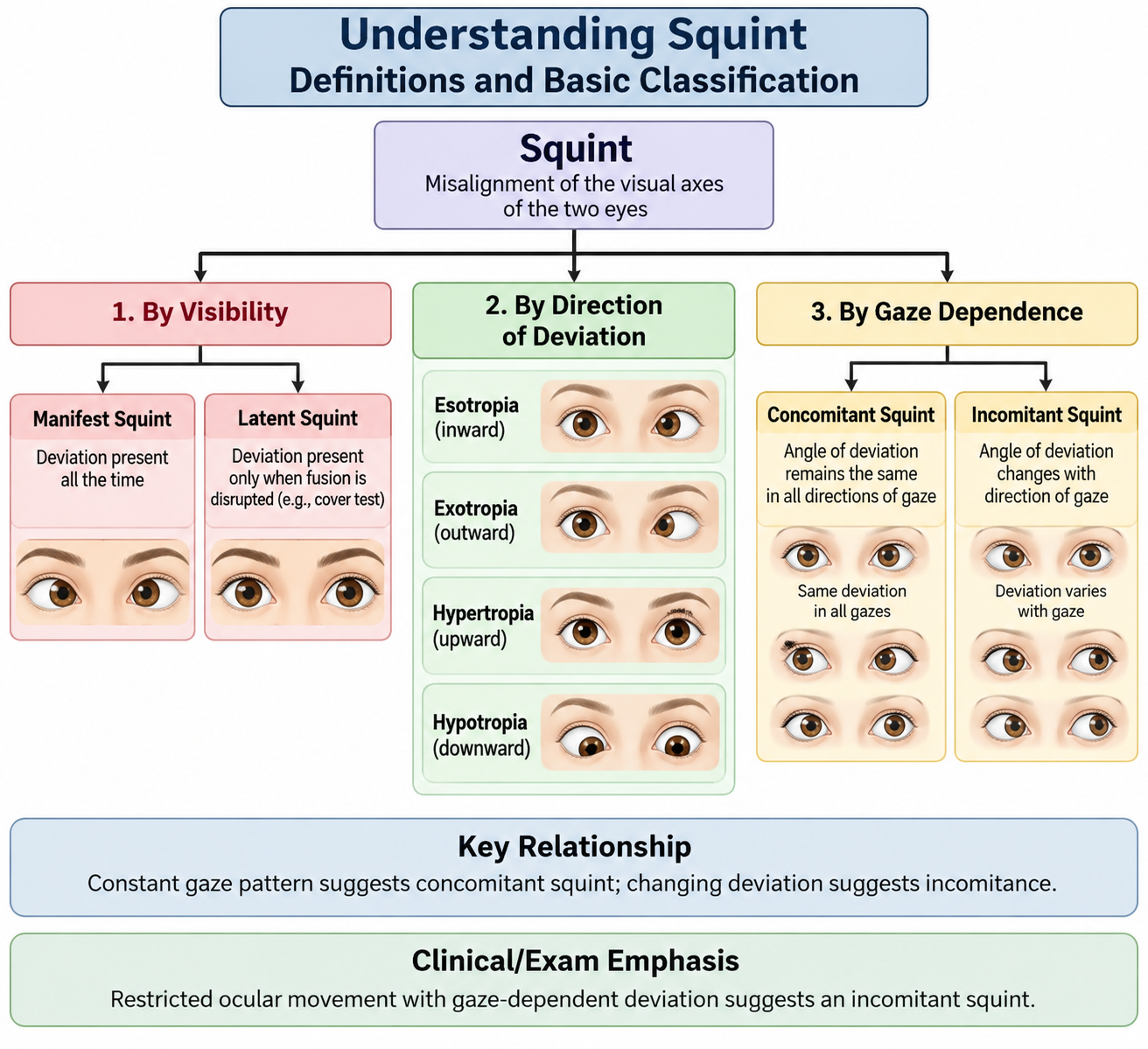
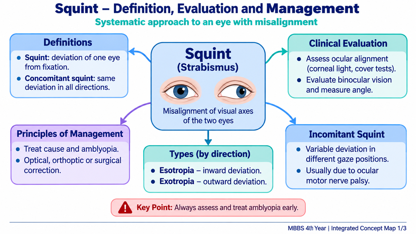
A squint, also called strabismus, is a condition in which the visual axes of the two eyes are not simultaneously directed toward the object of regard. One eye may be fixing the object while the other deviates. The deviation may be constantly present or appear only when normal binocular fusion is interrupted.
Manifest and Latent Deviation
A heterotropia is a manifest squint. The deviation is present while both eyes are open and attempting to view normally. It can therefore be demonstrated by a cover-uncover test.
A heterophoria is a latent tendency for the eyes to deviate that is normally controlled by binocular fusion. When fusion is interrupted, for example by covering one eye, the covered eye may drift from its normal position. The deviation becomes apparent particularly during alternate cover testing.
Classification According to Direction
- Esotropia: one eye deviates inward.
- Exotropia: one eye deviates outward.
- Hypertropia: one eye is vertically higher than the other.
- Hypotropia: one eye is vertically lower than the other.
- Cyclotropia: abnormal torsional rotation of an eye around its visual axis.
Concomitant Squint
A concomitant or comitant squint is one in which the angle of deviation remains approximately the same in different directions of gaze. Ocular movements are generally full because no individual extraocular muscle is significantly paralyzed. This pattern is particularly common in childhood squint.
Because childhood brains have considerable sensory adaptation, a child with a longstanding concomitant squint may not complain of diplopia. Instead, the brain may suppress the image from the deviating eye. Persistent suppression during visual development can interfere with binocular vision and can contribute to amblyopia.
Incomitant Squint
An incomitant or non-comitant squint is one in which the amount of deviation changes with the direction of gaze. It usually indicates an imbalance in ocular movement caused by an ocular motor nerve palsy, extraocular muscle disease or mechanical restriction. The deviation is greatest in the direction in which the weak or restricted muscle should normally act.
Acquired incomitant squint frequently causes binocular diplopia because the previously aligned visual system suddenly receives images at different retinal locations. The patient may adopt an abnormal head position to place the eyes in a direction of gaze where the separation of the images is smaller.
B. Clinical Evaluation of Squint and Principles of Management
Evaluation of squint has two main purposes: first, to define the type and size of ocular deviation; and second, to determine whether an underlying visual, muscular or neurological disorder is responsible. In a child, assessment must also determine whether visual development and binocular function have been affected.
History
The history should establish when the deviation began, whether it is constant or intermittent, which eye deviates, and whether the deviation has changed. An acute onset has a different significance from a stable childhood squint.
- Age at onset: very early onset favors childhood strabismus, whereas sudden adult onset raises concern for an acquired ocular motor disorder.
- Constant or intermittent: intermittent deviation indicates that fusional control is sometimes maintained.
- Diplopia: important in acquired squint; its direction and gaze dependence help localize the problem.
- Abnormal head posture: may represent an attempt to reduce diplopia or improve binocular alignment.
- Visual history: reduced vision in one eye may cause sensory strabismus.
- Previous spectacles: particularly important in esotropia associated with hypermetropia.
- Trauma or neurological symptoms: may suggest an acquired cranial nerve palsy.
- Systemic disease: vascular risk factors may be relevant in acquired ocular motor nerve palsies.
Visual Acuity and Refraction
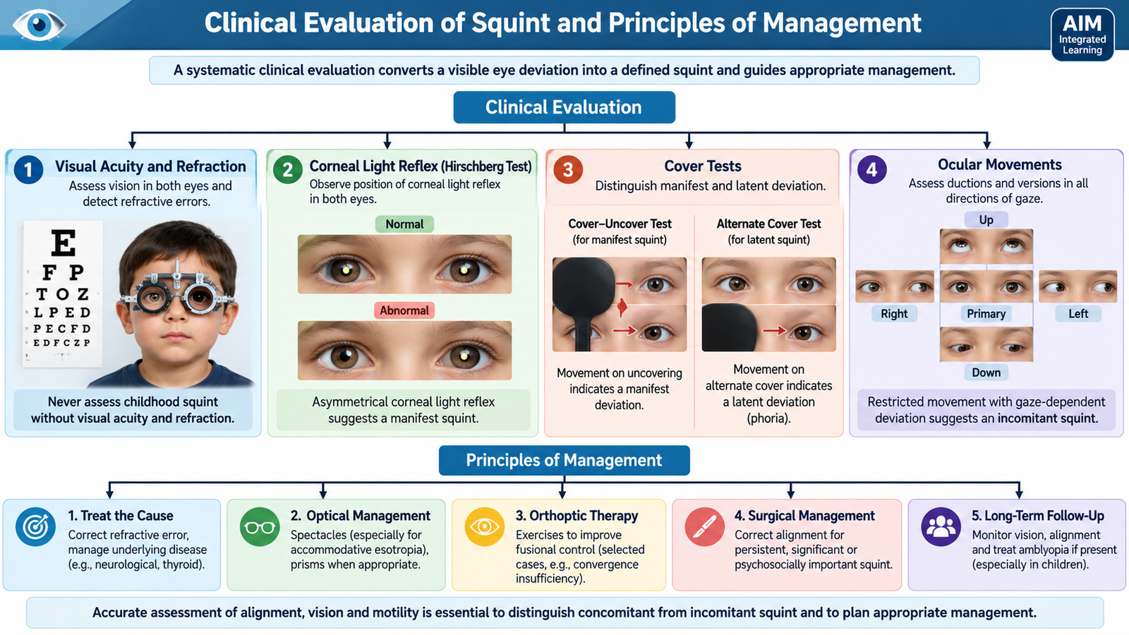
Visual acuity must be measured separately in each eye. Unequal visual acuity may indicate amblyopia or an ocular lesion causing sensory strabismus. In children, refractive assessment is particularly important because uncorrected refractive errors can contribute directly to squint.
Cycloplegic refraction temporarily relaxes accommodation and is especially useful in children. It allows hidden hypermetropia to be measured accurately. This is essential in suspected accommodative esotropia because correction of the hypermetropic refractive error may substantially reduce or eliminate the deviation.
Inspection and Corneal Light Reflex
Observe the resting position of the eyes, facial symmetry, ptosis and any compensatory head posture. The corneal light reflex test (Hirschberg test) gives a rapid estimate of ocular alignment. A light directed at both eyes normally produces symmetrical corneal reflections. Asymmetry of the reflections suggests a manifest deviation.
The test is particularly useful in young children who cannot cooperate with more formal measurements, but it is an estimate rather than a precise measurement of the angle of squint.
Cover Tests
The cover-uncover test detects a manifest deviation. While the patient fixes a target, one eye is covered and the uncovered eye is observed. If the uncovered eye makes a corrective movement to take up fixation, it had been deviated before the other eye was covered.
The alternate cover test repeatedly transfers the cover from one eye to the other. This interrupts binocular fusion and allows the full deviation, including any latent component, to appear.
A prism cover test uses prisms to neutralize the corrective movement and therefore measures the angle of deviation more accurately.
| Test | Main Purpose | Interpretation |
|---|---|---|
| Hirschberg test | Rapid assessment of alignment | Asymmetrical corneal reflex suggests manifest deviation |
| Cover-uncover test | Detect manifest squint | Corrective movement of uncovered eye identifies a tropia |
| Alternate cover test | Break fusion and reveal total deviation | Demonstrates manifest plus latent components |
| Prism cover test | Quantify deviation | Prism strength neutralizing movement estimates the angle |
Ocular Movements
Both ductions, which are movements of one eye, and versions, which are coordinated movements of both eyes, should be assessed. Full movements support a concomitant deviation. Limitation of a particular movement suggests muscle weakness, nerve palsy or mechanical restriction.
The examiner should note whether the angle of deviation changes in different gaze positions. This is essential when differentiating a childhood concomitant squint from an acquired paralytic or restrictive squint.
Other Important Examination Components
- Pupils: abnormal pupil size or reactivity may be especially important in third nerve palsy.
- Ptosis: may accompany third nerve dysfunction.
- Anterior segment and fundus: help identify ocular disease causing poor vision and sensory strabismus.
- Binocular function and stereopsis: assess how effectively the two eyes work together.
- Neurological examination: becomes important when the squint is acute, incomitant or associated with neurological symptoms.
Principles of Management
Management is directed at achieving the best possible vision in each eye, correcting important refractive errors, improving binocular alignment where possible and treating any underlying disease. The order of these steps matters: a child should not simply undergo alignment treatment while reduced visual acuity or an important refractive error remains unaddressed.
- Correct refractive error: particularly important in accommodative esotropia.
- Treat amblyopia when present: appropriate optical correction is essential; occlusion or pharmacological penalization of the better eye may be used under ophthalmic supervision.
- Treat the underlying cause: essential in sensory, neurological or restrictive squint.
- Prisms: may reduce troublesome binocular diplopia in selected patients.
- Orthoptic treatment: has a role in selected disorders of binocular control, rather than being a universal treatment for all squints.
- Strabismus surgery: may be required when a clinically important deviation remains despite appropriate optical and medical management.
C. Esotropia
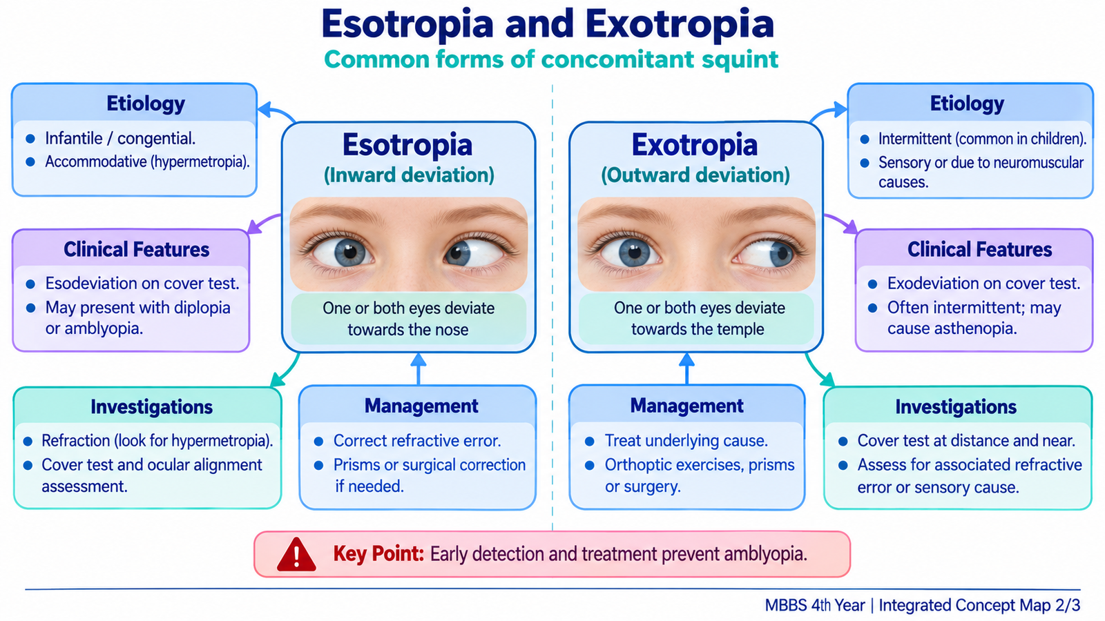
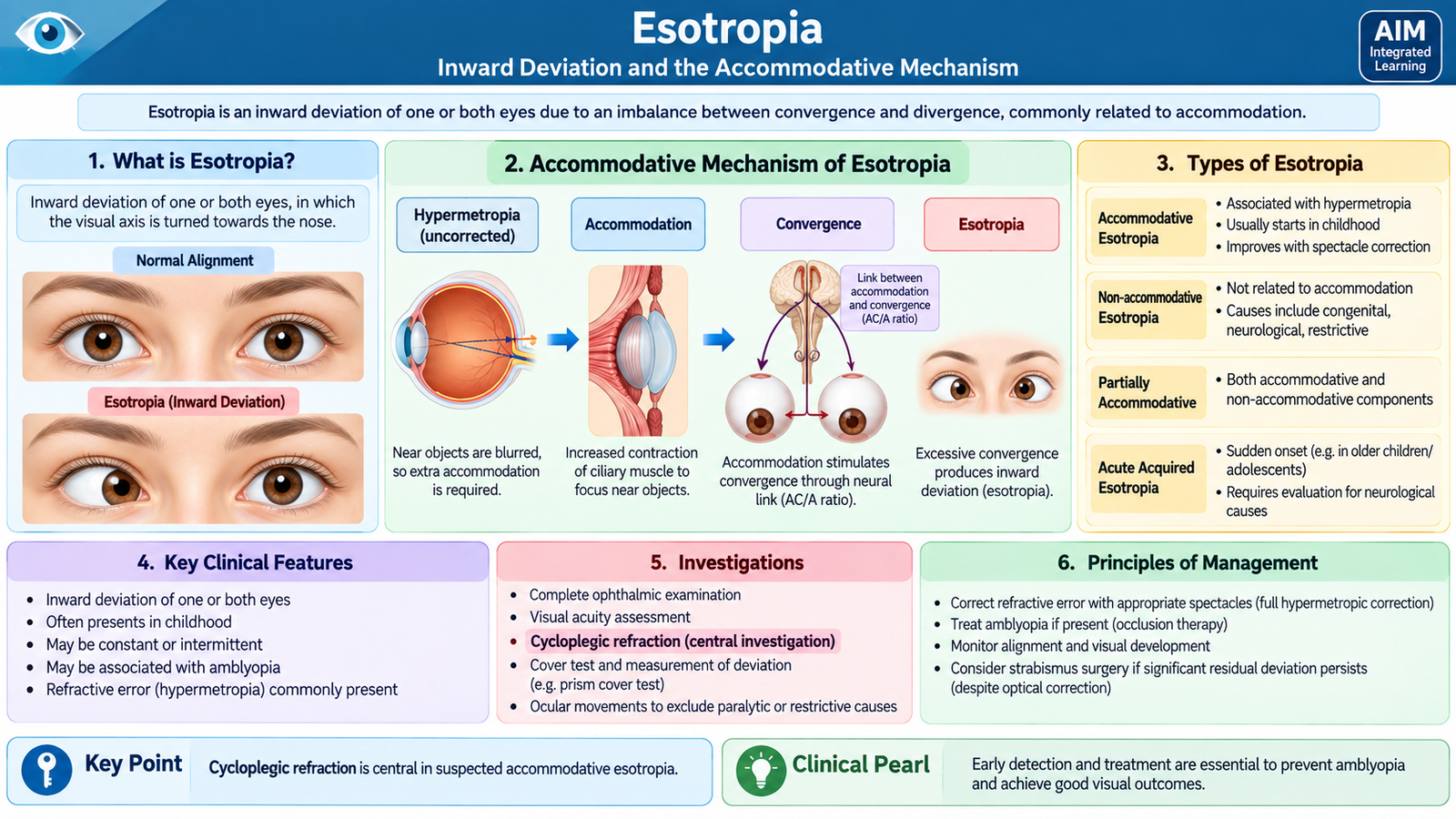
Esotropia is a manifest convergent squint in which one eye turns inward. It is an important childhood strabismus because persistent misalignment during visual development can interfere with binocular vision and may be associated with amblyopia. The cause is not the same in every child; therefore, correct classification and refractive assessment are central to management.
Etiology and Important Types
Esotropia can arise because normal binocular alignment fails during early childhood, because excessive accommodative convergence pulls the eyes inward, because one eye has poor vision, or as part of an acquired ocular motor disorder.
- Infantile esotropia: a large-angle esotropia beginning in early infancy. It is not primarily caused by accommodation.
- Accommodative esotropia: commonly associated with hypermetropia. Excessive accommodation required for clear vision is accompanied by increased accommodative convergence, producing inward deviation.
- Partially accommodative esotropia: refractive correction improves the deviation but a residual esotropia remains.
- Non-accommodative esotropia: the inward deviation is not explained adequately by accommodation.
- Sensory esotropia: occurs when impaired vision in one eye disrupts normal binocular fusion.
Hypermetropia → increased accommodation for clear vision → increased accommodative convergence → inward deviation of the eyes.
Clinical Features
The visible feature is an inward deviation of one eye or alternating inward deviation of both eyes. The deviation may be constant or intermittent depending on the type.
- Parents commonly notice that one eye turns inward.
- In accommodative esotropia, the deviation is related to accommodative effort and may improve with appropriate hypermetropic correction.
- Children often do not complain of diplopia because the visual system suppresses the image from the deviating eye.
- Persistent unilateral deviation can be associated with amblyopia.
- Binocular vision and stereopsis may be reduced, especially with early and constant misalignment.
Why is diplopia often absent? A young brain can suppress the unwanted image produced by the deviating eye. This protects the child from troublesome double vision, but prolonged suppression may impair normal binocular visual development.
Investigations and Ophthalmic Assessment
The investigations are aimed at establishing the angle and pattern of deviation, identifying refractive error, measuring vision in each eye and excluding ocular disease.
- Visual acuity: identifies reduced vision and possible amblyopia.
- Cycloplegic refraction: determines whether significant hypermetropia is driving accommodative convergence.
- Cover and prism cover tests: confirm and quantify the esotropia.
- Ocular motility: confirms whether movements are full and helps distinguish a concomitant esotropia from an abduction deficit such as sixth nerve palsy.
- Anterior segment and fundus examination: exclude ocular pathology responsible for sensory esotropia.
- Binocular vision assessment: evaluates fusion and stereopsis where cooperation permits.
Management
Treatment depends on the type of esotropia and should protect visual development before concentrating solely on cosmetic alignment.
Accommodative esotropia: appropriate correction of hypermetropia is the key treatment because reducing the accommodative requirement reduces the associated convergence. The child should be reassessed while wearing the correct prescription.
Amblyopia: if visual acuity is reduced in one eye because of amblyopia, it must be treated alongside refractive correction. This helps achieve useful vision in the deviating eye.
Residual or non-accommodative deviation: significant esotropia that remains despite appropriate optical treatment may require strabismus surgery to improve ocular alignment. The student should understand the indication rather than detailed operative technique.
Sensory esotropia: the underlying ocular cause of reduced vision should be identified and treated where possible. Alignment treatment can then be considered according to visual potential and the clinical situation.
D. Exotropia
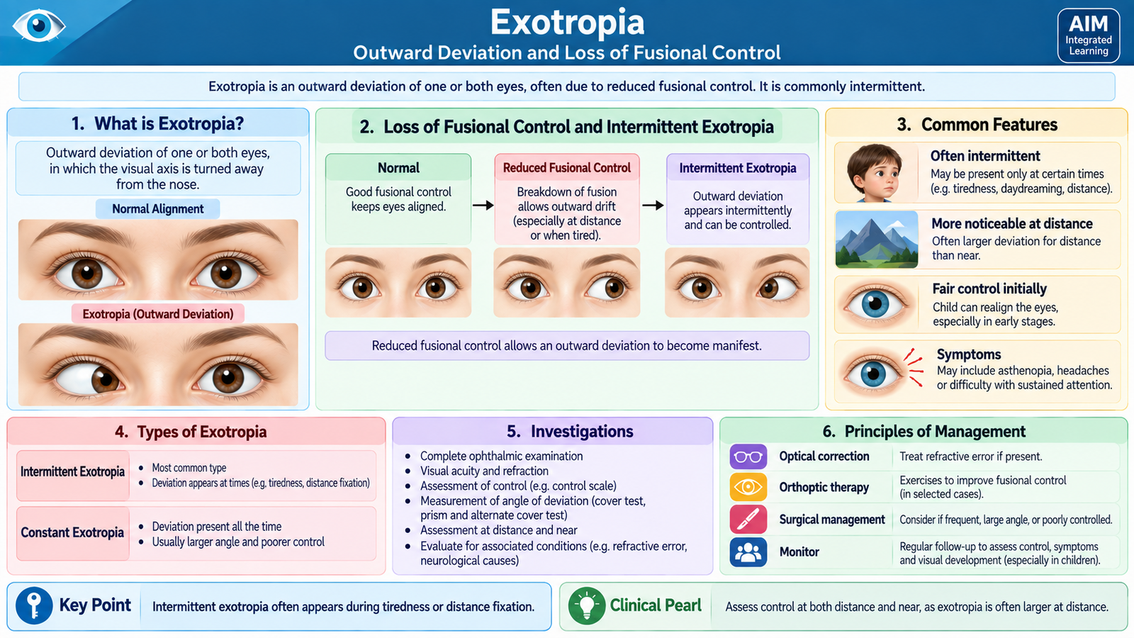
Exotropia is a manifest divergent squint in which one eye turns outward. The deviation may be intermittent, occurring when binocular control is reduced, or constant. Intermittent exotropia is an important clinical pattern because the eyes may appear well aligned at some times and clearly divergent at others.
Etiology and Important Types
Exotropia develops when the mechanisms maintaining convergent binocular alignment are unable to control an outward tendency of one eye. The cause and clinical significance vary with age and visual function.
- Intermittent exotropia: the eyes are aligned during periods of good fusional control but one eye drifts outward when control is lost.
- Constant exotropia: the outward deviation remains manifest.
- Sensory exotropia: may develop when poor vision in one eye prevents normal binocular fusion.
- Secondary exotropia: may occur following disruption of previously established ocular alignment.
Clinical Features
Intermittent exotropia is often more obvious when the patient is tired, inattentive or looking into the distance because fusional control is reduced under these circumstances.
- Intermittent or constant outward deviation of one eye.
- Parents may report that the deviation appears only at certain times.
- Some children close or cover one eye in bright light.
- Binocular function may be maintained when the eyes are aligned but becomes impaired during the manifest deviation.
- Longstanding constant deviation may be associated with reduced stereopsis.
- Amblyopia may occur, particularly when one eye consistently deviates or has poor vision, although it is less characteristic of alternating intermittent exotropia than of a persistent unilateral squint.
Investigations and Assessment
Assessment determines how frequently the deviation becomes manifest, how well the patient controls it, whether visual acuity is equal and whether the deviation is truly concomitant.
- Visual acuity: detects amblyopia or an ocular cause of sensory exotropia.
- Refraction: identifies refractive errors that should be appropriately corrected.
- Cover testing: demonstrates the outward deviation and determines whether it is manifest or latent.
- Prism cover testing: measures the size of the deviation.
- Distance and near assessment: establishes whether the deviation differs with fixation distance.
- Ocular motility: ensures there is no important limitation suggesting an incomitant disorder.
- Binocular function and stereopsis: help assess the functional impact and control of intermittent exotropia.
- Ocular examination: identifies a cause of visual loss when sensory exotropia is suspected.
Management
The need for treatment depends on the frequency and size of the deviation, the patient’s control, visual acuity and binocular function.
- Appropriate refractive correction: optimizes visual acuity and binocular control.
- Amblyopia treatment: is required if unequal vision and amblyopia are present.
- Observation: may be appropriate for selected patients with a small, infrequent and well-controlled deviation while binocular function is satisfactory.
- Orthoptic measures: may be useful in selected patients, particularly where convergence control is relevant.
- Surgery: may be considered when exotropia becomes frequent or constant, control deteriorates, binocular function is threatened or the deviation is clinically significant.
- Sensory exotropia: requires assessment and treatment of the underlying visual disorder where possible.
E. Diplopia: Causes, Clinical Evaluation and Cause-Directed Management
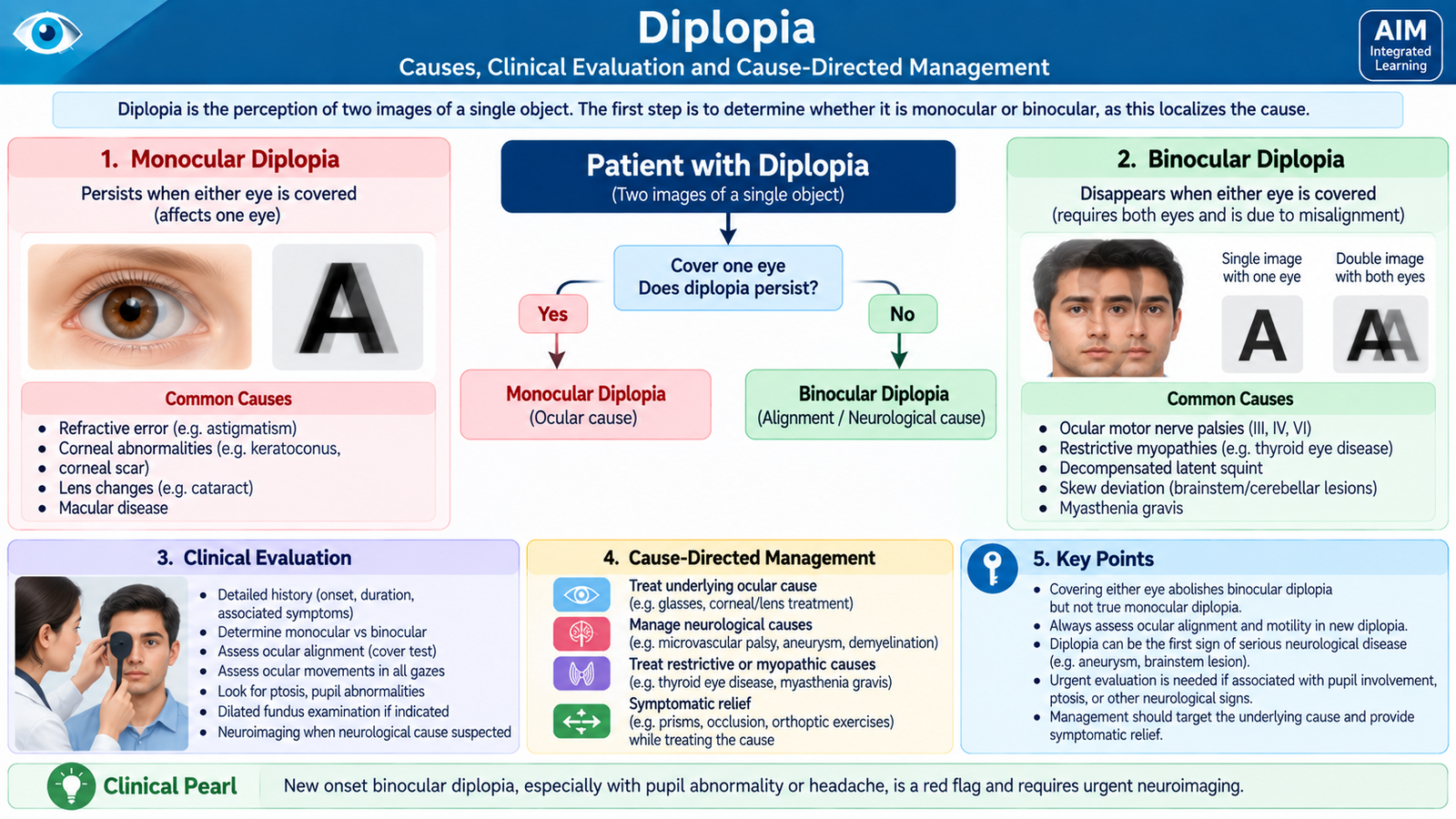
Diplopia means seeing two images of a single object. The most useful first distinction is between monocular diplopia and binocular diplopia. This simple clinical step immediately separates many optical or ocular causes from disorders of ocular alignment.
Monocular versus Binocular Diplopia
Monocular diplopia persists when the unaffected eye is covered and the symptomatic eye remains open. It usually results from an optical abnormality within that eye rather than misalignment between the two eyes.
Important ocular causes include:
- uncorrected or irregular refractive error;
- corneal surface or shape abnormalities;
- tear-film disturbance;
- lens opacity such as cataract;
- abnormal lens position or other optical distortion.
Binocular diplopia disappears when either eye is covered. It occurs because the eyes are misaligned, so the same object forms images on non-corresponding retinal points.
Important Causes of Binocular Diplopia
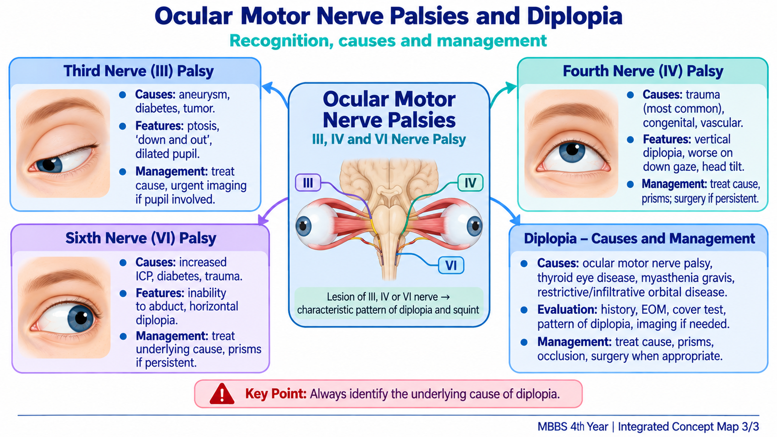
- Third, fourth or sixth cranial nerve palsy causing weakness of specific extraocular muscles.
- Decompensated heterophoria or acquired strabismus when fusion can no longer control a pre-existing tendency to deviate.
- Neuromuscular junction disease, especially when ocular weakness is variable.
- Restrictive orbital disease, in which an extraocular muscle cannot move normally because of mechanical restriction.
- Orbital trauma with muscle entrapment or damage.
- Brainstem, cavernous sinus or orbital disease affecting ocular motor pathways.
How the Pattern of Diplopia Helps Localization
The separation between the two images becomes greatest when the patient looks in the direction in which the weak muscle should normally act. This is why examination in different gaze positions is much more informative than simply asking whether diplopia is present.
- Horizontal diplopia: strongly suggests a disorder affecting horizontal alignment, especially sixth nerve palsy in the appropriate setting.
- Vertical or torsional diplopia: is characteristic of disorders such as fourth nerve palsy.
- Diplopia with marked ptosis and an abnormal “down and out” eye: suggests third nerve palsy.
- Variable diplopia that changes during the day: raises the possibility of a disorder of neuromuscular transmission rather than a fixed single-nerve palsy.
- Restricted movement with orbital signs: suggests mechanical or orbital disease.
Clinical Evaluation
Start by determining whether diplopia is monocular or binocular. For binocular diplopia, define the direction of image separation, the gaze in which it becomes greatest, the onset and associated features.
Important questions include whether onset was sudden, whether pain or headache is present, whether trauma occurred, and whether there are other neurological symptoms. Examination should assess visual acuity, pupils, eyelid position, ocular alignment, ductions and versions, and the presence of proptosis or other orbital findings. The fundus and optic discs should be examined when intracranial pressure or neurological disease is a concern.
Investigations
There is no single investigation for all diplopia. Testing is selected according to the localization suggested by the history and ocular examination.
- Refraction and slit-lamp/ocular examination: used when monocular diplopia suggests an optical or ocular cause.
- Formal ocular alignment and motility assessment: identifies the pattern of muscle weakness and helps localize binocular diplopia.
- Fundus examination: is especially important when sixth nerve palsy could be related to raised intracranial pressure because papilledema may provide an important clue.
- Neuroimaging: is indicated when an intracranial, orbital or compressive lesion is suspected from the clinical presentation.
- Vascular imaging: is urgently relevant when an acute painful pupil-involving third nerve palsy raises concern for an intracranial aneurysmal compression.
- Targeted systemic investigations: are selected when vascular, metabolic, neuromuscular or other systemic disease is clinically suspected.
Management According to the Underlying Cause
Diplopia is a symptom, not a final diagnosis. Management therefore depends on identifying and treating its cause while reducing the patient’s visual disturbance when necessary.
| Underlying Problem | Management Principle |
|---|---|
| Refractive or optical cause | Correct the refractive error or treat the responsible corneal, tear-film or lens disorder. |
| Ocular motor nerve palsy | Identify and treat the cause; temporary occlusion or selected prisms can relieve diplopia while recovery is assessed. |
| Decompensated binocular deviation | Correct optical problems; consider prism, appropriate orthoptic management or alignment surgery according to the disorder. |
| Restrictive orbital disorder | Treat the underlying orbital disease; mechanical restriction cannot be managed as simple nerve weakness. |
| Neuromuscular disorder | Confirm and treat the systemic disorder responsible for variable ocular misalignment. |
F. Third Cranial Nerve Palsy
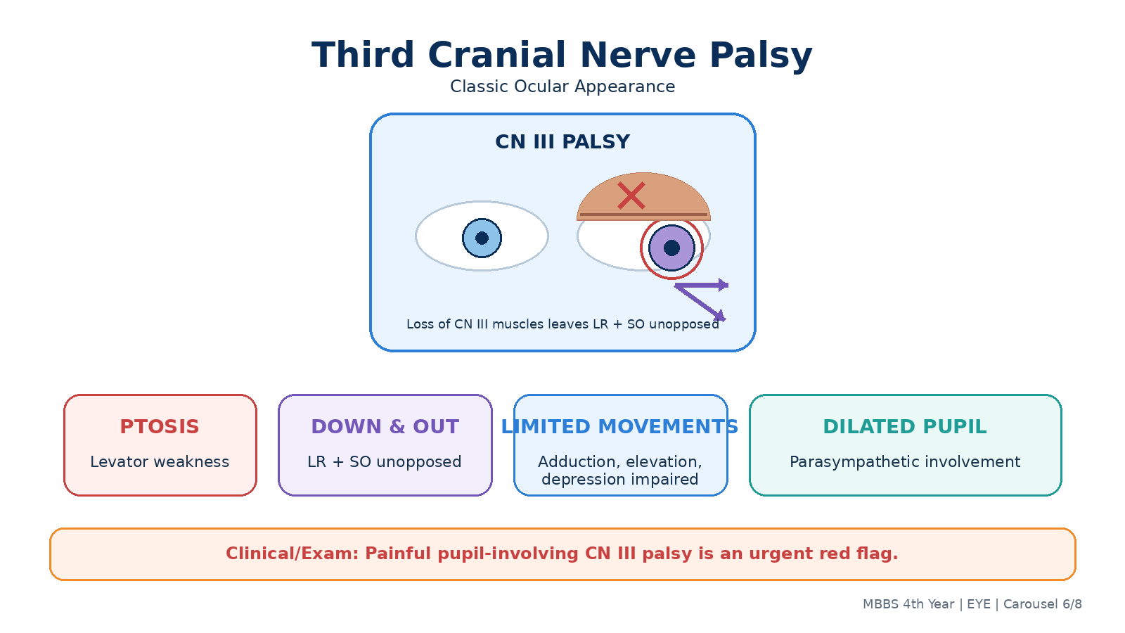
The oculomotor nerve (cranial nerve III) supplies most of the extraocular muscles, the levator muscle of the upper eyelid and parasympathetic fibers controlling pupillary constriction. A complete third nerve palsy therefore produces a distinctive combination of ocular deviation, ptosis and sometimes pupillary dilatation.
Applied Anatomy and Mechanism
Cranial nerve III supplies the medial rectus, superior rectus, inferior rectus and inferior oblique, as well as the levator palpebrae superioris. Its parasympathetic fibers travel to the pupil constrictor pathway.
If these muscles are paralyzed, the actions of the intact lateral rectus and superior oblique are relatively unopposed. The eye therefore rests in a characteristic “down and out” position.
Etiology
- Microvascular ischemia: may occur in patients with vascular risk factors such as diabetes mellitus and hypertension.
- Compressive lesions: an intracranial aneurysm, particularly involving the posterior communicating artery region, is an important cause because compression can affect superficial pupillary fibers.
- Trauma.
- Midbrain lesions.
- Cavernous sinus or orbital lesions, particularly when other cranial nerves or orbital structures are also involved.
Clinical Features
- Ptosis: caused by weakness of levator palpebrae superioris.
- Down-and-out eye: results from relatively unopposed lateral rectus and superior oblique action.
- Diplopia: occurs because the visual axes are no longer aligned.
- Impaired adduction: due to medial rectus weakness.
- Impaired elevation: because superior rectus and inferior oblique are affected.
- Impaired depression: due principally to inferior rectus involvement, although some depression by superior oblique remains.
- Dilated poorly reactive pupil: may occur when parasympathetic pupillary fibers are involved.
Why Pupil Examination Matters
The pupillary fibers lie superficially within the peripheral part of the third nerve and may be affected by external compression. Therefore, an acute painful third nerve palsy with a dilated or poorly reactive pupil is a major warning sign for a compressive lesion and requires urgent investigation.
Microvascular ischemic palsies are often pupil-sparing because ischemic injury may predominantly affect the central somatic fibers while the superficial pupillary fibers remain relatively preserved. However, pupil sparing does not remove the need for proper clinical evaluation of a new third nerve palsy.
Investigations
The investigation strategy is determined by onset, pupil involvement, pain, age, vascular risk and associated neurological signs.
- Ocular motility examination: confirms the characteristic limitation of third-nerve-controlled movements.
- Pupil assessment: helps identify a potentially compressive pattern.
- Neurological examination: looks for associated deficits that may localize the lesion beyond the isolated nerve.
- Neuroimaging: is used when intracranial or orbital pathology is suspected.
- Urgent vascular imaging: is required when clinical features raise concern for aneurysmal compression, particularly an acute painful pupil-involving palsy.
- Assessment of systemic vascular factors: is appropriate when microvascular ischemia is suspected.
Management
The first priority is to identify and treat the underlying cause. A new third nerve palsy should not simply be treated as a strabismus without considering neurological disease.
- Suspected compressive third nerve palsy: requires urgent neurological/ophthalmological assessment and appropriate vascular imaging.
- Microvascular palsy: systemic vascular risk factors should be assessed and managed while ocular recovery is followed.
- Temporary diplopia relief: occlusion of one eye or a selected prism may be useful depending on the pattern.
- Persistent stable deviation: may later require strabismus management or surgery after the neurological condition and potential for spontaneous recovery have been appropriately assessed.
G. Fourth Cranial Nerve Palsy
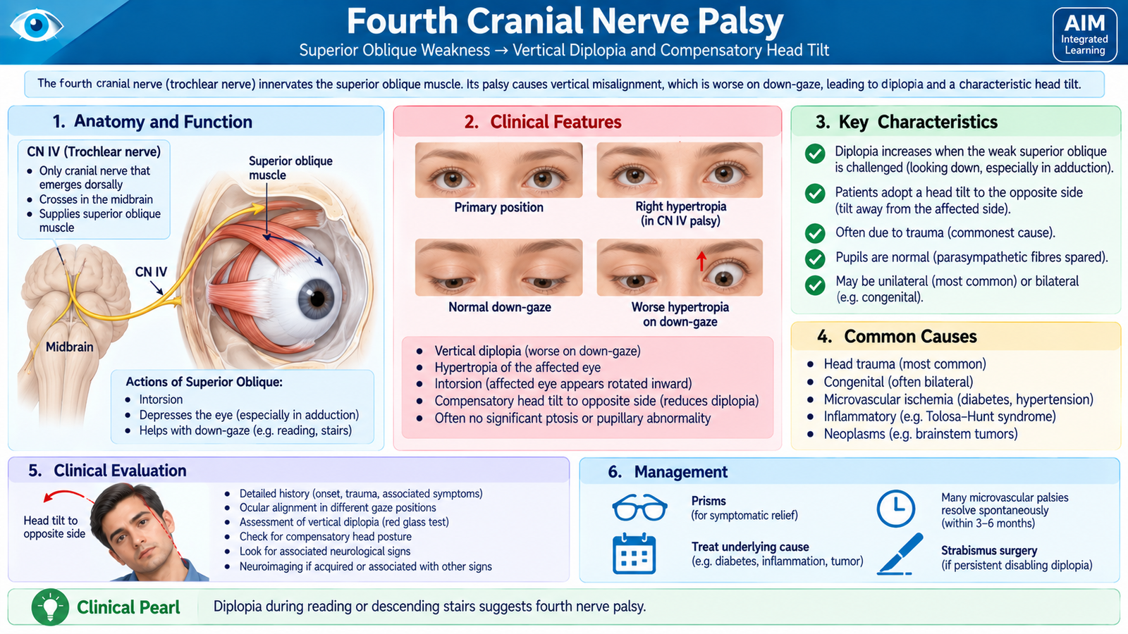
The trochlear nerve (cranial nerve IV) supplies the superior oblique muscle. Its palsy commonly produces vertical and torsional diplopia, particularly during activities requiring depression of an adducted eye, such as reading or walking downstairs.
Applied Function of the Superior Oblique
The superior oblique contributes to intorsion and also depresses the eye particularly when the eye is adducted. Therefore, weakness becomes most obvious when the patient attempts to look downward with the affected eye turned inward.
Etiology
- Congenital fourth nerve palsy: may remain compensated for years before becoming clinically apparent.
- Head trauma: is an important acquired cause because of the long intracranial course of the trochlear nerve.
- Microvascular ischemia: may occur in adults with vascular risk factors.
- Intracranial lesions: should be considered when the palsy is acquired, atypical or accompanied by other neurological findings.
Clinical Features
- Vertical diplopia: the images are displaced mainly vertically.
- Torsional component: images may appear tilted because the superior oblique normally contributes to intorsion.
- Worse on looking down: reading and descending stairs may be particularly troublesome.
- Hypertropia of the affected eye: becomes more apparent in gaze positions that require superior oblique action.
- Compensatory head tilt: the patient often tilts the head away from the affected side to reduce vertical/torsional separation of the images.
Parks-Bielschowsky Principle
In a unilateral superior oblique palsy, the hypertropia becomes greater in characteristic positions, including head tilt toward the affected side. The head-tilt response is useful because tilting the head normally activates muscles responsible for ocular torsional compensation. A weak superior oblique cannot provide the required response, increasing vertical misalignment.
The pattern should be interpreted together with ocular motility rather than used as an isolated diagnostic sign.
Investigations
- Cover testing in different gaze positions: demonstrates the incomitance and identifies where hypertropia increases.
- Ocular motility examination: demonstrates deficient superior oblique function.
- Head-tilt testing: helps support localization to the superior oblique/trochlear pathway.
- Neurological assessment and imaging: are considered in acquired or atypical cases or when other neurological features are present.
Management
Management depends on the cause and whether spontaneous recovery is expected. The underlying cause is treated where possible. Temporary symptomatic treatment may include occlusion or a suitable prism for troublesome diplopia. A persistent, stable and clinically significant deviation may require strabismus surgery after appropriate assessment.
H. Sixth Cranial Nerve Palsy
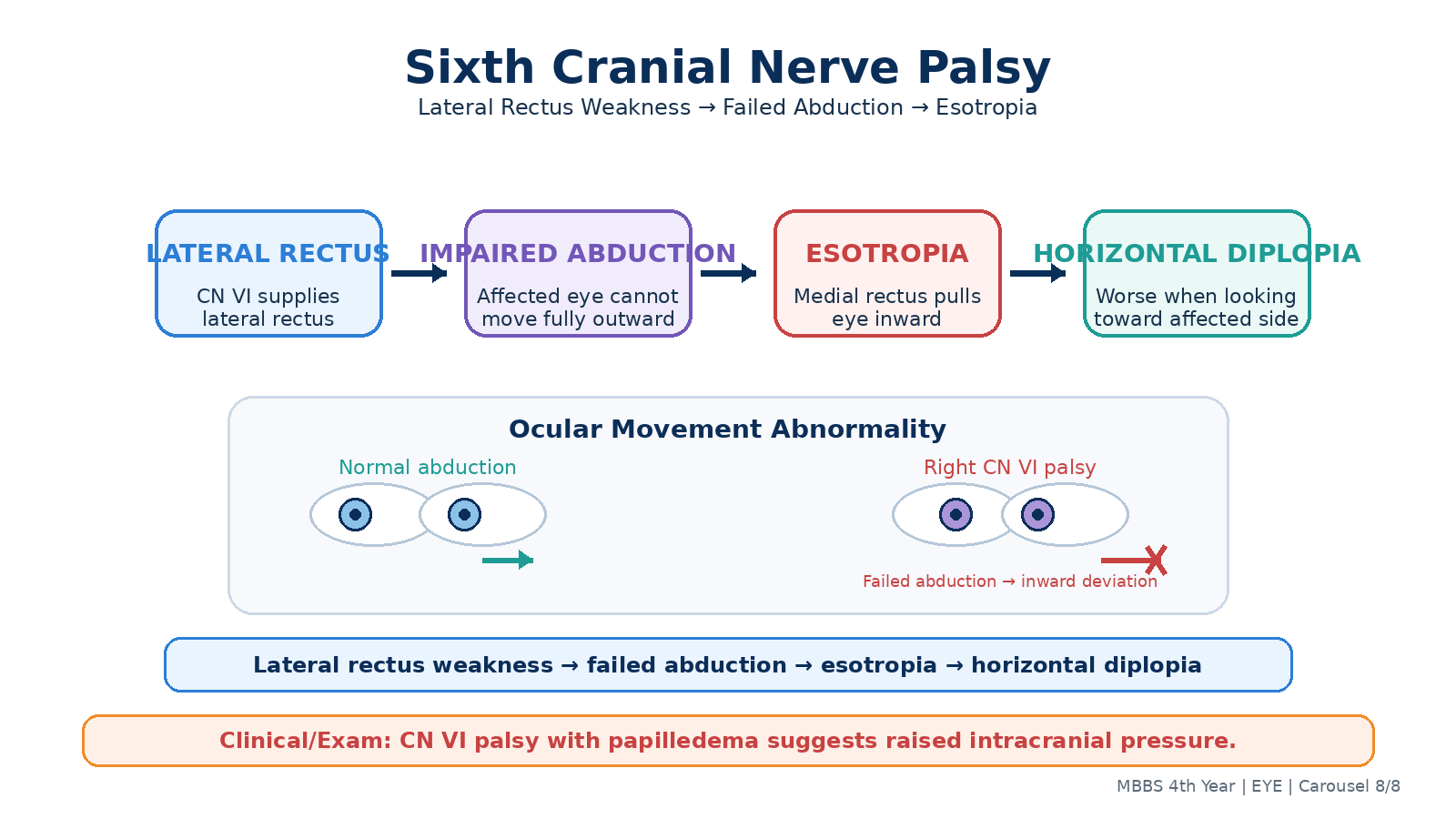
The abducens nerve (cranial nerve VI) supplies the lateral rectus muscle. The lateral rectus abducts the eye. A sixth nerve palsy therefore causes impaired abduction, producing an incomitant esotropia and characteristic horizontal diplopia.
Mechanism
When the lateral rectus is weak, medial rectus action is relatively unopposed and the affected eye turns inward. The misalignment becomes greatest when the patient attempts to look toward the affected side because this is the direction in which the weak lateral rectus is required most strongly.
Etiology
The abducens nerve has a long intracranial course and can be affected by disease at several anatomical levels.
- Microvascular ischemia: particularly in adults with vascular risk factors.
- Raised intracranial pressure: may stretch or distort the sixth nerve along its intracranial course. For this reason, sixth nerve palsy can act as a false-localizing sign.
- Head trauma.
- Brainstem lesions.
- Cavernous sinus disease.
- Orbital lesions affecting the nerve or lateral rectus.
- Intracranial disease in children or younger patients: requires careful evaluation because an acquired sixth nerve palsy should not automatically be assumed to be benign.
Clinical Features
- Esotropia: the affected eye turns inward because the medial rectus is relatively unopposed.
- Impaired abduction: the affected eye cannot move normally toward the temporal side.
- Horizontal diplopia: images are separated horizontally.
- Diplopia worse at distance: divergence is more important for distant fixation.
- Diplopia worse on gaze toward the affected side: because lateral rectus action is required in that direction.
- Compensatory face turn toward the affected side: moves the required gaze away from the weak muscle’s field of action and can reduce diplopia.
Investigations
The presence of an abduction deficit establishes the ocular motor pattern, but further evaluation is needed to determine why the sixth nerve is affected.
- Ocular motility examination: confirms limitation of abduction and demonstrates incomitance.
- Cover/prism cover assessment: documents the esotropia and its variation with gaze.
- Fundus and optic disc examination: looks for papilledema when raised intracranial pressure is a possible cause.
- Neurological examination: looks for brainstem or other cranial nerve abnormalities.
- Neuroimaging: is required when the clinical context suggests an intracranial, cavernous sinus or orbital cause, or when the presentation is atypical.
- Systemic assessment: is appropriate when a microvascular cause is suspected.
Management
The cause determines management. A patient with signs of raised intracranial pressure, other neurological abnormalities or concerning intracranial features requires urgent assessment of the underlying disorder.
- Treat the underlying neurological, vascular, traumatic or orbital cause.
- Address modifiable vascular risk factors when microvascular ischemia is suspected.
- Temporary occlusion may relieve troublesome diplopia.
- Prisms can help selected patients when image separation is suitable for prism correction.
- Persistent stable deviation may eventually require strabismus surgery after the underlying condition and potential for spontaneous recovery have been assessed.
Integrated Mechanism Flow
Squint, nerve palsy or restriction
Images fall on non-corresponding retinal points
Child: suppression • Adult: diplopia
Cover test + ocular movements + pupils
Concomitant, paralytic, restrictive or optical
Protect vision, relieve symptoms, treat cause
Important Comparison
Concomitant versus Incomitant Squint
| Feature | Concomitant Squint | Incomitant Squint |
|---|---|---|
| Deviation in different gazes | Approximately similar | Varies with direction of gaze |
| Ocular movements | Usually full | May be limited |
| Typical setting | Common in childhood | Nerve palsy, muscle disease or restriction |
| Diplopia | Often absent in longstanding childhood squint because of suppression | Common when acquired |
| Abnormal head posture | Less characteristic | Common compensation for diplopia |
Third, Fourth and Sixth Cranial Nerve Palsies
| Feature | CN III Palsy | CN IV Palsy | CN VI Palsy |
|---|---|---|---|
| Main muscle pattern | Most extraocular muscles affected | Superior oblique weak | Lateral rectus weak |
| Characteristic eye position/finding | Down and out; often ptosis | Hypertropia in characteristic gaze positions | Esotropia with impaired abduction |
| Diplopia pattern | Complex separation | Vertical/torsional; worse down gaze | Horizontal; worse toward affected side |
| Head posture | Variable | Tilt away from affected side | Face turn toward affected side |
| Key associated clue | Pupil involvement may indicate compression | Difficulty reading or descending stairs | Consider raised intracranial pressure |