
Topic 6 — Ocular Trauma, Globe Injuries & Visual Rehabilitation
1. Topic Introduction
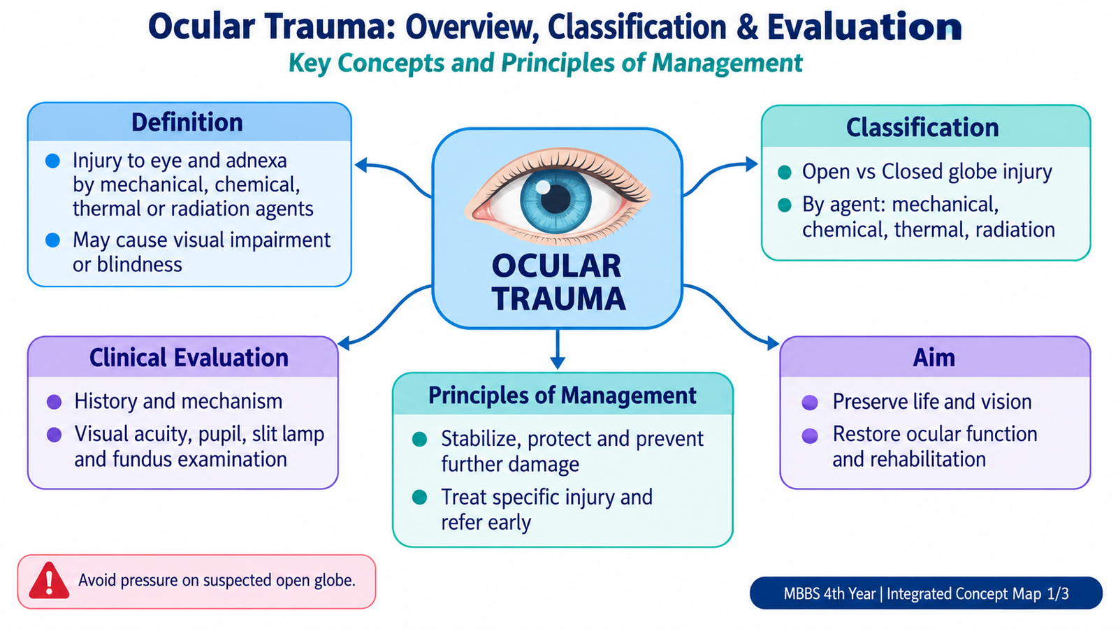
Ocular trauma means injury to the eye or its surrounding structures caused by mechanical force, foreign material, chemicals, heat or radiation. Some injuries affect only the conjunctiva or superficial cornea, while others disrupt the full thickness of the globe and threaten permanent visual loss. The most important skill is to recognize which injuries are minor and which require immediate protection of the eye and urgent ophthalmic management. In this chapter you will learn how ocular trauma is classified and evaluated, how open and closed globe injuries differ, how foreign bodies and ocular burns are managed, why sympathetic ophthalmia may follow penetrating injury, and how visual and blindness rehabilitation support patients when normal vision cannot be completely restored.
2. Core Learning Material
A. Foundations, Classification, Clinical Evaluation & Initial Management of Ocular Injury
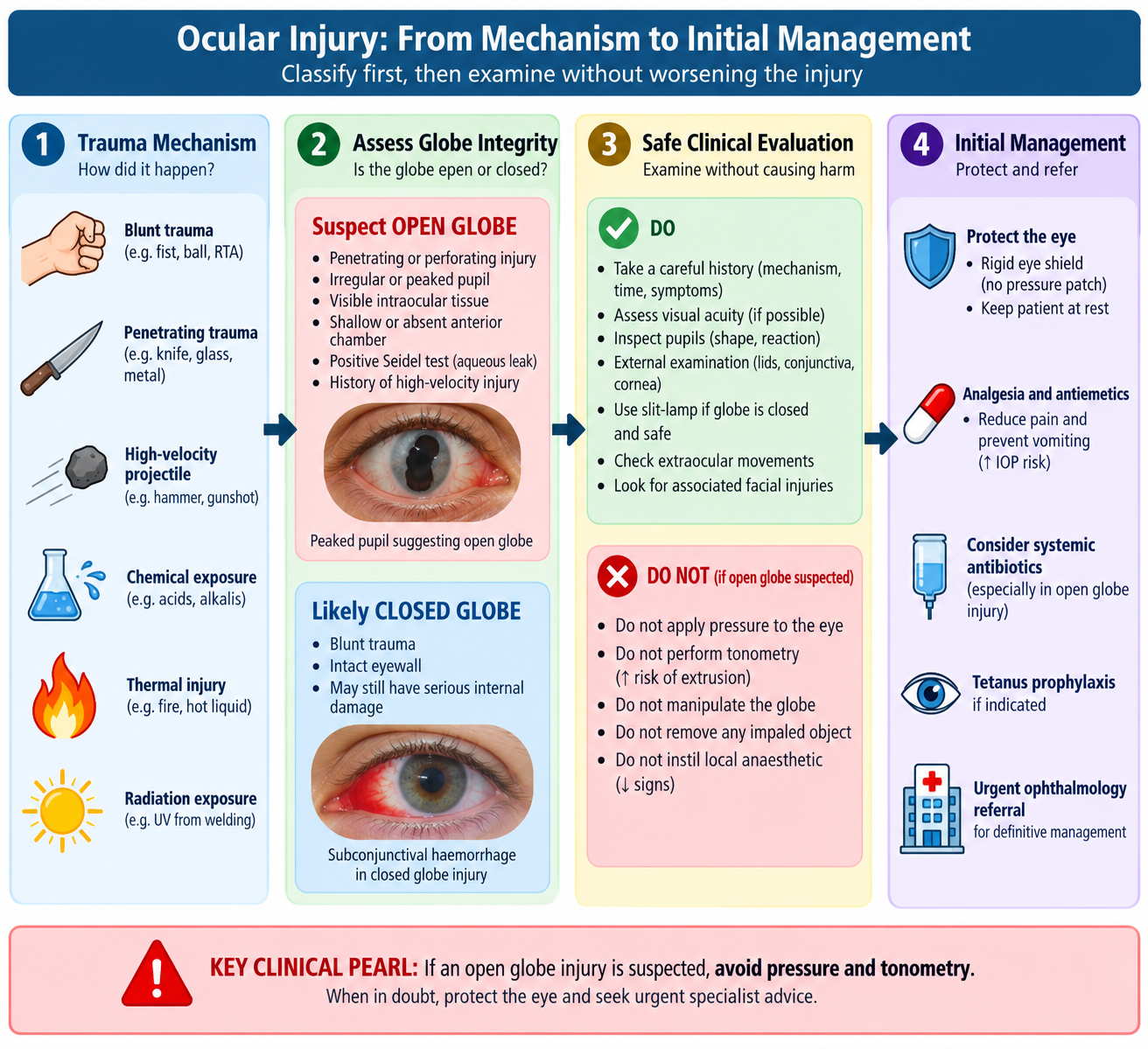
Ocular injury should first be approached by asking a simple question: has the wall of the globe remained intact? The eyewall consists of the cornea and sclera. A full-thickness wound of either structure creates an open globe injury; if no full-thickness eyewall wound exists, the globe injury is closed. This distinction is critical because manipulation or pressure on an open globe can worsen prolapse of intraocular tissues and further damage vision.
Classification
Ocular injuries may be considered according to the mechanism and the tissue involved:
- Mechanical injury: blunt or sharp trauma affecting the globe.
- Open globe injury: full-thickness wound of the cornea or sclera.
- Closed globe injury: trauma without a full-thickness eyewall wound.
- Superficial foreign-body injury: material lodged in or on the conjunctiva or cornea without entering the globe.
- Chemical injury: exposure to acids, alkalis or other damaging chemicals.
- Thermal injury: damage caused by heat, hot liquids, flame or molten material.
- Radiation injury: damage from ultraviolet, infrared or ionizing radiation.
Clinical evaluation
Assessment begins with the mechanism of injury. A small external wound does not always mean a minor injury. High-velocity hammering, grinding or metal-on-metal work can produce a tiny entry wound while leaving an intraocular foreign body deep inside the eye.
- Time and exact mechanism
- Blunt versus sharp trauma
- High-velocity projectile
- Material involved
- Chemical or heat exposure
- Contact-lens use where relevant
- Previous ocular disease or surgery
- Change in vision after injury
- Pain or foreign-body sensation
- Reduced visual acuity
- Photophobia
- Watering or bleeding
- Diplopia
- Flashes, floaters or field loss
Visual acuity should be checked in each eye as early as safely possible because it provides an important baseline and helps estimate the severity of injury. The examiner then inspects the eyelids, conjunctiva and cornea, observes the anterior chamber and pupil, and assesses ocular movements where appropriate. Pupillary examination is important because a relative afferent pupillary defect (RAPD) may suggest severe retinal or optic-nerve dysfunction. Slit-lamp examination and fluorescein staining help identify corneal epithelial defects and some leaking wounds. The anterior chamber is examined for blood, altered depth or inflammation. Fundus examination is performed when it is safe and the ocular media permit visualization.
Principles of initial management
Management depends on the mechanism, but the first aim is always to prevent additional ocular damage while definitive assessment is arranged.
- Protect a suspected open globe with a rigid eye shield rather than a pressure patch.
- Do not remove an object protruding from the globe.
- Provide appropriate analgesia and control vomiting because sudden rises in pressure may worsen an open injury.
- Arrange urgent ophthalmology assessment for suspected penetrating injury, severe visual loss or other sight-threatening findings.
- Systemic antimicrobial prophylaxis and tetanus-related measures are considered in open, penetrating or contaminated wounds according to the clinical situation.
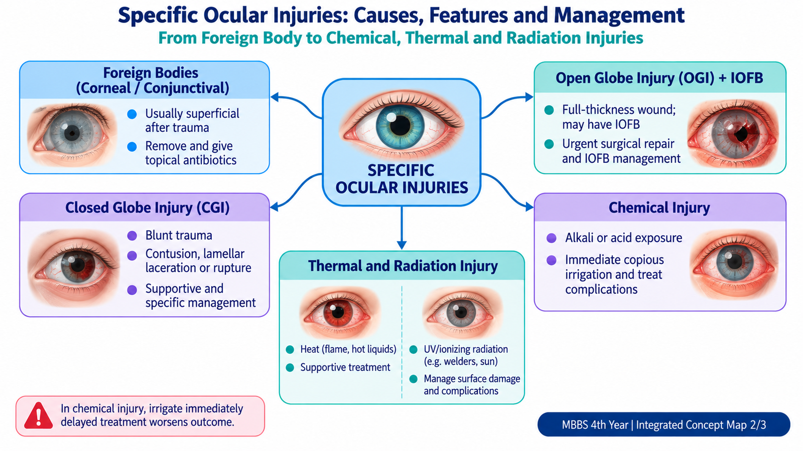
- Chemical injury is an important exception to the usual examination sequence: immediate irrigation takes priority over detailed history and examination.
B. Corneal & Conjunctival Foreign Bodies
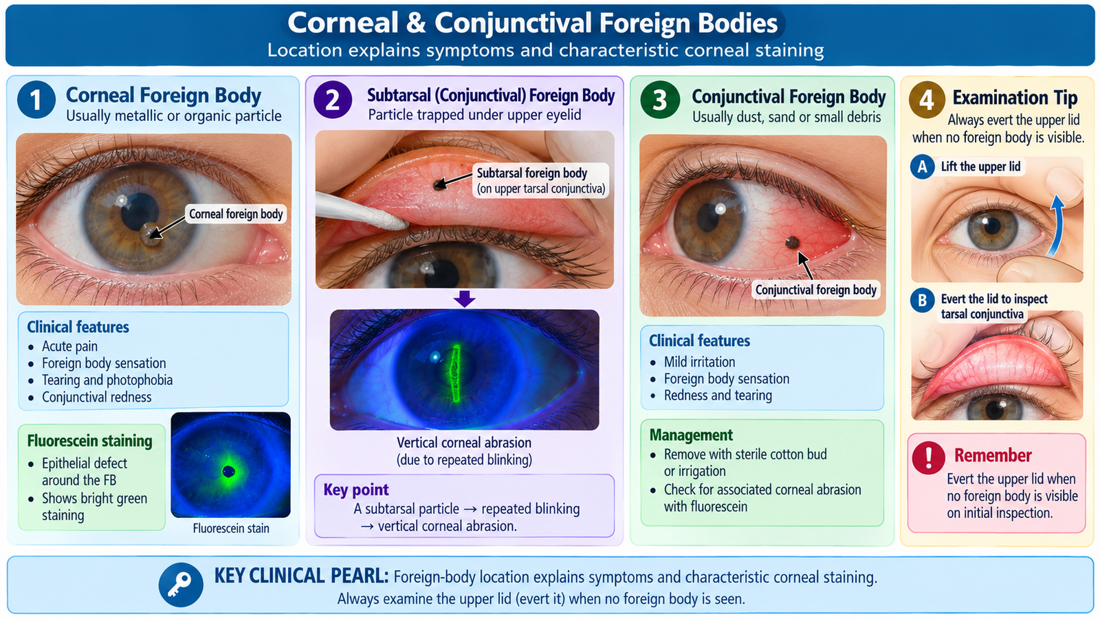
A superficial ocular foreign body is material lying on or embedded in the conjunctival or corneal surface without entering the globe. Dust, sand, metal particles, vegetable material and workplace debris are common causes. Corneal foreign bodies are usually more painful than conjunctival foreign bodies because the cornea is richly supplied with sensory nerves.
Clinical features
- Sudden foreign-body sensation
- Pain, often worse with blinking
- Watering
- Photophobia
- Redness
- Blepharospasm
- Blurred vision when the central cornea is involved
A foreign body trapped beneath the upper eyelid repeatedly scratches the cornea during blinking. This can produce vertical linear corneal abrasions. Therefore, when symptoms strongly suggest a foreign body but none is visible on the exposed ocular surface, the upper eyelid should be everted carefully.
Examination and investigations
Visual acuity is documented first. The conjunctiva and cornea are examined with good illumination or slit lamp. Fluorescein staining outlines an epithelial defect because the dye enters areas where corneal epithelium has been lost. The depth and position of the foreign body are important: a deep, central or high-velocity foreign body raises concern for penetration. A metallic corneal foreign body may leave a rust ring. Organic or soil-contaminated material requires particular caution because it increases the possibility of microbial keratitis.
Treatment
Loose superficial particles may be removed by gentle irrigation. A superficial embedded foreign body may be removed under magnification by a trained clinician after topical anesthesia. Residual corneal epithelial injury is then treated as an abrasion, commonly with lubrication and appropriate topical antimicrobial cover according to clinical circumstances.
- Do not attempt blind or forceful removal of a deeply embedded object.
- Do not remove material when globe penetration is suspected.
- Topical anesthetic may assist examination and removal but is not routinely given for repeated home use because misuse can delay healing and damage the cornea.
- Persistent pain, reduced vision, central corneal involvement, a retained rust ring, suspected infection or possible penetration requires ophthalmic review.
C. Open Globe Injury & Intraocular Foreign Body
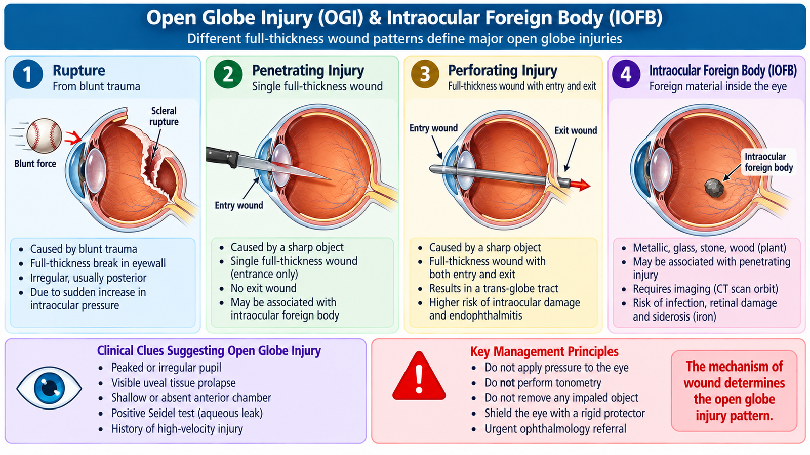
An open globe injury (OGI) is a full-thickness wound of the cornea, sclera or both. It is an ophthalmic emergency because the protective wall of the eye has been breached and intraocular structures may be damaged, contaminated or lost. Open globe injuries are broadly divided into rupture and laceration.
Classification of open globe injury
1. Rupture
A rupture results from blunt trauma. The globe is suddenly compressed, intraocular pressure rises sharply, and the eyewall breaks at its weakest point. The wound may therefore occur away from the exact site of impact. 2. Laceration A laceration is caused by a sharp object or projectile cutting from outside toward the interior of the globe. It includes:
- Penetrating injury: one full-thickness entry wound without an exit wound.
- Perforating injury: separate entry and exit wounds produced by the same object.
- Intraocular foreign body (IOFB): a penetrating injury in which foreign material remains inside the eye.
Etiology
Common mechanisms include sharp objects, broken glass, occupational tools, road or domestic trauma and high-velocity metal fragments. Hammering metal against metal or grinding is particularly important because a small fast-moving fragment may penetrate the globe with little external evidence.
Clinical features
The appearance depends on the wound site and structures damaged. Severe visual loss may result from direct injury to the cornea, lens, vitreous, retina or optic nerve, or from hemorrhage and contamination.
- Reduced visual acuity
- Ocular pain and redness
- Visible corneal or scleral wound
- Marked subconjunctival hemorrhage, particularly when extensive
- Shallow, irregular or abnormally deep anterior chamber
- Hyphema
- Irregular or peaked pupil, sometimes pointing toward the wound
- Prolapse of iris, vitreous or other intraocular tissue
- Lens disruption or traumatic cataract
- Vitreous hemorrhage
- Positive aqueous leakage through a corneal wound when safely demonstrable
A peaked pupil may occur because iris tissue is pulled toward or trapped within a wound. This makes the pupil an important visual clue to occult penetration.
Clues to an intraocular foreign body
An IOFB should be suspected after high-velocity injury even if the entry wound is tiny. Possible findings include a corneal or scleral entry wound, iris defect, focal lens opacity, vitreous hemorrhage or retinal injury.
Investigations
CT of the orbits without contrast is an important investigation when an IOFB or occult open globe is suspected. It helps identify many radiopaque foreign bodies and provides information about their location and associated orbital injury. MRI must be avoided when a metallic IOFB is possible because a magnetic field may move ferromagnetic material and cause further ocular damage. Ocular ultrasonography can demonstrate posterior-segment pathology when the media are opaque, but it is avoided or used only with specialist caution when an unrepaired open globe is suspected because pressure from the probe may worsen the injury.
Management
- Place a rigid protective shield over the injured eye.
- Avoid pressure patches and ocular manipulation.
- Do not remove a protruding object.
- Do not perform tonometry.
- Provide analgesia and antiemetic treatment as required.
- Keep the patient prepared for possible operative management.
- Provide systemic antimicrobial prophylaxis in penetrating/open injuries according to specialist management.
- Address tetanus protection when clinically indicated.
- Definitive treatment usually requires primary closure of the eyewall, with management or removal of an IOFB and associated intraocular damage according to its location and nature.
Important complications include endophthalmitis, traumatic cataract, retinal detachment, proliferative scarring, secondary glaucoma and severe permanent visual loss. Penetrating trauma is also an important setting for development of sympathetic ophthalmia.
D. Closed Globe Injury
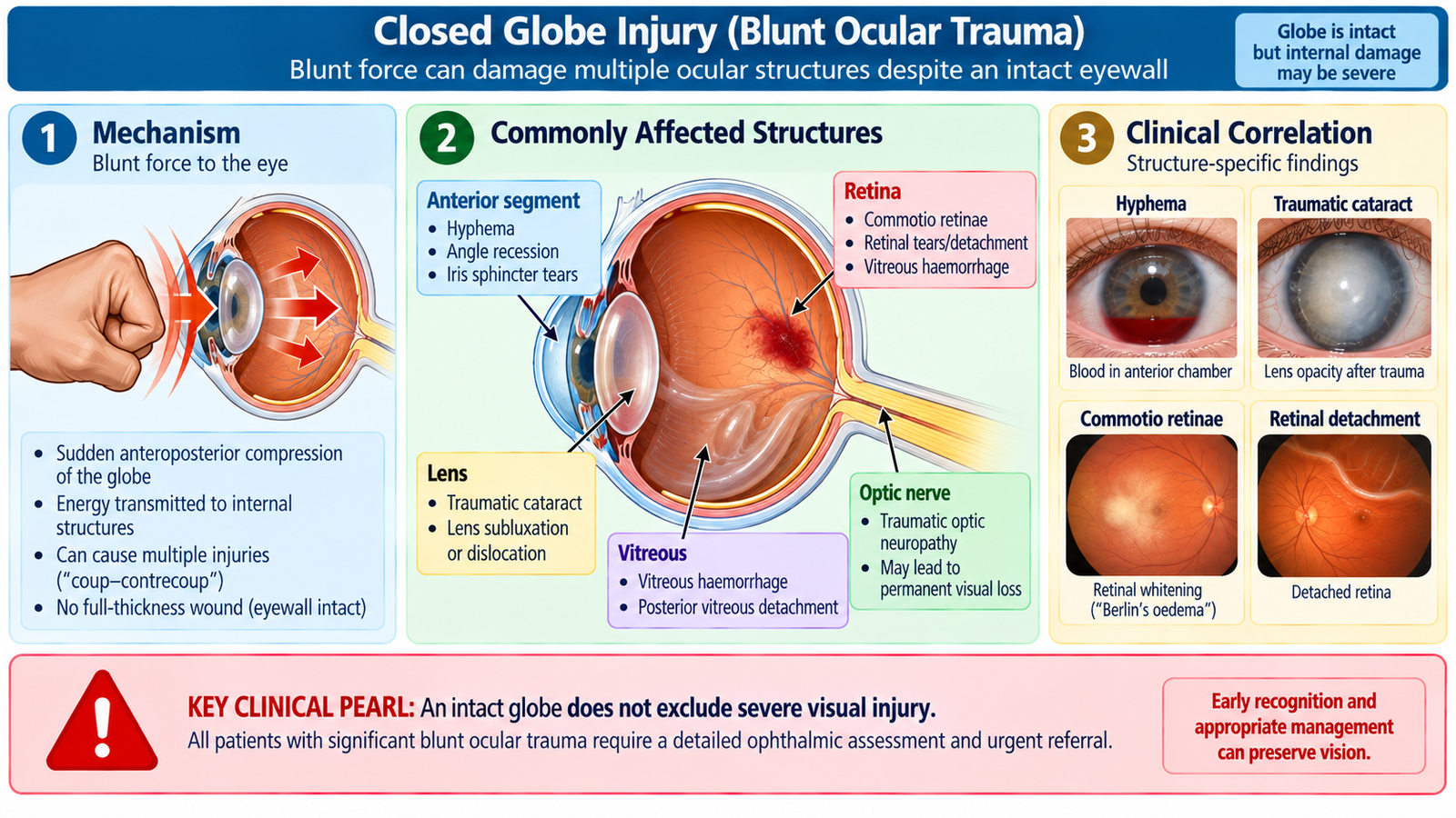
A closed globe injury (CGI) means that trauma has occurred without a full-thickness wound of the cornea or sclera. Although the eyewall remains intact, blunt impact can transmit considerable force through the eye and damage the anterior chamber, lens, vitreous, retina, choroid or optic nerve. Therefore, an intact globe does not necessarily mean a minor injury.
Classification
- Contusion: blunt trauma produces internal ocular damage without a full-thickness wound.
- Lamellar laceration: a partial-thickness wound of the cornea or sclera that does not pass completely through the eyewall.
How blunt trauma damages the eye
When the eye is struck from the front it is briefly compressed in the front-to-back direction and expands at the equator. This rapid deformation and transmission of energy can tear delicate intraocular structures even though the external wall remains closed.
Clinical features
The findings depend on which structure receives the transmitted force:
- Cornea: abrasion or edema may cause pain, photophobia and blurred vision.
- Anterior chamber: bleeding from damaged iris or ciliary-body vessels produces hyphema.
- Iris: sphincter tears may cause traumatic mydriasis; separation at the iris root causes iridodialysis.
- Lens: zonular disruption may produce lens subluxation or dislocation; lens injury may later cause traumatic cataract.
- Vitreous: hemorrhage may obscure the fundus and produce floaters or marked visual reduction.
- Retina: commotio retinae produces transient retinal whitening after blunt trauma; tears may subsequently lead to retinal detachment.
- Choroid: choroidal rupture may cause permanent central visual impairment when the macular region is affected.
- Optic nerve: traumatic optic neuropathy may produce severe visual loss and an RAPD despite limited visible anterior-segment injury.
Investigations
After an open globe has been excluded, examination may include visual acuity, slit-lamp assessment, pupillary testing, intraocular pressure measurement and dilated fundus examination. Gonioscopy may later identify angle recession, which is important because damaged angle structures can lead to delayed secondary glaucoma. Posterior-segment imaging such as OCT or ocular ultrasonography is used when clinically indicated. CT is useful when associated orbital fracture or significant deeper trauma is suspected.
Management
There is no single treatment for every closed globe injury. Management is directed toward the damaged structure and prevention of complications.
- Corneal abrasions receive appropriate surface treatment.
- Hyphema requires ophthalmic assessment, protection from further trauma and monitoring for pressure-related complications.
- Lens damage may eventually require surgical management if it significantly affects vision.
- Retinal tears or detachment require prompt retinal assessment and treatment.
- Patients with angle injury may require long-term monitoring for secondary glaucoma.
- Marked visual loss, RAPD, severe pain, retinal symptoms or significant hyphema are important reasons for urgent specialist assessment.
E. Sympathetic Ophthalmia
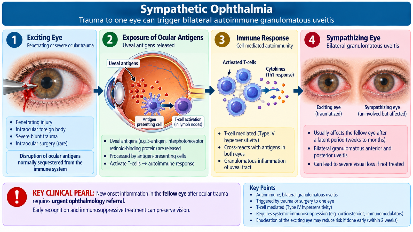
Sympathetic ophthalmia is a rare bilateral granulomatous uveitis that may follow penetrating ocular trauma or other disruption of the internal ocular tissues. The originally injured eye is traditionally called the exciting eye, while inflammation occurring in the fellow eye involves the sympathizing eye. The clinical importance is that trauma to one eye can therefore threaten useful vision in the other eye.
Etiology and pathogenesis
Ocular trauma can expose normally sequestered ocular antigens to the immune system. An autoimmune cellular response may then develop against melanocyte-associated and other ocular antigens, producing inflammation in the uveal tissues of both eyes.
Clinical features
The interval after injury is variable. The patient may develop symptoms in the injured eye and, importantly, new symptoms in the previously unaffected fellow eye.
- Blurred or reduced vision
- Photophobia
- Ocular pain and redness
- Floaters
- Bilateral granulomatous anterior uveitis
- Vitritis
- Choroidal inflammation
- Multiple yellow-white lesions at the level of the choroid or retinal pigment epithelium
- Exudative retinal detachment in more severe disease
Investigations
The diagnosis is primarily clinical and is supported by the history of ocular trauma together with bilateral intraocular inflammation. Slit-lamp and dilated fundus examination determine the extent of anterior and posterior inflammation. OCT can demonstrate subretinal fluid and inflammatory retinal or choroidal changes. Additional retinal or choroidal imaging may be used by ophthalmologists when required.
Management
Sympathetic ophthalmia requires urgent specialist treatment because uncontrolled inflammation can cause permanent bilateral visual loss. Systemic corticosteroids are used to suppress the inflammatory response. Patients with severe, persistent or recurrent disease may require additional immunosuppressive therapy under specialist supervision. Modern management aims to preserve a potentially useful traumatized eye whenever possible. Removal of an irreversibly damaged blind eye is an individualized specialist decision rather than a routine treatment for every penetrating injury.
F. Chemical, Thermal & Radiation Injuries
Non-mechanical ocular injuries may damage the eyelids, conjunctiva, cornea and deeper ocular structures without a penetrating wound. Their severity depends on the damaging agent, concentration or energy, duration of exposure and how deeply the injury penetrates. Chemical burns deserve special priority because immediate irrigation can directly change the visual outcome.
Chemical injury
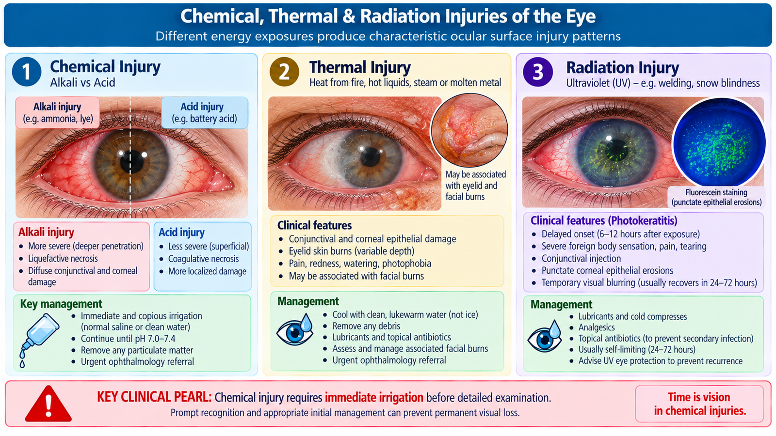
Chemical burns commonly result from household, industrial or laboratory exposure. They are divided broadly into alkali and acid injuries. Alkalis are particularly dangerous because they penetrate ocular tissues rapidly. They damage cell membranes and stromal tissue and may continue penetrating after the initial exposure. Acids tend to produce coagulation of surface proteins, which can limit deeper penetration in many acid injuries.
Clinical features
- Severe pain, although very severe alkali injury may reduce corneal sensation
- Redness and watering
- Photophobia
- Blepharospasm
- Corneal epithelial defect
- Corneal haze or opacity in deeper injury
- Conjunctival damage
- Limbal ischemia, seen as blanching of the normally vascular limbal region
- Reduced vision according to severity
Limbal ischemia is important because the limbus contains stem cells needed for renewal of the corneal epithelium. Severe limbal damage therefore increases the risk of poor epithelial healing and later corneal surface failure.
Assessment and investigations
Initial management must not be delayed for a complete examination. Irrigation is started immediately with available clean water or an appropriate irrigating fluid. After adequate irrigation, ocular-surface pH is reassessed and the eyelids are everted to remove retained particulate material. Visual acuity, fluorescein staining, corneal clarity, epithelial loss, limbal perfusion and anterior-segment findings are then evaluated.
Management
- Immediate, prolonged and generous irrigation is the most important first step.
- Do not delay irrigation while trying to identify the exact chemical.
- Remove particles trapped in the fornices after lid eversion.
- Do not attempt chemical neutralization with another reactive chemical.
- After irrigation, medical management may include lubrication, topical antimicrobial protection, cycloplegia and specialist-directed anti-inflammatory treatment.
- Severe burns require urgent ophthalmic care because corneal ulceration, melting, scarring, glaucoma and permanent visual loss may develop.
Thermal injury
Thermal injuries occur after exposure to flame, hot liquids, steam, hot metal or other intense heat. The blink reflex and rapid eye closure often protect the globe, so eyelid and superficial ocular-surface burns are common. However, severe heat or molten particles can damage the conjunctiva and cornea.
Clinical features and assessment
- Eyelid erythema, edema or burns
- Conjunctival redness
- Corneal epithelial defects
- Pain, watering and photophobia
- Reduced vision when the cornea or deeper structures are involved
- Retained hot particles or foreign material in some injuries
Examination determines the depth of eyelid injury and whether the cornea or conjunctiva has been damaged. Fluorescein is useful for identifying corneal epithelial loss.
Management
The heat source is removed and the ocular surface is cooled and irrigated when required. Debris is removed carefully, corneal injury is assessed and superficial ocular-surface injury is treated with appropriate lubrication and other ophthalmic therapy. Deep corneal, extensive conjunctival or severe eyelid burns require urgent specialist care.
Radiation injury
Different forms of radiation affect different ocular tissues. The common acute undergraduate example is ultraviolet photokeratitis, which may occur after unprotected welding-arc exposure or intense ultraviolet reflection. Ultraviolet radiation injures corneal epithelial cells, but symptoms may begin only after a delay because epithelial injury develops after the exposure.
Clinical features of ultraviolet photokeratitis
- Usually bilateral severe pain
- Foreign-body sensation
- Photophobia
- Watering
- Blepharospasm
- Diffuse superficial punctate corneal epithelial staining with fluorescein
Other radiation forms may cause deeper or delayed injury. Repeated or intense infrared exposure may affect ocular structures including the lens, while ionizing radiation can produce delayed lens or retinal damage depending on the exposure.
Investigations and management
Assessment includes visual acuity, slit-lamp examination and fluorescein staining for suspected photokeratitis. Management of uncomplicated ultraviolet epithelial injury is supportive, including protection from further exposure, lubrication and appropriate analgesia. Persistent visual reduction, unusual unilateral findings or evidence of deeper damage requires ophthalmic evaluation. Prevention depends on suitable protective eyewear during welding, ultraviolet exposure and radiation-related work.
G. Visual Rehabilitation After Ocular Trauma
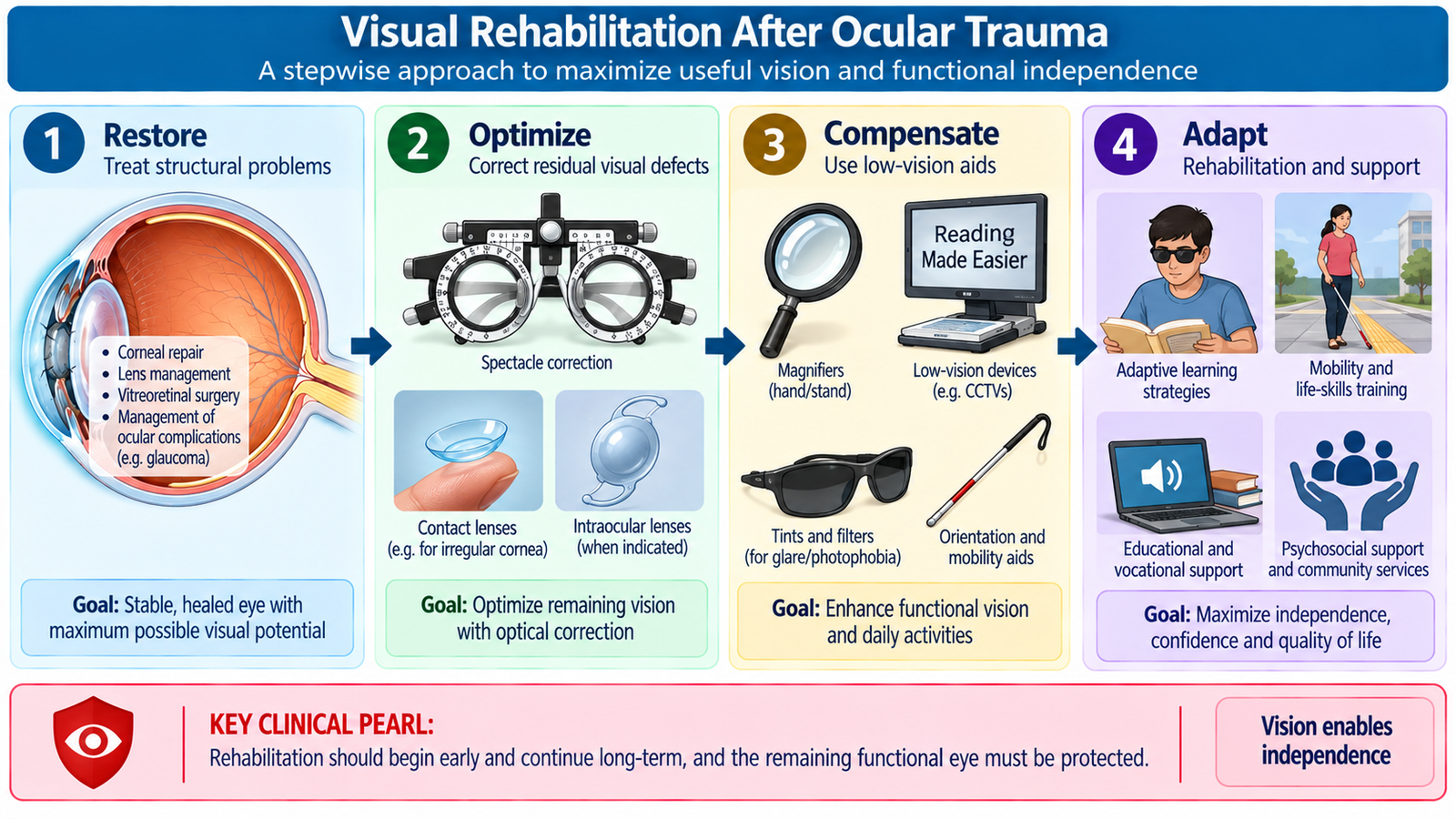
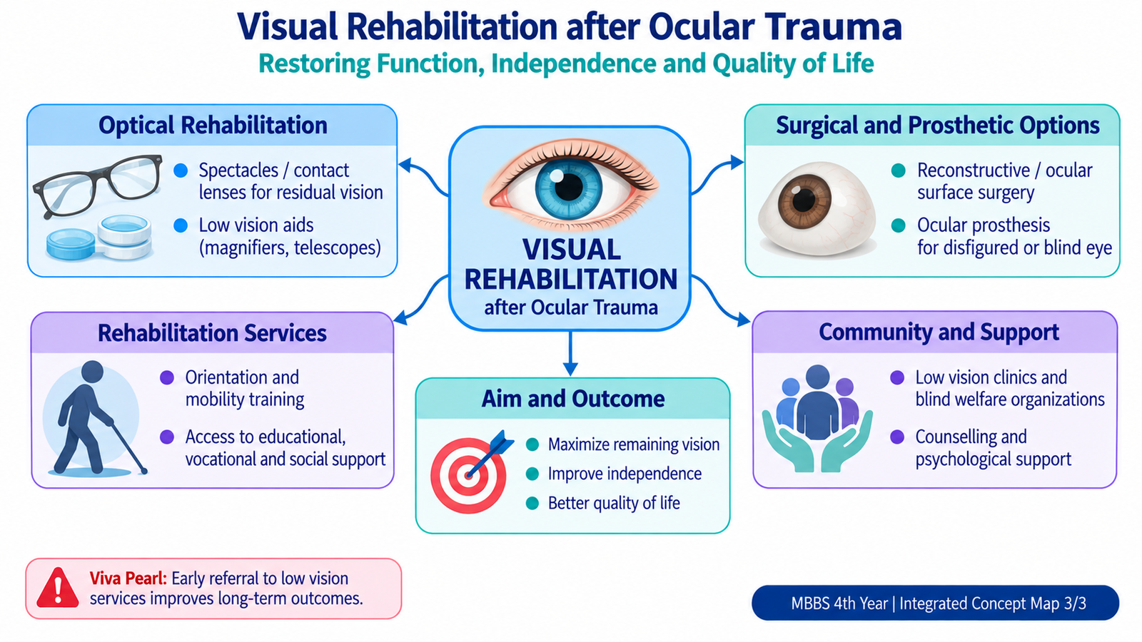
Visual rehabilitation begins when the acute injury has been stabilized. Its purpose is not limited to restoring normal visual acuity. It aims to obtain the best possible useful vision and functional independence from the patient’s remaining visual potential. The appropriate option depends on whether vision can be restored medically or surgically, improved optically, or compensated for with low-vision and functional rehabilitation.
1. Correction of residual refractive error
Trauma may change corneal shape, alter the lens or leave significant refractive error. Spectacles or contact lenses may improve vision when the ocular structures remain capable of forming a useful image. Contact lenses may be particularly useful in selected cases of irregular corneal optics or major refractive differences between the two eyes.
2. Surgical visual rehabilitation
Surgery may improve vision when trauma has produced a correctable structural problem. Examples include management of a traumatic cataract, selected corneal scarring, retinal injury or other reconstructable ocular damage. The principle is to correct the structure responsible for the visual deficit while considering the overall visual potential of the eye.
3. Low-vision rehabilitation
When conventional treatment cannot restore normal vision but some useful vision remains, low-vision aids can help the patient use that vision more effectively.
- High-powered spectacles
- Hand or stand magnifiers
- Telescopic devices for distance tasks
- Electronic magnification systems
- Improved lighting and contrast
- Large-print and digital accessibility tools
4. Cosmetic and prosthetic rehabilitation
A severely damaged blind eye may have major cosmetic and psychological effects. Depending on the condition, cosmetic contact lenses or an ocular prosthesis may improve appearance after loss or removal of the eye. Prosthetic rehabilitation does not restore sight but can considerably improve facial symmetry and social confidence.
5. Functional rehabilitation
Patients with major residual visual loss may need training to perform daily activities safely. Orientation and mobility training, environmental modification, assistive technology and occupational adaptation help the patient remain independent.
H. Rehabilitation Services for Blind People in Our Setup
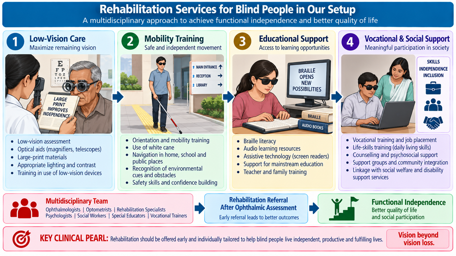
When vision cannot be restored sufficiently for ordinary daily activities, rehabilitation becomes a continuing part of patient care. Blindness rehabilitation is multidisciplinary. The ophthalmology team identifies the degree of remaining vision, treats preventable causes and connects the patient with services that improve education, mobility, communication, employment and independence. The exact availability of individual services varies between hospitals, districts and rehabilitation organizations, so referral should be based on locally accessible facilities.
Low-vision and ophthalmic rehabilitation services
- Assessment of residual visual function
- Prescription and training in low-vision devices
- Advice on illumination, contrast and reading techniques
- Follow-up of the better or remaining functional eye
Orientation and mobility services
These services teach a visually impaired person to move safely and independently. Training may include environmental orientation, safe navigation techniques and appropriate use of a mobility cane when required.
Educational and communication support
- Braille instruction when appropriate
- Large-print educational material for people with residual vision
- Screen-reading and text-to-speech technology
- Accessible computer and smartphone use
- Special-education support for children
Vocational and social rehabilitation
Vocational training can help visually impaired adults develop skills suitable for employment or self-employment. Social-welfare and disability-support pathways may assist with educational, occupational and social needs where such services are locally available.
Psychological and family support
Sudden visual loss after trauma may affect mood, independence, employment and family roles. Counseling, family education and peer or community support help patients adapt to a new level of vision and use rehabilitation services effectively.
Important Comparison — Open versus Closed Globe Injury
| Feature | Open Globe Injury | Closed Globe Injury |
|---|---|---|
| Eyewall | Full-thickness corneal or scleral wound | No full-thickness wound |
| Main classification | Rupture or laceration | Contusion or lamellar laceration |
| Typical mechanism | Sharp penetration or severe blunt rupture | Usually blunt trauma or partial-thickness wound |
| Important clues | Wound, peaked pupil, tissue prolapse, abnormal chamber depth | Hyphema, traumatic mydriasis, lens/retinal injury with intact wall |
| Tonometry | Avoid when OGI is suspected | May be performed after globe integrity is confirmed |
| Immediate principle | Rigid shield and urgent surgical ophthalmic assessment | Identify and treat the damaged ocular structure |
3. Integrated Mechanism Flow
⭐ AIM High-Yield Review
corneal trauma and foreign bodies • blunt ocular injury and hyphema • open-globe injury • chemical burns • sight-threatening trauma