
Topic 3 — Eyelid Lumps, Tumors & Malpositions
Topic Introduction
Eyelid disorders are common and are often recognized by careful inspection before any special investigation is required. A patient may present with a localized lump, an abnormal lid position, drooping of the upper lid, or eyelashes rubbing against the eye. Most eyelid lumps are benign or inflammatory, but some malignant tumors initially resemble harmless lesions. Similarly, eyelid malpositions can disturb tear drainage or allow lashes to damage the cornea. This chapter explains how to classify and assess lid bumps, understand chalazion and stye, recognize important eyelid tumors, and assess and manage ptosis, trichiasis, entropion and ectropion at an appropriate undergraduate level.
A. Approach to Eyelid Lumps and Lid-Bump Assessment
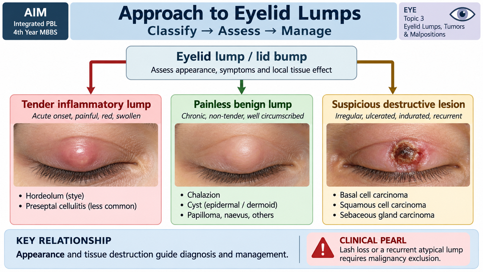
A lid bump is a localized swelling or mass arising from the eyelid skin, glands, hair follicles, tarsal plate or other eyelid tissues. The first clinical task is not to name every possible lesion. It is to decide whether the lesion behaves like an acute inflammatory process, a chronic benign lump, or a potentially malignant tumor. The time course, tenderness and effect on normal lid structures are particularly useful.
Main Causes of Lid Bumps
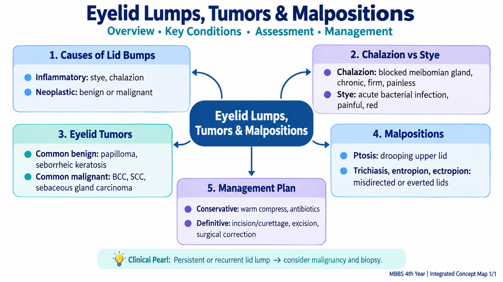
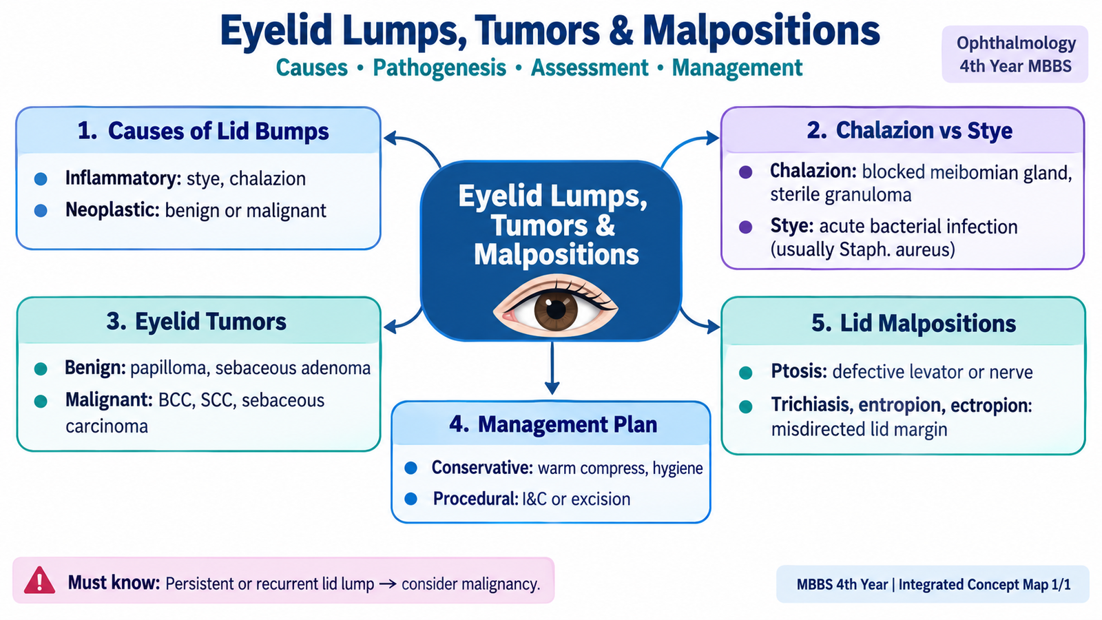
Lid bumps can be grouped according to the process producing the swelling. This gives the student a useful framework before considering individual diagnoses.
- Inflammatory or infective lesions: hordeolum or stye and inflamed eyelid glands.
- Blocked gland lesions: chalazion, caused by retained meibomian gland secretion followed by granulomatous inflammation.
- Cystic and other benign lesions: small retention cysts, papillomatous lesions and melanocytic nevi.
- Malignant tumors: particularly basal cell carcinoma, squamous cell carcinoma and sebaceous gland carcinoma.
History
The history helps establish whether the process is acute, recurrent or progressively enlarging. Acute painful swelling favors inflammation or infection, whereas a slowly enlarging painless lesion requires careful examination for benign or malignant features.
- Ask when the lump appeared and whether it is enlarging.
- Ask about pain, tenderness, redness or discharge.
- Determine whether similar lesions have occurred previously.
- Ask about bleeding, crusting or persistent ulceration.
- Note a history of previous eyelid surgery, chronic inflammation or significant sun exposure when relevant.
Ophthalmic Examination
Examination should determine where the lesion arises and whether it preserves or destroys normal eyelid architecture. A benign cystic lesion usually displaces surrounding structures without destroying them. A malignant lesion may infiltrate the lid margin, destroy lash follicles or ulcerate the surface.
- Visual acuity: provides a basic record and helps detect associated ocular involvement.
- Inspection: assess site, size, surface, pigmentation, ulceration, crusting and surrounding inflammation.
- Lid margin and lashes: loss of lashes, irregularity or destruction of the margin is suspicious for malignancy.
- Palpation: assess tenderness, consistency, mobility and fixation to deeper tissues.
- Upper-lid eversion: may reveal a tarsal or conjunctival component that is not visible externally.
- Regional examination: lymph nodes should be assessed when a malignant lesion is suspected.
General Management Plan
Management follows the clinical category. Clearly inflammatory lesions are treated according to their cause. Typical benign lesions may be observed or removed when symptomatic. Persistent or suspicious lesions require ophthalmic assessment and, when appropriate, tissue diagnosis.
Clinical pathway: identify the lesion pattern → look for inflammatory versus destructive features → examine the lid margin and ocular surface → treat a typical benign or inflammatory lesion appropriately → biopsy or refer when the diagnosis is uncertain or malignancy is suspected.
B. Chalazion and Stye: Pathophysiology, Recognition and Management
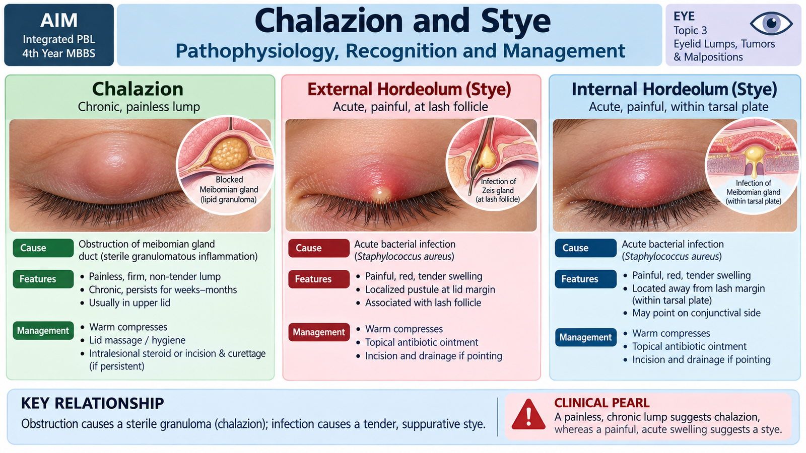
Chalazion and stye are both common eyelid lumps, but their underlying processes are different. A chalazion is mainly a sterile inflammatory reaction to retained lipid material from a meibomian gland. A stye or hordeolum is an acute infective and suppurative process involving an eyelid gland or lash follicle. This explains the major clinical distinction: chalazion is usually a chronic, relatively painless lump, whereas a stye is usually acute, red and tender.
Chalazion
The meibomian glands lie within the tarsal plate and normally release oily secretion onto the tear film. When the duct becomes obstructed, lipid material is retained and may leak into surrounding tissue. The body reacts to this material with a lipogranulomatous inflammatory response. Because this is primarily a chronic inflammatory process rather than an acute bacterial abscess, marked pain is usually absent.
A chalazion usually appears as a slowly developing, localized, firm swelling within the lid. It may initially be mildly inflamed, but the established lesion is usually minimally tender or painless. A large chalazion can mechanically distort the eyelid and may press on the globe.
Management
Initial management aims to improve drainage of the obstructed gland. Warm compresses and lid hygiene are therefore appropriate for an uncomplicated lesion. Persistent or troublesome lesions may require ophthalmic treatment such as incision and curettage. Intralesional corticosteroid is another specialist option in selected cases.
A lesion that is recurrent in the same location, unusually firm, associated with lash loss, or otherwise atypical should be reconsidered. In particular, sebaceous gland carcinoma can mimic a recurrent chalazion, so suspicious tissue may require histopathological examination.
Stye or Hordeolum
A hordeolum is an acute infection of an eyelid gland, commonly associated with staphylococcal infection. The inflammatory response produces vascular dilatation, edema and pus formation. These changes explain the sudden pain, redness and localized tenderness.
- External hordeolum: develops near an eyelash follicle and associated superficial glands, producing a tender swelling close to the lid margin.
- Internal hordeolum: arises from a meibomian gland and produces a deeper, painful swelling within the tarsal plate.
Warm compresses encourage spontaneous drainage. Associated lid-margin disease should be addressed when present. Antimicrobial treatment is used when clinically indicated, particularly when infection extends beyond a small localized lesion. A lesion that does not behave like an uncomplicated hordeolum should be reassessed.
| Feature | Chalazion | Stye / Hordeolum | Suspicious Tumor |
|---|---|---|---|
| Basic process | Blocked gland with granulomatous inflammation | Acute gland or follicle infection | Neoplastic tissue growth |
| Typical course | Chronic or slowly developing | Acute | Persistent or progressive |
| Pain | Usually painless when established | Usually painful and tender | Often painless initially |
| Concerning clue | Recurrent or atypical lesion | Spreading inflammation | Ulceration, lash loss or lid distortion |
C. Eyelid Tumors and Their Pathogenesis
Eyelid tumors may arise from the epidermis, hair follicles, glands, melanocytes or other tissues within the lid. Most eyelid lesions encountered clinically are benign, but malignant tumors are important because an apparently small lesion can progressively destroy the lid margin and surrounding structures. Recognition depends more on the lesion’s behavior and appearance than on memorizing long lists of tumors.
Benign Eyelid Lesions
Benign lesions generally grow slowly and preserve the surrounding eyelid architecture. Examples include papillomatous lesions, melanocytic nevi and cystic lesions. Their surfaces may be smooth, papillomatous or pigmented depending on the tissue of origin.
A benign appearance does not remove the need for assessment. Change in size, shape, color, surface or symptoms should lead to reconsideration of the diagnosis.
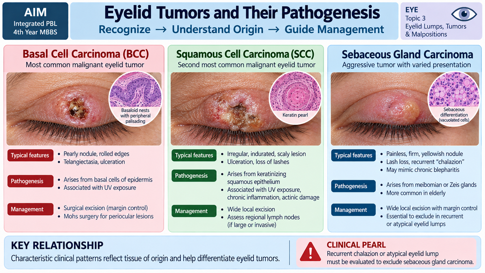
Basal Cell Carcinoma
Basal cell carcinoma is an important malignant eyelid tumor associated with chronic ultraviolet exposure. It commonly affects sun-exposed eyelid skin, particularly the lower lid and medial canthal region. The tumor grows by local infiltration and progressively destroys surrounding tissue.
A typical lesion may appear as a slowly enlarging pearly or nodular lesion with surface telangiectatic vessels. Ulceration can develop as the tumor outgrows and destroys central tissue. Destruction of lash follicles produces loss of lashes, an important warning feature. Basal cell carcinoma is mainly locally invasive; distant spread is uncommon.
Squamous Cell Carcinoma
Squamous cell carcinoma arises from squamous epithelium and is also related to cumulative ultraviolet injury. The lesion may appear scaly, thickened, indurated, nodular or ulcerated. Because malignant squamous cells invade deeper tissue, progressive destruction and fixation may occur.
Compared with basal cell carcinoma, squamous cell carcinoma has a greater tendency to spread to regional lymph nodes and therefore deserves careful regional assessment.
Sebaceous Gland Carcinoma
Sebaceous gland carcinoma arises from sebaceous glands of the eyelid, especially the meibomian glands. Its importance lies partly in its ability to masquerade as a benign inflammatory disorder. A localized tumor may resemble a persistent or recurrent chalazion, while more diffuse involvement may resemble chronic unilateral blepharoconjunctivitis.
Tumor cells can spread within the epithelium beyond the obvious mass, which makes early recognition important. A recurrent atypical chalazion, unexplained focal lid thickening or persistent unilateral inflammatory appearance should therefore raise suspicion.
Melanoma
Eyelid melanoma arises from melanocytes. It is less common than the carcinomas above but is clinically important because of its aggressive malignant potential. A changing pigmented lesion with irregular shape, variable pigmentation, growth or ulceration requires specialist assessment rather than simple observation.
Assessment and Management Principles
Suspicious eyelid tumors require careful examination of the lesion, lid margin, conjunctival surface and regional lymph nodes where appropriate. A tissue diagnosis is required when malignancy is suspected because clinical appearance alone cannot reliably determine tumor type.
Treatment is based on complete control of the tumor while preserving eyelid function and protecting the ocular surface. Localized malignant tumors are generally treated surgically with histological confirmation of adequate tumor removal, followed by reconstruction when required. More extensive or aggressive tumors require specialist oncological assessment.
D. Ptosis: Causes, Assessment and Management
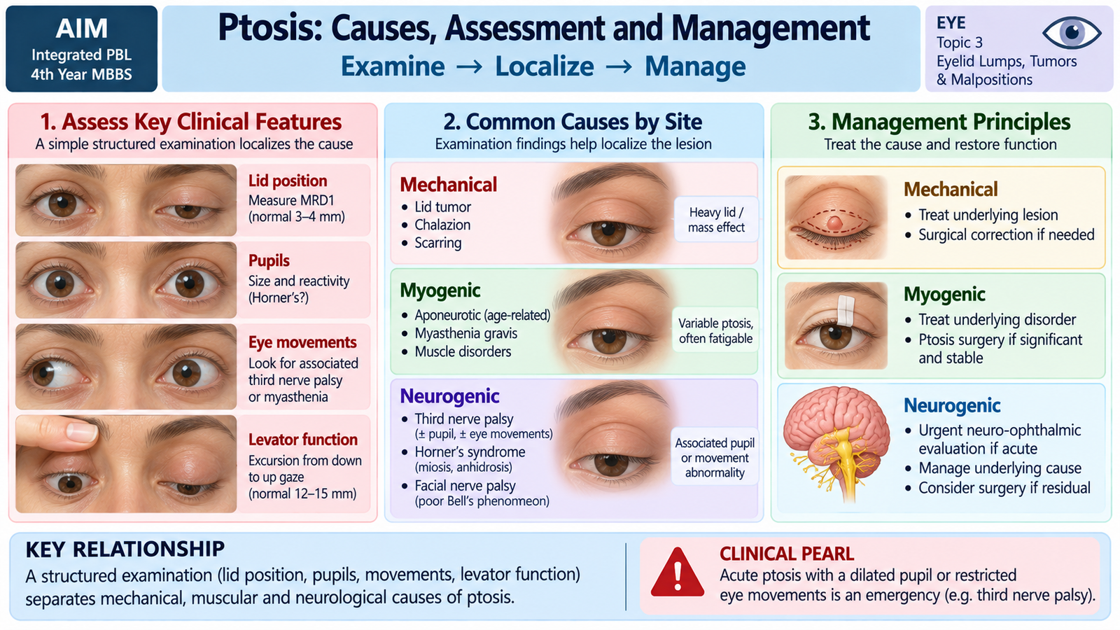
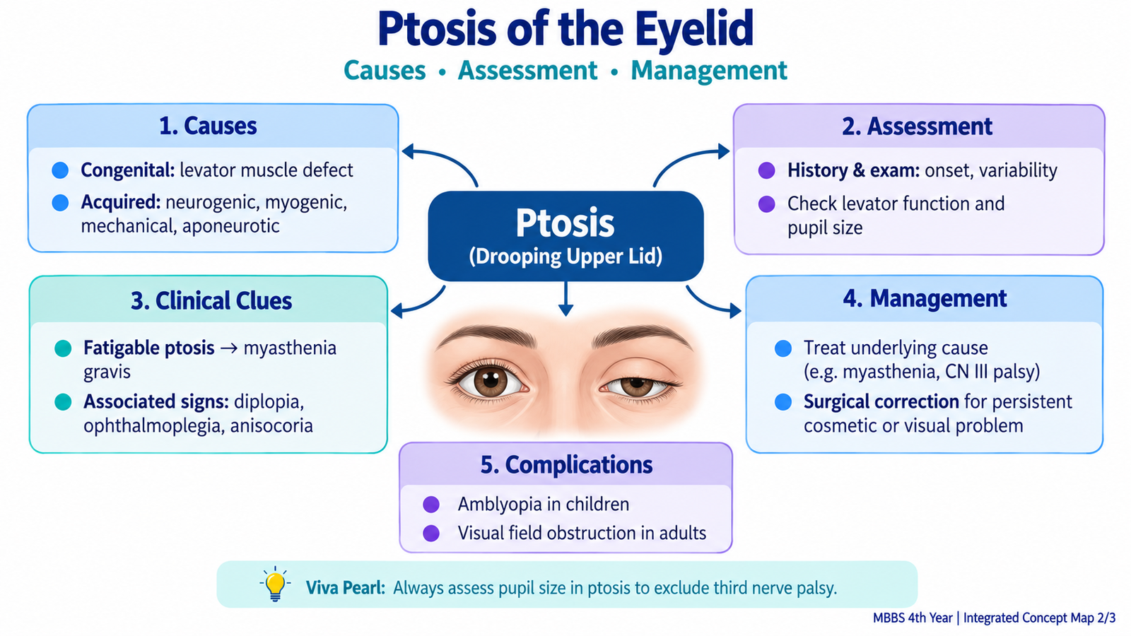
Ptosis is abnormal drooping of the upper eyelid. It results when the structures responsible for elevating or maintaining the upper lid fail to function normally, or when the lid becomes mechanically too heavy to elevate. Ptosis is therefore a clinical sign rather than a single disease. Correct assessment aims to determine the mechanism and, importantly, to identify neurological or visual problems that require urgent attention.
Relevant Functional Anatomy
The main elevator of the upper lid is the levator palpebrae superioris, supplied by the oculomotor nerve. Its aponeurosis transmits levator force to the eyelid. The superior tarsal or Müller muscle, supplied by sympathetic fibers, provides a smaller contribution to lid elevation. Disease affecting the muscle, its nerve supply, its aponeurosis or the mechanical weight of the lid can therefore produce ptosis.
Causes
Classifying ptosis by mechanism helps connect the clinical appearance with the underlying abnormality.
- Congenital: commonly due to abnormal development of the levator muscle, producing reduced levator function from childhood.
- Aponeurotic: stretching or dehiscence of the levator aponeurosis weakens transmission of levator force. This is common with aging and may also follow ocular surgery or trauma.
- Neurogenic: occurs when nerve supply to the lid elevators is impaired, for example in third cranial nerve palsy or Horner syndrome.
- Myogenic: results from impaired muscle or neuromuscular function, as in myasthenia gravis or certain myopathies.
- Mechanical: a heavy lid mass, edema, scarring or other physical abnormality prevents normal elevation.
- Traumatic: injury can affect the levator muscle, aponeurosis or nerve supply.
Assessment
Assessment begins by establishing onset and pattern. Sudden ptosis has a different significance from a stable lifelong droop. Variability during the day suggests a different mechanism from a fixed mechanical abnormality.
- History: onset, progression, variability, diplopia, headache, trauma, previous surgery and systemic neuromuscular symptoms.
- Visual acuity: especially important in children because severe ptosis may obstruct the visual axis and interfere with normal visual development.
- Lid position: compare both upper lids and document the relationship of the lid margin to the corneal light reflex, commonly using margin-reflex distance.
- Levator function: observe the amount of upper-lid movement from downgaze to upgaze while limiting frontalis compensation.
- Lid crease: its position and definition can provide clues to levator aponeurosis function.
- Pupils: anisocoria may indicate an associated neurological disorder.
- Ocular movements: limitation together with ptosis supports an oculomotor nerve disorder rather than isolated mechanical ptosis.
- Variability and fatigability: fluctuating ptosis raises suspicion of neuromuscular junction disease such as myasthenia gravis.
Management
Management depends on the cause and on whether vision or neurological function is threatened. An underlying neurological or neuromuscular disorder must be treated appropriately rather than simply correcting the lid position. Stable structural ptosis can be corrected surgically when function or vision is significantly affected.
In children, severe congenital ptosis can obstruct the visual axis and contribute to amblyopia. Protection of visual development therefore takes priority. Surgical planning is based broadly on the severity of ptosis and the amount of remaining levator function.
E. Trichiasis and Entropion
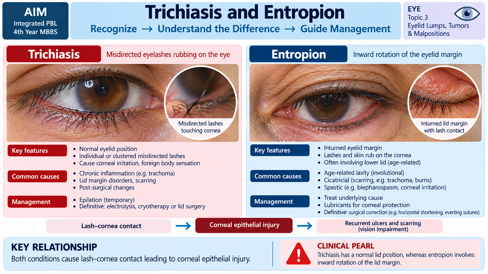
Trichiasis and entropion both cause eyelashes to rub against the ocular surface, but the anatomical abnormality is different. In trichiasis, one or more lashes are misdirected toward the eye while the overall lid margin remains in its normal position. In entropion, the eyelid margin itself rotates inward, carrying normally positioned eyelashes against the cornea and conjunctiva. This distinction is important because treatment must correct the actual source of the lash-cornea contact.
Trichiasis
Trichiasis develops when eyelash follicles become misdirected, commonly following chronic lid-margin inflammation or conjunctival scarring. Trauma or previous surgery can also alter follicle direction. Each blink then drags the abnormal lash across the ocular surface.
Examination should identify the abnormal lashes and determine whether the lid margin itself remains normally positioned. The cornea should be examined for epithelial damage. Fluorescein staining can demonstrate epithelial defects produced by repeated mechanical trauma.
Management
Removing the offending lash by epilation can provide temporary relief, but the lash commonly regrows. Recurrent localized trichiasis may require a procedure directed at the abnormal follicle. If trichiasis results from more extensive scarring or associated lid deformity, correction of the underlying eyelid abnormality is required.
Entropion
Entropion is inward rotation of the eyelid margin. It most commonly affects the lower lid in older adults because age-related lid laxity and changes in the lower-lid retractors allow the margin to turn inward. Other forms may result from conjunctival scarring, muscular spasm or congenital abnormalities.
- Involutional entropion: related to age-associated lid laxity and altered lower-lid support.
- Cicatricial entropion: scarring of the posterior eyelid surface pulls the margin inward.
- Spastic entropion: increased orbicularis activity contributes to inward rotation.
- Congenital entropion: results from developmental abnormalities of lid anatomy.
Patients complain of irritation, foreign-body sensation, watering, redness and sometimes photophobia. These symptoms occur because the lashes repeatedly scrape the corneal epithelium. Persistent contact can produce abrasion, keratitis, ulceration and ultimately threaten vision.
Assessment and Management
Examination confirms inward rotation of the lid margin and assesses the cornea for injury. Lubrication and temporary measures that keep the lid away from the globe can protect the ocular surface while definitive treatment is arranged. Persistent structural entropion generally requires surgical correction of the underlying lid malposition.
F. Ectropion: Causes, Assessment and Management
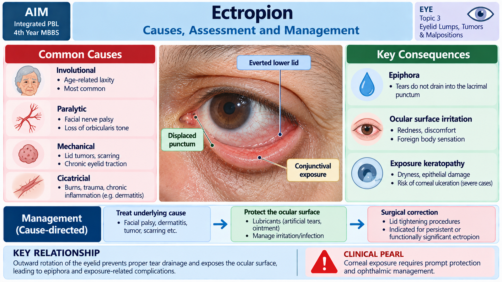
Ectropion is outward turning of the eyelid margin, most commonly involving the lower lid. The abnormal position separates the lid and punctum from the globe. This interferes with normal tear drainage and may leave the conjunctiva and cornea inadequately protected. The characteristic combination of watering and ocular-surface exposure follows directly from this altered anatomy.
Causes
Different mechanisms can pull or allow the lid to turn outward.
- Involutional ectropion: age-related horizontal lid laxity reduces normal apposition of the lower lid to the globe.
- Paralytic ectropion: facial nerve weakness reduces orbicularis tone, allowing the lower lid to sag outward.
- Cicatricial ectropion: shortening or scarring of anterior eyelid skin pulls the lid away from the globe.
- Mechanical ectropion: a mass or other physical weight displaces the eyelid outward.
- Congenital ectropion: results from developmental abnormality and is uncommon.
Clinical Presentation
Epiphora or watering is common because the lower punctum no longer sits normally against the tear lake, so tears do not enter the drainage system efficiently. Paradoxically, exposure-related irritation can stimulate additional reflex tear production, making watering even more noticeable.
The exposed palpebral conjunctiva becomes dry, congested and irritated. If lid closure becomes inadequate, the corneal surface may also dry, producing exposure keratopathy.
Assessment
Examination confirms outward rotation and identifies the mechanism responsible. The examiner should observe lid position, punctal position, ability to close the eye, facial nerve function and the state of the ocular surface. The cornea is particularly important because epithelial exposure converts a simple lid malposition into a potential threat to vision.
Management
Initial management protects the ocular surface. Lubrication and other protective measures reduce drying while the underlying cause is addressed. Facial nerve dysfunction requires assessment of the neurological cause and careful corneal protection when eyelid closure is impaired.
Persistent structural ectropion is usually corrected surgically by restoring appropriate lid position and apposition to the globe. The exact operation depends on whether laxity, paralysis, scarring or a mechanical factor is responsible.
| Feature | Trichiasis | Entropion | Ectropion |
|---|---|---|---|
| Main abnormality | Misdirected lashes | Lid margin turns inward | Lid margin turns outward |
| Lid position | Usually normal | Inverted | Everted |
| Main ocular effect | Direct lash-cornea trauma | Lashes rub ocular surface | Exposure and poor tear drainage |
| Typical symptom | Foreign-body sensation | Irritation and watering | Watering and exposure irritation |
| Definitive principle | Correct abnormal lashes or follicles | Correct inward lid rotation | Restore lid-globe apposition |
⭐ AIM High-Yield Review
Diseases of Eyelids — Chalazion, Stye, Ptosis & Tumors
Reinforce the key clinical features, mechanisms and differentiation of chalazion, hordeolum, eyelid tumors, ptosis, trichiasis, entropion and ectropion.