Topic 10 — Cataract: Types, Pathogenesis, Complications & Management
Topic Introduction
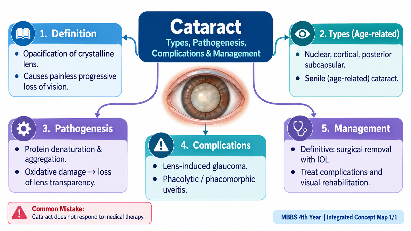
A cataract is loss of the normal transparency of the crystalline lens of the eye. The opaque lens scatters and blocks light before it reaches the retina, so vision gradually becomes blurred. Age-related cataract is particularly important in clinical ophthalmology and commonly develops in recognizable patterns involving the lens nucleus, cortex or posterior subcapsular region. Understanding cataract becomes much easier when the condition is viewed as a sequence: the transparent lens undergoes age-related structural and biochemical changes, these changes produce lens opacity, the opacity interferes with vision, and an advanced untreated cataract may produce additional ocular complications. Management is guided mainly by the degree to which cataract affects visual function or threatens the eye.
A. Cataract: Core Concept and Lens Transparency
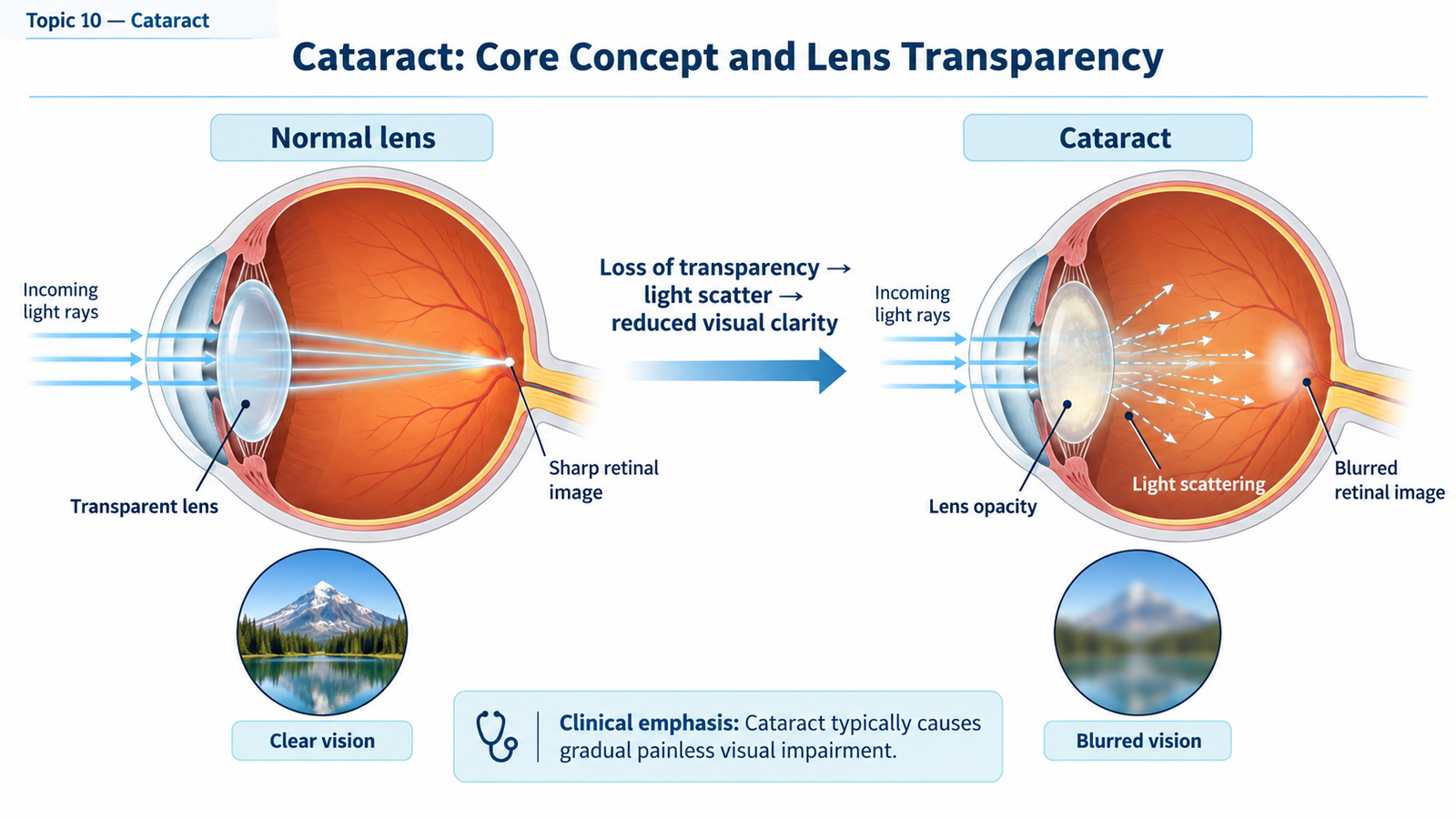
The crystalline lens is normally transparent so that light can pass through it and focus on the retina. Cataract develops when this transparency is lost. The opacity may be localized initially, but increasing involvement of the lens progressively scatters light and reduces the quality of the retinal image.
Definition
Cataract is an opacity or loss of transparency of the crystalline lens that interferes with the passage of light and may impair vision.
Why is the normal lens transparent?
Lens transparency depends on the highly organized arrangement of lens fibres, the regular distribution of lens proteins called crystallins, absence of blood vessels and careful control of water and electrolytes within the lens. These features allow light to pass with very little scattering.
When ageing alters lens proteins, fibres or hydration, the refractive index within different parts of the lens becomes irregular. Incoming light is then scattered rather than transmitted uniformly. This explains the characteristic gradual, painless reduction of vision associated with uncomplicated cataract.
Functional effects of lens opacity
The exact visual problem depends partly on the position and density of the opacity. Important effects may include:
- Gradual painless blurring of vision because the image reaching the retina becomes less clear.
- Glare, especially from bright lights, because irregular lens areas scatter light.
- Reduced contrast, making objects appear less distinct even when some visual acuity remains.
- Alteration of refraction, particularly a myopic shift in nuclear cataract.
- In some patients, monocular diplopia or multiple images may occur because light passes through lens regions with different refractive properties.
Ophthalmic recognition
Visual acuity helps determine the functional effect of cataract, while examination of the lens—particularly with slit-lamp illumination—shows the opacity itself. The normal red reflex may become reduced, irregular or absent as the cataract becomes denser because less light passes through the lens to and from the fundus.
B. Age-Related Cataract: Types and Recognition
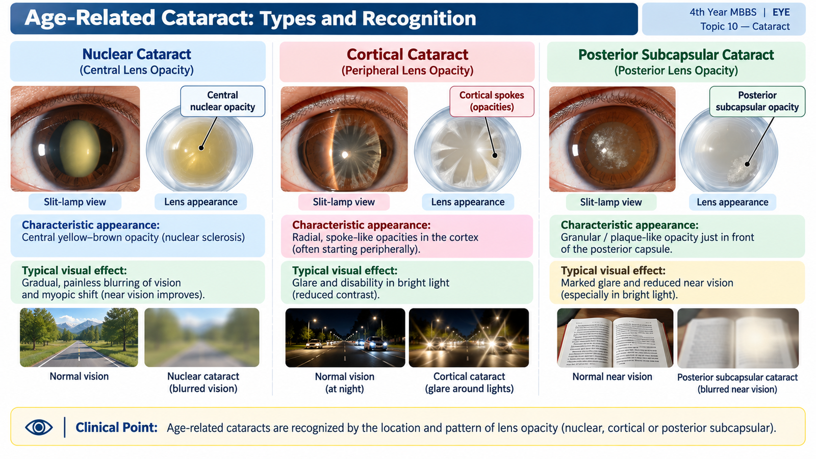
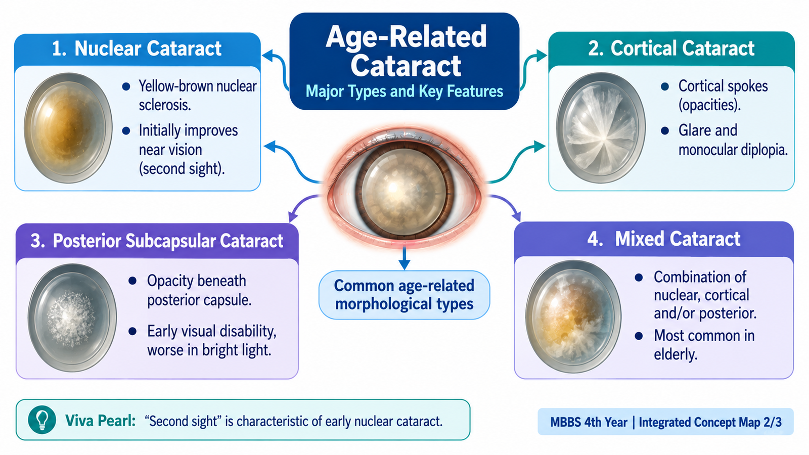
Age-related cataract is classified mainly according to the part of the lens in which the important opacity develops. The three major patterns are nuclear cataract, cortical cataract and posterior subcapsular cataract. More than one pattern may occur in the same lens, producing a mixed cataract.
1. Nuclear Cataract
Nuclear cataract involves the central nucleus of the lens. With ageing, the nucleus becomes progressively harder and more compact, a process known as nuclear sclerosis. At first it develops a yellow discoloration; with increasing severity it may become yellow-brown or brown.
The increasing density of the nucleus changes its refractive power. This may shift the eye toward myopia, so some patients temporarily find that their near vision improves despite deterioration in distance vision. This phenomenon is sometimes called “second sight.” It is temporary and does not represent recovery of the lens.
2. Cortical Cataract
Cortical cataract develops in the lens cortex surrounding the nucleus. Age-related disturbance of lens fibre membranes and electrolyte balance allows water to enter the cortical fibres. Small clefts and vacuoles form, followed by characteristic radial or spoke-like opacities.
The cortical spokes commonly extend from the peripheral lens toward the centre. When the opacities enter the visual axis, vision becomes more significantly affected. Irregular light scattering can make glare particularly troublesome.
3. Posterior Subcapsular Cataract
A posterior subcapsular cataract develops immediately in front of the posterior lens capsule, usually near the central visual axis. Because this opacity lies close to the nodal region through which important central rays pass, even a relatively small posterior subcapsular opacity may cause marked visual symptoms.
Patients often have significant glare and difficulty with near work. Vision may be particularly troublesome in bright illumination because the pupil constricts and light is forced through the central opacity.
Mature and Hypermature Cataract
Mature and hypermature describe advanced stages rather than separate anatomical types of age-related cataract. In a mature cataract, essentially the entire lens has become opaque. In a hypermature cataract, further degeneration occurs; the cortex may liquefy and the lens may shrink. These advanced changes are important because they can lead to lens-induced complications.
| Feature | Nuclear | Cortical | Posterior Subcapsular |
|---|---|---|---|
| Main site | Lens nucleus | Lens cortex | Immediately anterior to posterior capsule |
| Typical appearance | Yellow-brown central sclerosis | Radial spoke-like opacities | Central posterior granular/plaque opacity |
| Important visual effect | Myopic shift may occur | Glare from irregular light scatter | Marked glare and near-vision difficulty |
| Visual recognition | Dense central nucleus | Peripheral spokes extending centrally | Opacity near central posterior visual axis |
C. Pathogenesis: How Lens Opacity Develops
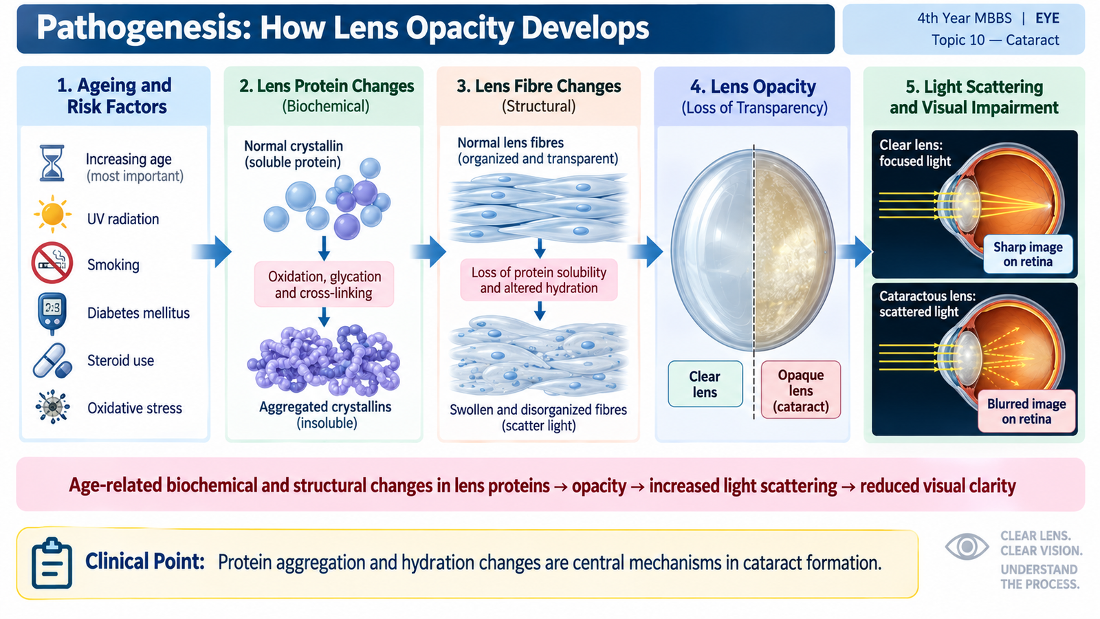
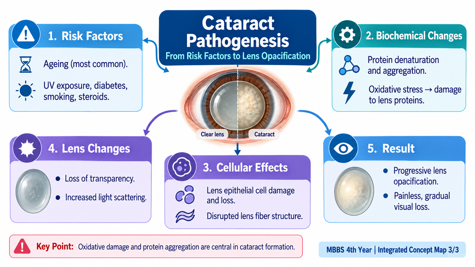
The lens remains transparent only while its proteins, fibres and water content remain precisely organized. Ageing progressively disturbs these systems. The most important underlying processes are oxidative damage, alteration and aggregation of lens proteins, changes in lens fibre structure, and disturbed water and electrolyte balance. These processes increase differences in refractive index within the lens and therefore increase light scattering.
Age-related change in lens proteins
Crystallins are the major structural proteins that help maintain lens transparency. With increasing age, lens proteins undergo chemical modification and become less soluble. Damaged proteins may aggregate into larger complexes. Instead of allowing light to pass uniformly, these aggregates scatter light and contribute to opacity.
Changes in the lens nucleus
The central lens fibres are the oldest fibres in the lens. Over many years they become increasingly compacted. Protein modification and accumulation of pigments cause the nucleus to become hard, yellow and eventually brown. Increasing nuclear density changes the refractive index of the lens, explaining both the opacity and the possible myopic shift seen in nuclear cataract.
Changes in the lens cortex
Normal lens fibres depend on controlled movement of ions and water across their membranes. Age-related membrane dysfunction disturbs this balance. Water accumulates within cortical lens fibres, producing hydration, clefts and vacuoles. As the affected fibres lose their orderly arrangement, radial cortical opacities become visible.
Posterior subcapsular change
In posterior subcapsular cataract, abnormal lens epithelial-derived cells and altered lens fibres accumulate beneath the posterior capsule. Because this change occurs close to the visual axis, it interferes strongly with the transmission of central light rays. This explains why a relatively small posterior opacity may produce substantial glare and visual difficulty.
Why does vision become blurred?
In a clear lens, light rays remain organized as they pass toward the retina. Cataract creates multiple areas with different refractive properties. Light is scattered in different directions, reducing the sharpness and contrast of the retinal image. As the opacity becomes denser or involves the visual axis, the visual impairment becomes greater.
D. Complications of Cataract
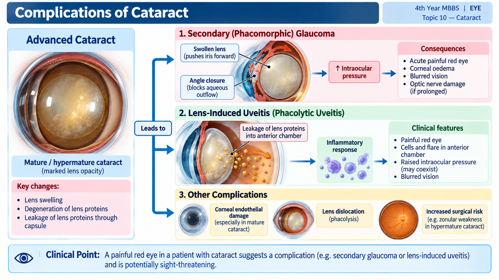
The main consequence of cataract is progressive loss of useful vision. If a cataract becomes advanced and remains untreated, however, the lens itself may produce secondary ocular disease. These complications occur because the lens may become swollen, its proteins may leak from a hypermature lens, or degenerative changes may provoke inflammation.
Progressive Visual Disability
Increasing lens opacity reduces visual acuity, contrast and the ability to function in everyday activities. A dense mature cataract can produce severe visual impairment because very little useful light passes through the lens.
Phacomorphic Glaucoma
An advanced cataract may become intumescent, meaning that the lens becomes swollen. The enlarged lens pushes the iris forward and makes the anterior chamber angle narrow. Obstruction of aqueous drainage may then cause a marked rise in intraocular pressure. This is called phacomorphic glaucoma.
Phacolytic Glaucoma
In a hypermature cataract, altered high-molecular-weight lens proteins may escape through an apparently intact lens capsule. Macrophages ingest these proteins, and protein-rich material together with macrophages can obstruct the trabecular meshwork. Aqueous outflow falls and intraocular pressure rises. This produces phacolytic glaucoma.
Lens-Induced Uveitis
Lens proteins are normally isolated from the intraocular immune environment by the lens capsule. When altered lens material escapes, it may provoke an inflammatory response. The patient may develop anterior uveitis with ocular pain, redness and inflammatory cells in the anterior chamber.
Changes in a Hypermature Lens
As cortical material degenerates and liquefies, the lens may shrink and the capsule may become wrinkled. Progressive degeneration can also be associated with weakness of the lens supporting structures. These changes help explain why a long-standing hypermature cataract is not simply a stable opacity but may become a source of additional ocular problems.
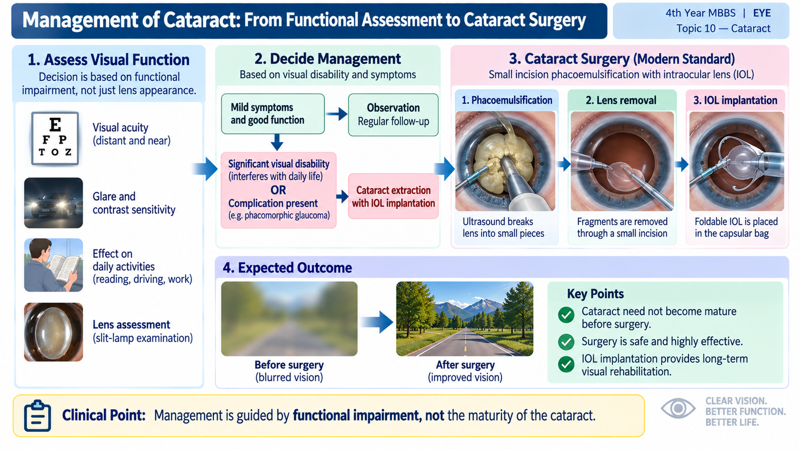
E. Management: From Functional Assessment to Cataract Surgery
Management of cataract is based primarily on the effect of the opacity on the patient’s vision and daily function, rather than on the mere presence of lens opacity. Early cataract with acceptable visual function may be observed, while visually significant cataract is treated surgically. There is no established medical treatment that can reliably restore transparency to an age-related cataract once it has formed.
Assessment Before Deciding Treatment
The ophthalmic assessment should establish that the cataract is responsible for the patient’s visual problem and determine whether the eye is likely to benefit from surgery.
- Visual acuity: measures the degree of visual impairment and allows comparison with the patient’s functional complaints.
- Refraction: determines whether part of the reduced vision can still be improved with an appropriate optical correction.
- Slit-lamp examination: identifies the type, site and density of lens opacity.
- Pupil and anterior-segment examination: helps identify other ocular abnormalities that could influence management.
- Fundus examination: is performed when the lens allows an adequate view, because retinal or optic-nerve disease may also limit vision.
A dense cataract may prevent adequate visualization of the fundus. In such a situation, posterior-segment evaluation is performed as clinically appropriate so that significant pathology is not overlooked before surgery.
Non-Surgical Management
When cataract is mild and visual function remains satisfactory, treatment is directed at improving usable vision and monitoring progression. Updating spectacles or refraction and improving illumination may provide temporary functional benefit. These measures do not remove the cataract.
When is Surgery Indicated?
Cataract surgery is considered when the benefit of removing the opaque lens outweighs the risks of continued observation. Important indications include:
- Visual impairment affecting daily activities, such as reading, mobility or other necessary tasks.
- Significant glare or loss of visual quality despite apparently moderate reduction in measured acuity.
- Lens-induced ocular complications, such as phacomorphic or phacolytic glaucoma or lens-induced inflammation.
- A cataract that prevents necessary examination or treatment of important posterior-segment disease.
Therefore, there is no need to wait for every cataract to become “mature” before operating. The decision is individualized according to visual needs, ocular findings and complications.
Principle of Cataract Surgery
The aim of cataract surgery is simple: remove the opaque crystalline lens and restore the eye’s focusing power with an artificial intraocular lens (IOL). Before surgery, ocular measurements are used to select an appropriate IOL power.
Phacoemulsification
In phacoemulsification, the cataractous lens nucleus is broken into small fragments using ultrasound energy and removed through a small incision. The posterior part of the lens capsule is generally retained to support a posterior chamber IOL. The artificial lens is then implanted into the capsular bag.
For an undergraduate student, the key point is not the detailed operative technique but the concept that the opaque lens contents are removed while a stable capsular support is preserved for IOL implantation.
Small-Incision and Extracapsular Cataract Surgery
In manual small-incision cataract surgery, the lens nucleus is removed through a self-sealing incision without phacoemulsification, followed by IOL implantation. In conventional extracapsular cataract extraction, the lens nucleus and cortex are removed while the posterior capsule is left behind to support the IOL. These procedures follow the same fundamental therapeutic principle: remove the opaque lens material and restore optical power with an intraocular lens.
Visual Rehabilitation
The implanted IOL replaces much of the refractive power lost when the natural lens is removed. Final visual outcome also depends on the health of the cornea, retina, macula and optic nerve. Therefore, cataract surgery can remove the optical obstruction caused by the lens, but it cannot correct visual loss arising from unrelated retinal or optic-nerve disease.