
Topic 4 — Proptosis, Orbital Disorders & Neuro-Ophthalmic Mimics
1. Topic Introduction
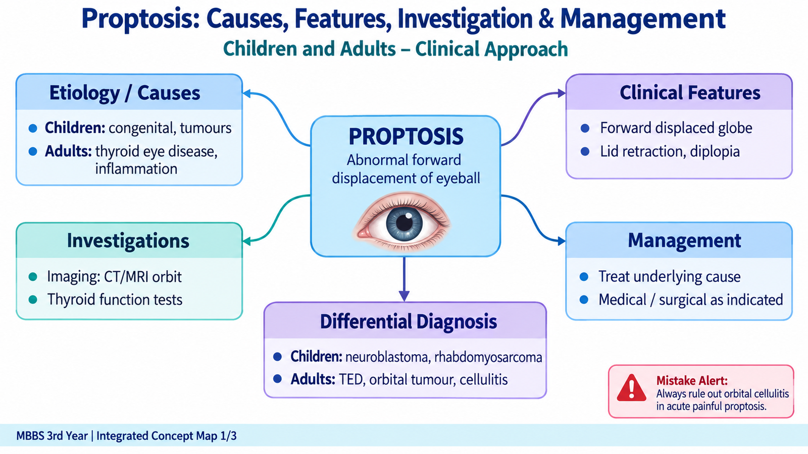
Proptosis means abnormal forward displacement of the eyeball from the orbit. It occurs when the volume of orbital contents increases, when venous drainage is impaired, or when a lesion pushes the globe forwards. Because the orbit is a confined bony cavity, even a relatively small increase in orbital volume may affect ocular movements, the cornea or the optic nerve. The pattern of proptosis therefore provides important diagnostic information. This topic also includes thyroid eye disease, an important cause of adult proptosis, and two conditions that may produce confusing visual or ocular symptoms without true orbital enlargement: myasthenia gravis and migraine. :contentReference[oaicite:0]{index=0}
A. Proptosis: Core Concept and Orbital Localization
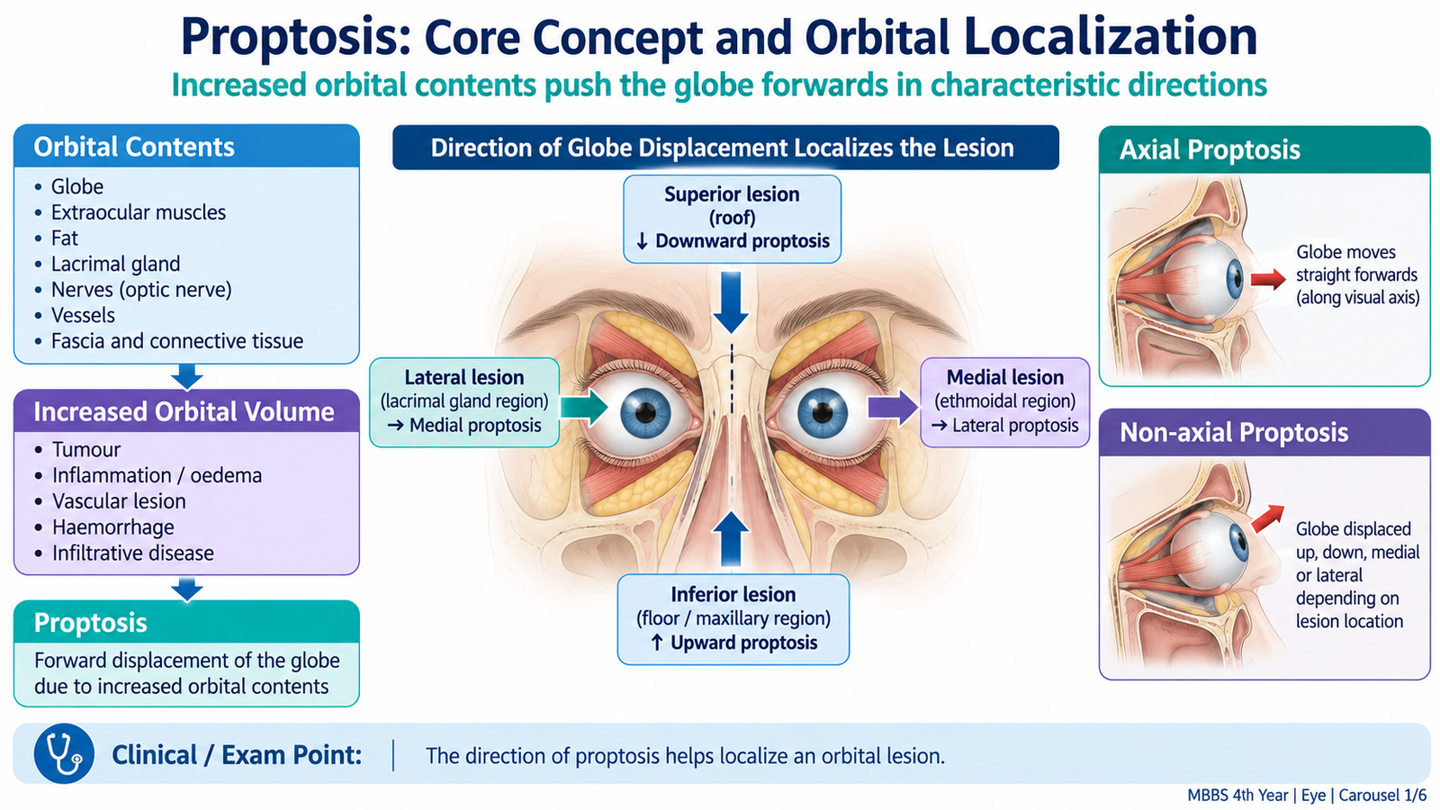
Proptosis is a clinical sign rather than a single disease. It represents forward displacement of the globe beyond its normal position within the orbit. Understanding the anatomy of the orbit helps explain why different orbital lesions produce different patterns of displacement.
Core Concept
The bony orbit contains the globe, extraocular muscles, optic nerve, orbital fat, blood vessels, lacrimal gland and other soft tissues. Because the bony walls cannot expand, an increase in the volume of these contents tends to displace the globe.
Axial and Non-Axial Proptosis
Axial proptosis occurs when the lesion lies within the muscle cone or directly behind the globe. The eye moves mainly forwards along its normal visual axis. Lesions involving the optic nerve or central intraconal orbit commonly produce this pattern.
Non-axial proptosis occurs when an eccentric orbital lesion pushes the globe forwards and away from the lesion. The direction of globe displacement therefore helps localize the abnormality.
Other Useful Descriptions
- Unilateral or bilateral: bilateral disease suggests a systemic or symmetrical orbital process such as thyroid eye disease, although exceptions occur.
- Acute or gradual: acute onset suggests inflammation, infection, hemorrhage or a vascular event; gradual progression is more typical of many tumors or chronic inflammatory disorders.
- Pulsatile: suggests transmission of arterial pulsation or an abnormal vascular connection.
- Reducible: may occur in some venous orbital lesions when pressure or body position changes.
True Proptosis and Pseudoproptosis
In true proptosis, the globe is genuinely displaced forwards by an orbital process. In pseudoproptosis, the eye appears prominent without an increase in orbital contents. Examples include a large eye in high axial myopia, lid retraction, facial asymmetry or apparent prominence caused by a relatively sunken opposite eye.
B. Proptosis in Children and Adults: Causes and Clinical Features
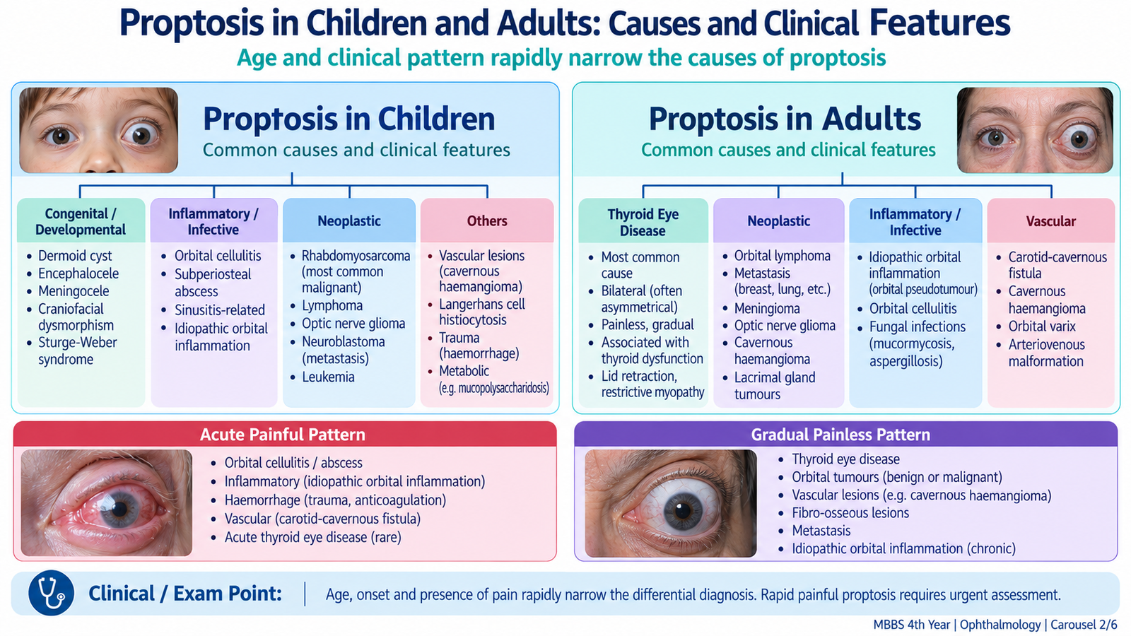
The likely cause of proptosis depends strongly on the patient’s age, speed of onset, whether one or both eyes are involved, and the presence of pain, fever, trauma, pulsation or systemic disease. The purpose of clinical assessment is therefore not simply to confirm that the eye is prominent, but to identify the pattern that points toward the underlying cause.
Important Causes in Children
In children, rapidly developing unilateral proptosis requires particular attention because infection and rapidly growing tumors may progress quickly. Congenital and developmental orbital lesions also occur.
- Orbital cellulitis: painful, acute proptosis with fever, eyelid swelling and restricted eye movements.
- Rhabdomyosarcoma: an important rapidly growing malignant orbital tumor that may cause progressive unilateral proptosis.
- Retinoblastoma with orbital extension: advanced intraocular malignancy may extend into the orbit and produce proptosis.
- Optic nerve glioma: may produce slowly progressive axial proptosis, often with visual impairment.
- Dermoid cyst: usually presents as a slowly growing, painless orbital or peri-orbital mass.
- Vascular or lymphatic malformations: may enlarge intermittently or after infection or hemorrhage.
Important Causes in Adults
In adults, thyroid eye disease is a major cause, particularly when proptosis is bilateral. Unilateral adult proptosis requires careful assessment for orbital inflammation, tumor, infection, vascular abnormality or hemorrhage.
- Thyroid eye disease: commonly associated with lid retraction, restrictive ocular movement and bilateral proptosis.
- Orbital tumors: may cause slowly progressive, usually painless proptosis.
- Idiopathic orbital inflammation: often produces painful proptosis with inflammatory signs.
- Orbital cellulitis: causes acute painful proptosis with systemic and local inflammatory features.
- Carotid-cavernous fistula: may produce pulsatile proptosis, conjunctival vascular congestion, bruit and raised orbital venous pressure.
- Retrobulbar hemorrhage: may follow trauma or surgery and can cause sudden painful proptosis with rapid visual compromise.
- Paranasal sinus or adjacent lesions: may extend into or displace the orbit.
Clinical Assessment
The history should establish when the prominence began and how rapidly it progressed. Acute painful proptosis suggests a different process from slowly progressive painless proptosis. Associated systemic symptoms may provide an important clue.
- Pain and fever: suggest infection or active inflammation.
- Rapid progression: raises concern for infection, hemorrhage, vascular disease or an aggressive tumor.
- Diplopia: indicates altered extraocular muscle function or mechanical restriction.
- Reduced visual acuity: may indicate corneal exposure, optic nerve compression or severe orbital disease.
- Redness and chemosis: occur with orbital inflammation or venous congestion.
- Pulsation or bruit: suggests a vascular cause.
- Thyroid symptoms: support thyroid eye disease but their absence does not exclude it.
Ophthalmic Examination
Both eyes should be examined systematically. Visual acuity is assessed first because loss of vision changes the urgency of management. The examiner then looks for asymmetry, lid position, conjunctival congestion, corneal exposure and the direction of globe displacement.
- Visual acuity: detects functional impairment.
- Pupils: an abnormal relative afferent pupillary response may indicate optic nerve dysfunction.
- Colour vision: reduction may be an early clue to optic neuropathy.
- Ocular movements: restriction may result from inflammation, enlarged muscles or mechanical compression.
- Cornea: look for exposure-related epithelial damage when lid closure is incomplete.
- Optic disc: may appear swollen or, later, pale if the optic nerve is compromised.
- Visual fields: may be abnormal when the optic nerve or orbital apex is involved.
Globe prominence can be quantified by exophthalmometry. The value is interpreted together with asymmetry between the two eyes and the overall clinical picture rather than in isolation.
C. Proptosis: Investigation, Differential Diagnosis and Management
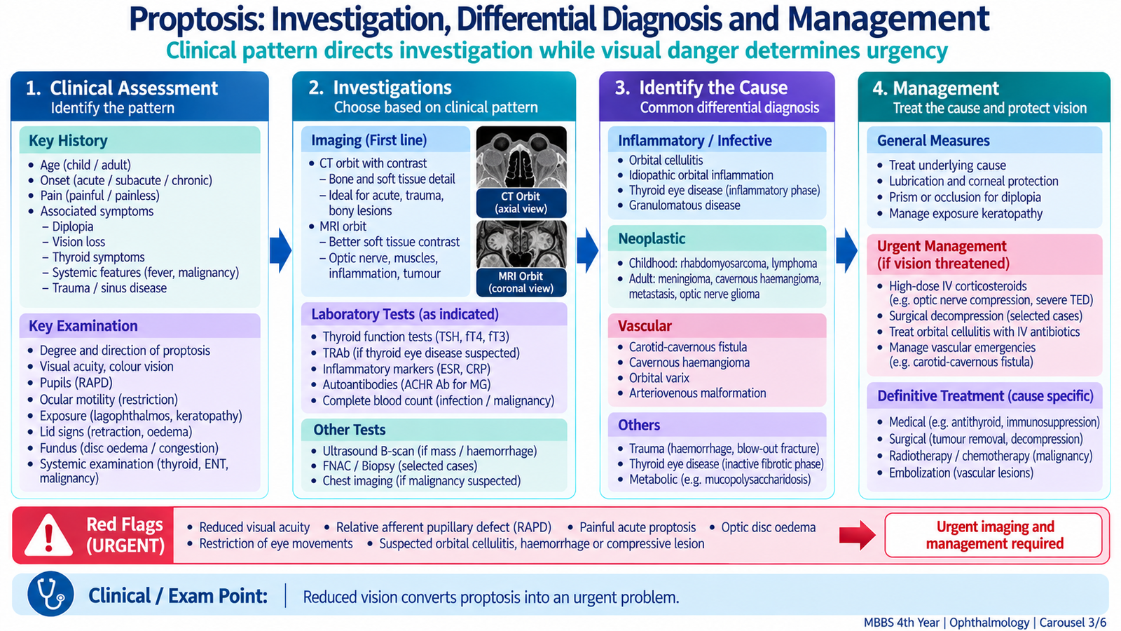
Investigations are chosen according to the suspected cause. The aim is to determine what occupies or affects the orbit, whether the optic nerve or cornea is threatened, and whether the process is inflammatory, infective, vascular or neoplastic. Management then targets the underlying disease while protecting vision.
Investigation Approach
Orbital imaging is particularly important when the cause is not obvious clinically, when a mass is suspected or when deep orbital structures must be assessed.
- CT orbit and paranasal sinuses: demonstrates the orbit, bony walls, paranasal sinuses, many masses, extraocular muscle enlargement and traumatic abnormalities. It is especially useful when sinus disease, trauma or orbital cellulitis is suspected.
- MRI: gives better soft-tissue contrast and is useful for the optic nerve, orbital apex, cavernous sinus and selected soft-tissue lesions.
- Thyroid investigations: thyroid function tests and relevant thyroid autoantibodies support assessment when thyroid eye disease is suspected.
- Inflammatory or infective testing: blood count and inflammatory markers may support suspected orbital infection or inflammation.
- Vascular imaging: is required when findings suggest a carotid-cavernous fistula or another vascular lesion.
- Histopathological assessment: biopsy may be required when an orbital tumor or infiltrative lesion cannot be diagnosed confidently by clinical and imaging findings alone.
How the Pattern Narrows the Differential Diagnosis
| Clinical Pattern | Important Possibilities | Reasoning Clue |
|---|---|---|
| Acute painful proptosis with fever | Orbital cellulitis | Infection causes orbital edema and inflammation. |
| Sudden painful proptosis after trauma | Retrobulbar hemorrhage | Rapid orbital pressure rise may threaten the optic nerve. |
| Bilateral proptosis with lid retraction | Thyroid eye disease | Extraocular muscle and orbital soft-tissue enlargement. |
| Pulsatile congested eye with bruit | Carotid-cavernous fistula | Abnormal arterial flow raises orbital venous pressure. |
| Slow painless progressive unilateral proptosis | Orbital mass | Gradually enlarging lesion displaces orbital contents. |
Management Principles
Treatment depends on the cause. However, two priorities apply to every patient: protect visual function and treat the orbital process before irreversible corneal or optic nerve damage develops.
- Corneal protection: lubricating preparations and measures to improve eyelid closure are used when exposure occurs.
- Orbital cellulitis: requires urgent systemic antimicrobial treatment and ophthalmic assessment; drainage may be necessary when a significant orbital collection is present.
- Orbital hemorrhage with visual compromise: is an ophthalmic emergency requiring immediate pressure-relieving intervention by trained clinicians.
- Orbital tumors: management depends on the lesion and may involve observation, biopsy, excision or oncological treatment.
- Vascular lesions: suspected carotid-cavernous fistula requires specialist neurovascular assessment and treatment.
- Thyroid eye disease: management depends on activity, severity, exposure and presence of optic neuropathy.
D. Thyroid Eye Disease
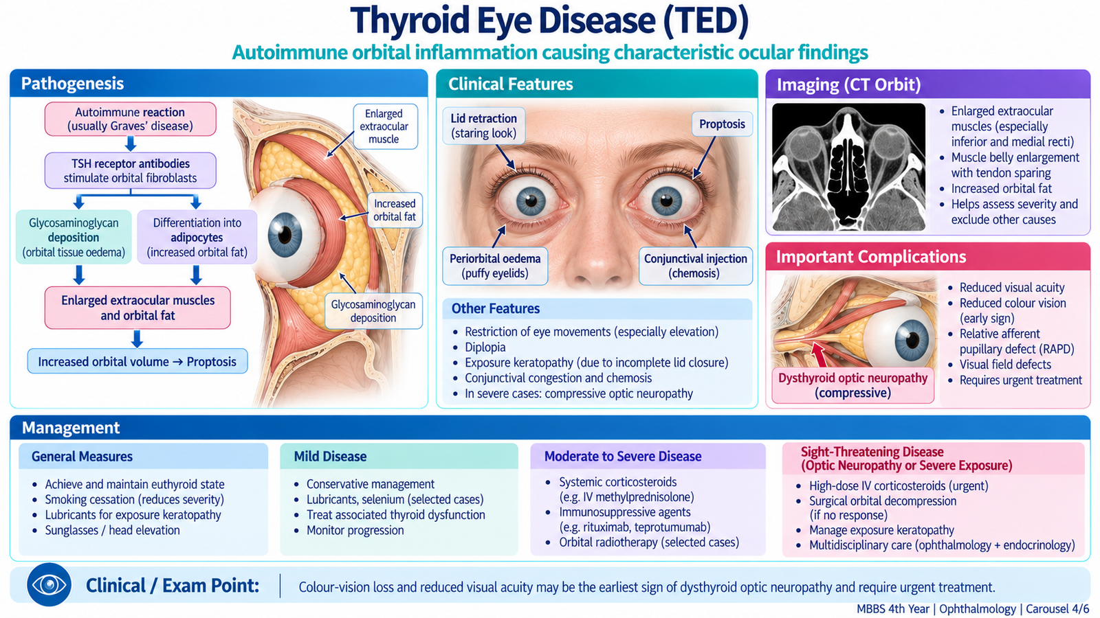
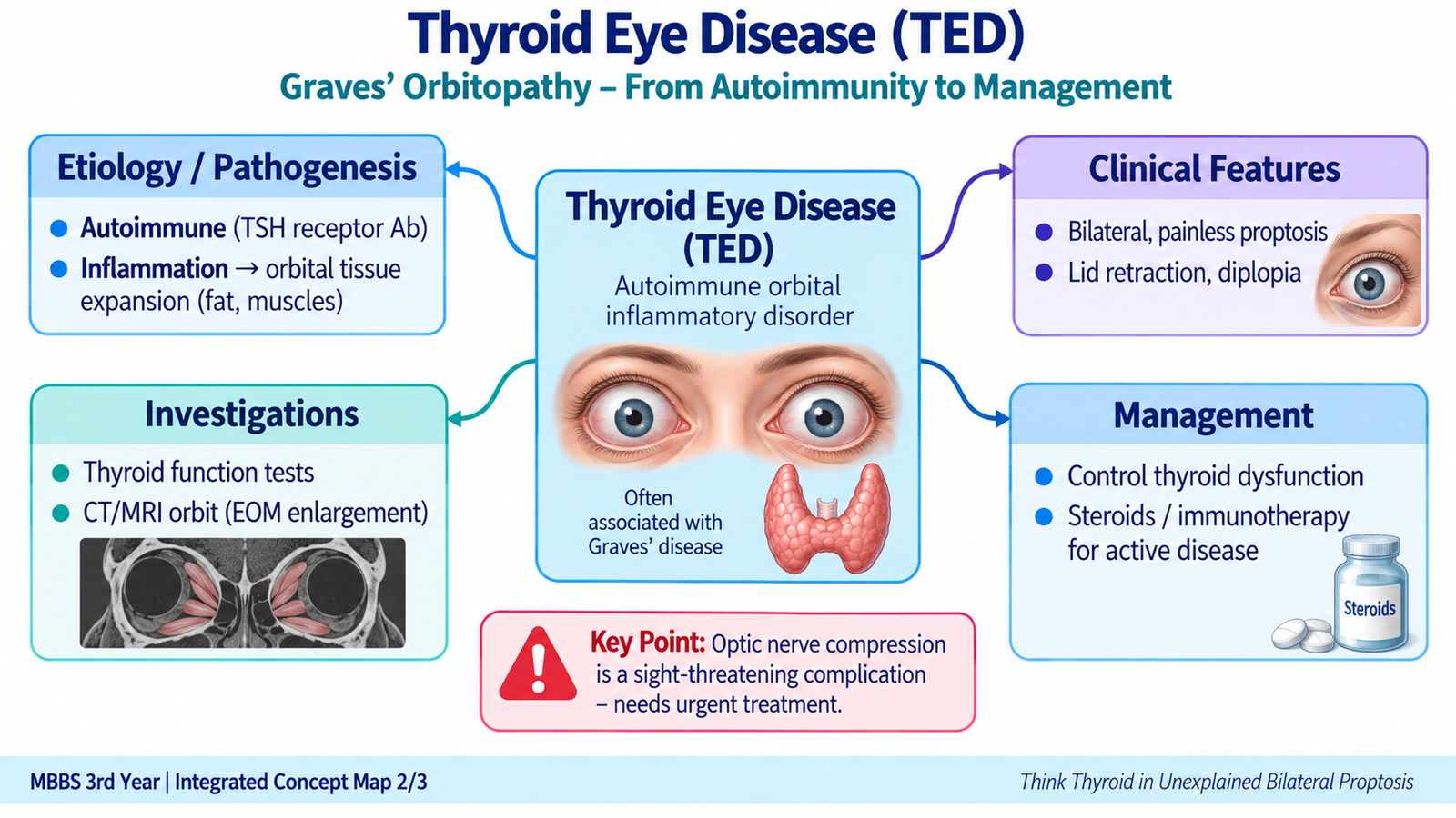
Thyroid eye disease (TED), also called thyroid-associated orbitopathy, is an autoimmune inflammatory disorder of the orbit. It is most strongly associated with Graves disease but may also occur in patients who are euthyroid or have other thyroid dysfunction. The orbital disease and the thyroid hormone state are related but are not identical processes.
Etiology and Pathogenesis
Autoimmune activity targets orbital fibroblasts and tissues within the orbit. Inflammatory cells release mediators that stimulate fibroblasts and increase production of hydrophilic glycosaminoglycans. These molecules attract water, producing edema. Orbital fat may enlarge and extraocular muscles become swollen.
Smoking is an important modifiable factor associated with more severe thyroid eye disease. Maintaining stable thyroid function is also important because thyroid dysfunction may worsen the clinical course.
Clinical Features
TED often affects both eyes, although the degree may be asymmetrical. Lid abnormalities may be the earliest visible findings.
- Lid retraction: the upper lid sits unusually high, producing a staring appearance.
- Lid lag: during downward gaze, the upper lid does not follow the globe normally.
- Proptosis: enlargement of orbital tissues pushes the globe forwards.
- Conjunctival redness and chemosis: result from inflammation and orbital venous congestion.
- Exposure symptoms: dryness, foreign-body sensation and watering occur when the exposed corneal surface dries.
- Diplopia: enlarged and later fibrotic extraocular muscles restrict ocular movement.
- Orbital pressure or discomfort: may occur during active inflammation.
Restrictive Ophthalmopathy
TED produces mechanical restriction rather than a primary nerve palsy. The inferior and medial rectus muscles are commonly involved. When the inferior rectus is tight, elevation becomes limited. When the medial rectus is involved, abduction may be restricted. This mechanical pattern helps differentiate TED from neurological causes of diplopia.
Investigations
Investigations establish thyroid status, characterize orbital involvement and identify complications.
- Thyroid function tests: determine whether the patient is hyperthyroid, hypothyroid or euthyroid.
- Thyroid autoantibodies: may support an autoimmune thyroid disorder.
- CT or MRI of the orbit: typically demonstrates enlargement of extraocular muscle bellies, often with relative sparing of the tendinous insertions. Imaging also assesses crowding at the orbital apex.
- Visual acuity, colour vision, pupils and visual fields: help detect optic nerve dysfunction.
- Corneal examination: identifies exposure keratopathy.
Activity and Severity
It is useful to distinguish active inflammatory disease from inactive residual disease. Active disease is characterized by inflammatory symptoms and signs such as pain, redness, swelling and progressive change. Inactive disease may leave persistent proptosis, diplopia or lid abnormalities after inflammation has settled. Management differs because anti-inflammatory treatment is most useful during active disease, while corrective surgery is generally planned after stability.
Management
Treatment is determined by severity and whether the disease is active. Mild disease often requires conservative ocular protection, while active moderate or severe disease requires specialist-directed anti-inflammatory treatment.
- Smoking cessation: should be strongly encouraged.
- Control of thyroid dysfunction: stable thyroid status is an important part of care.
- Lubrication: protects the ocular surface in exposure and dry-eye symptoms.
- Active moderate-to-severe disease: may require systemic glucocorticoid therapy and other specialist-directed immunomodulatory treatment.
- Persistent inactive disease: selected patients may require orbital, strabismus or eyelid surgery for functional or rehabilitative correction.
E. Myasthenia Gravis
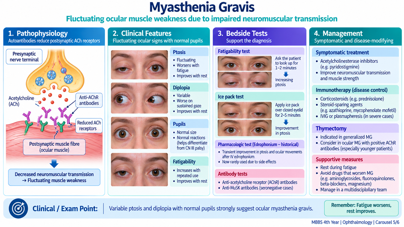
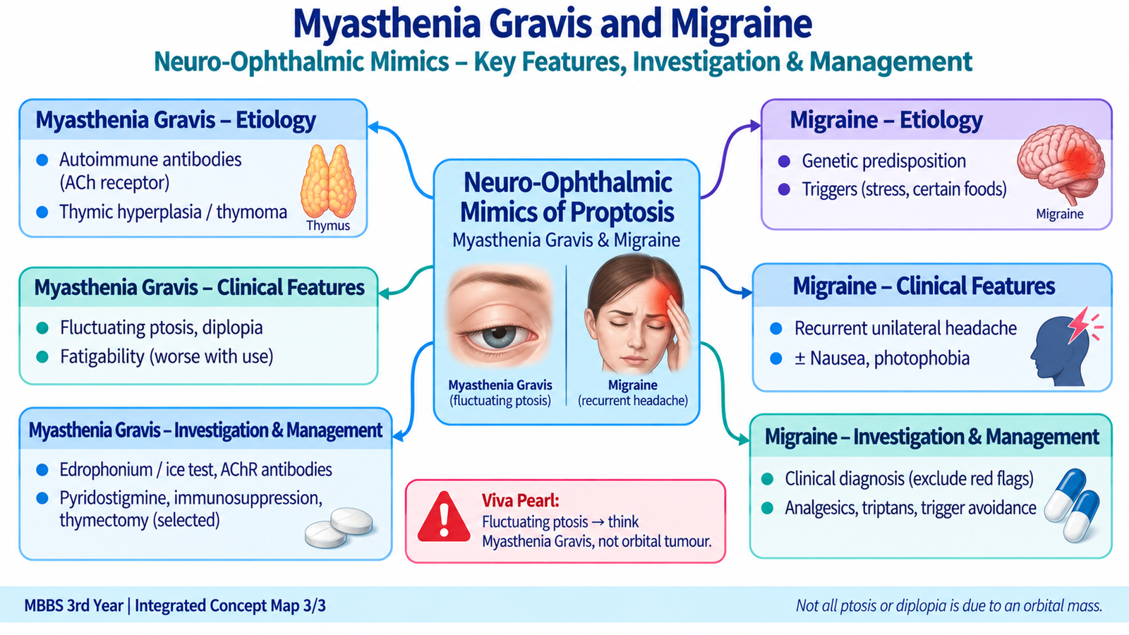
Myasthenia gravis (MG) is an autoimmune disorder of the neuromuscular junction. It causes fluctuating weakness of voluntary muscles because neuromuscular transmission becomes less effective. Ocular muscles are frequently affected, so ptosis and diplopia may be the first manifestations. This can mimic an ocular motor nerve disorder or other orbital disease, but MG does not itself cause true proptosis.
Etiology and Mechanism
In most patients, antibodies are directed against components of the postsynaptic neuromuscular junction, most commonly the acetylcholine receptor. This reduces the efficiency with which acetylcholine activates the muscle membrane.
Because repeated activity places greater demand on an already inefficient neuromuscular junction, weakness typically becomes more obvious with sustained use and improves after rest.
Ocular Clinical Features
- Ptosis: may be unilateral, bilateral or alternating and often varies during the day.
- Diplopia: results from variable weakness of extraocular muscles and may not follow the pattern of one cranial nerve palsy.
- Fluctuation: ocular findings often worsen after prolonged activity and improve with rest.
- Pupils: remain normal because pupillary muscles are not supplied through the somatic neuromuscular junction affected in MG.
- Cogan lid twitch: may be observed when the upper lid briefly overshoots after the patient looks from down gaze to primary position.
Generalized Features
The disease may remain limited to the eyes or involve other skeletal muscles. Facial weakness, difficulty chewing, dysarthria, dysphagia and weakness of the limbs or neck may occur. Respiratory muscle weakness is particularly important because it can produce a myasthenic crisis.
Investigations
The diagnosis is supported by demonstrating fatigable weakness and confirming impaired neuromuscular transmission or relevant antibodies.
- Ice-pack test: cooling may temporarily improve myasthenic ptosis, providing a useful bedside clue in suitable patients.
- Acetylcholine receptor antibodies: support autoimmune MG when positive.
- Other antibody testing: may be considered when routine antibody testing is negative but clinical suspicion remains strong.
- Repetitive nerve stimulation: may demonstrate impaired neuromuscular transmission.
- Single-fibre electromyography: is a sensitive physiological test of neuromuscular transmission.
- Chest imaging: is performed to assess the thymus because thymic abnormalities, including thymoma, are associated with MG.
Management
Management aims to improve neuromuscular transmission, control the autoimmune process and identify patients at risk of respiratory or bulbar deterioration.
- Acetylcholinesterase inhibition: pyridostigmine improves the availability of acetylcholine at the neuromuscular junction and provides symptomatic benefit.
- Immunosuppression: corticosteroids and other immunomodulatory treatments are used when symptoms require immune control.
- Thymectomy: is required for thymoma and may be beneficial in selected patients with generalized autoimmune MG.
- Myasthenic crisis: respiratory or severe bulbar weakness requires urgent hospital care, respiratory support when necessary and rapid specialist treatment such as intravenous immunoglobulin or plasma exchange.
F. Migraine and Neuro-Ophthalmic Differentiation
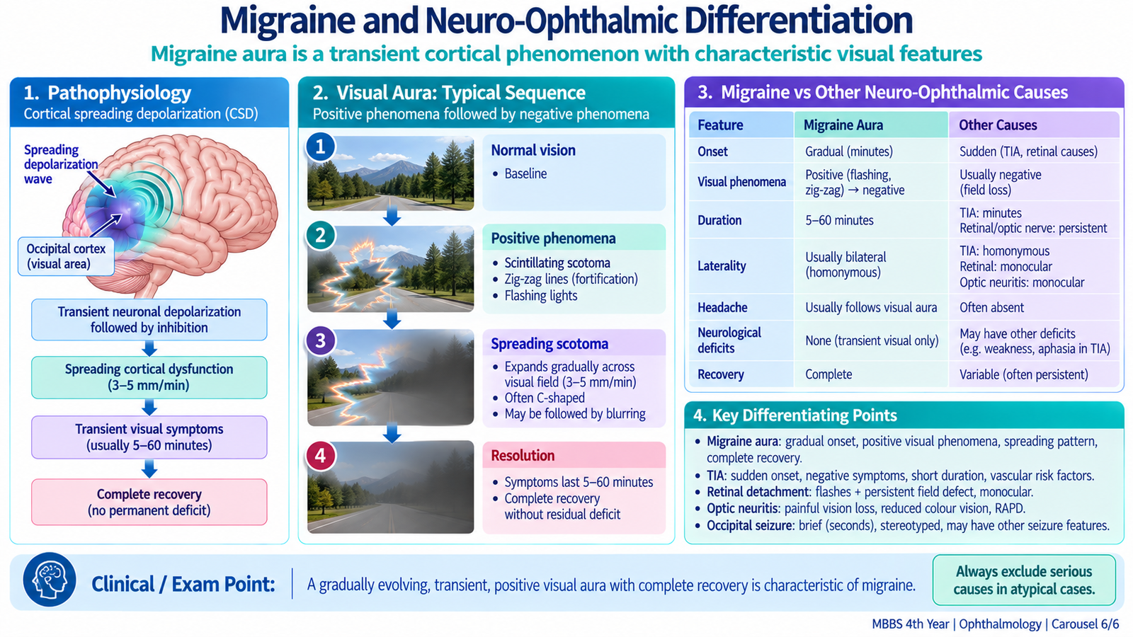
Migraine is a recurrent neurological disorder characterized by episodic headache and associated neurological symptoms. Visual symptoms are common and may lead patients to seek ophthalmic assessment. The key ophthalmic task is to recognize the typical reversible visual pattern while identifying features that suggest retinal, optic-nerve, vascular or other neurological disease instead.
Etiology and Mechanism
Migraine results from altered excitability and activation of pain-processing pathways within the nervous system. In migraine with aura, a temporary wave of altered cortical neuronal activity can affect the visual cortex. This explains why typical visual aura usually involves the visual field of both eyes rather than a structural abnormality in one eye.
Clinical Features
Migraine headache is commonly recurrent and may be unilateral and pulsating. Physical activity can aggravate it, and nausea, photophobia or phonophobia may accompany the attack. Not every patient has an aura.
A typical visual aura develops gradually and may contain positive visual phenomena such as flashing lights, shimmering lines or zig-zag patterns. A scotoma or area of reduced vision may accompany these positive symptoms. The disturbance usually resolves completely.
Why the Visual History Matters
Asking whether the disturbance affects one eye or both visual fields is important. A patient may describe the symptom as affecting “one side,” while the actual defect may involve the same half of the visual field in both eyes because the origin is cortical.
- Gradual development of shimmering or zig-zag phenomena: supports migraine aura.
- Transient binocular field disturbance: supports a posterior visual pathway origin.
- Persistent monocular visual loss: is not typical of ordinary visual aura and requires ocular or vascular evaluation.
- New neurological deficit: requires assessment for causes other than uncomplicated migraine.
Investigations
Typical recurrent migraine with a characteristic neurological examination often does not require extensive investigation. Investigation becomes important when the presentation is new, atypical or accompanied by concerning neurological or ophthalmic findings. Neuroimaging is considered when secondary headache or structural neurological disease is suspected.
Ophthalmic examination is particularly important when visual symptoms are persistent, truly monocular, associated with a red painful eye, or accompanied by abnormal visual acuity, pupils, optic disc or retina.
Management
Management depends on the frequency and severity of attacks. General measures include recognizing individual triggers, maintaining regular sleep and meals, and avoiding excessive use of acute headache medication.
- Acute attacks: appropriate analgesics or non-steroidal anti-inflammatory drugs may be used; triptans are used in suitable patients when migraine-specific therapy is required.
- Frequent or disabling migraine: preventive treatment may be considered using established preventive drug classes according to the patient’s clinical circumstances.
- Atypical visual or neurological symptoms: should not simply be attributed to migraine without appropriate assessment.
3. Integrated Mechanism Flow
4. Important Comparison
Thyroid eye disease, ocular myasthenia and migraine can all present with ocular or visual complaints, but the mechanism and examination findings are very different.
| Feature | Thyroid Eye Disease | Myasthenia Gravis | Migraine |
|---|---|---|---|
| Main problem | Autoimmune orbital inflammation | Neuromuscular-junction weakness | Neurological headache disorder |
| True proptosis | Common | Absent | Absent |
| Ptosis | Not characteristic; lid retraction is typical | Characteristic and fluctuating | Not typical |
| Diplopia | Restrictive and relatively fixed | Variable and fatigable | Not a usual defining feature |
| Pupils | Usually normal unless optic nerve compromised | Normal | Usually no persistent abnormality in uncomplicated migraine |
| Typical visual clue | Proptosis + lid retraction | Variable ptosis/diplopia | Reversible spreading visual aura |
| Key investigation | Thyroid assessment + orbital imaging when indicated | Antibody and neuromuscular testing | Mainly clinical; investigate atypical/red-flag presentations |