
Topic 9 — Angle-Closure, Secondary Glaucoma & Treatment
Topic Introduction
Glaucoma is a group of disorders in which the optic nerve may be damaged, usually in association with an intraocular pressure that is too high for that eye. In this topic, the central problem is obstruction of aqueous humor outflow. In primary angle closure, the peripheral iris blocks access to the trabecular meshwork. Secondary glaucomas develop because another ocular disorder, such as retinal ischemia or an abnormal lens, interferes with aqueous drainage. Some forms develop gradually, while acute angle closure can produce severe pain and rapid visual damage. Understanding the mechanism makes the clinical findings, investigations and treatment much easier: first identify why the pressure has risen, then lower the pressure and correct the underlying cause whenever possible.
A. Primary Angle-Closure Disease: Mechanism and Stages
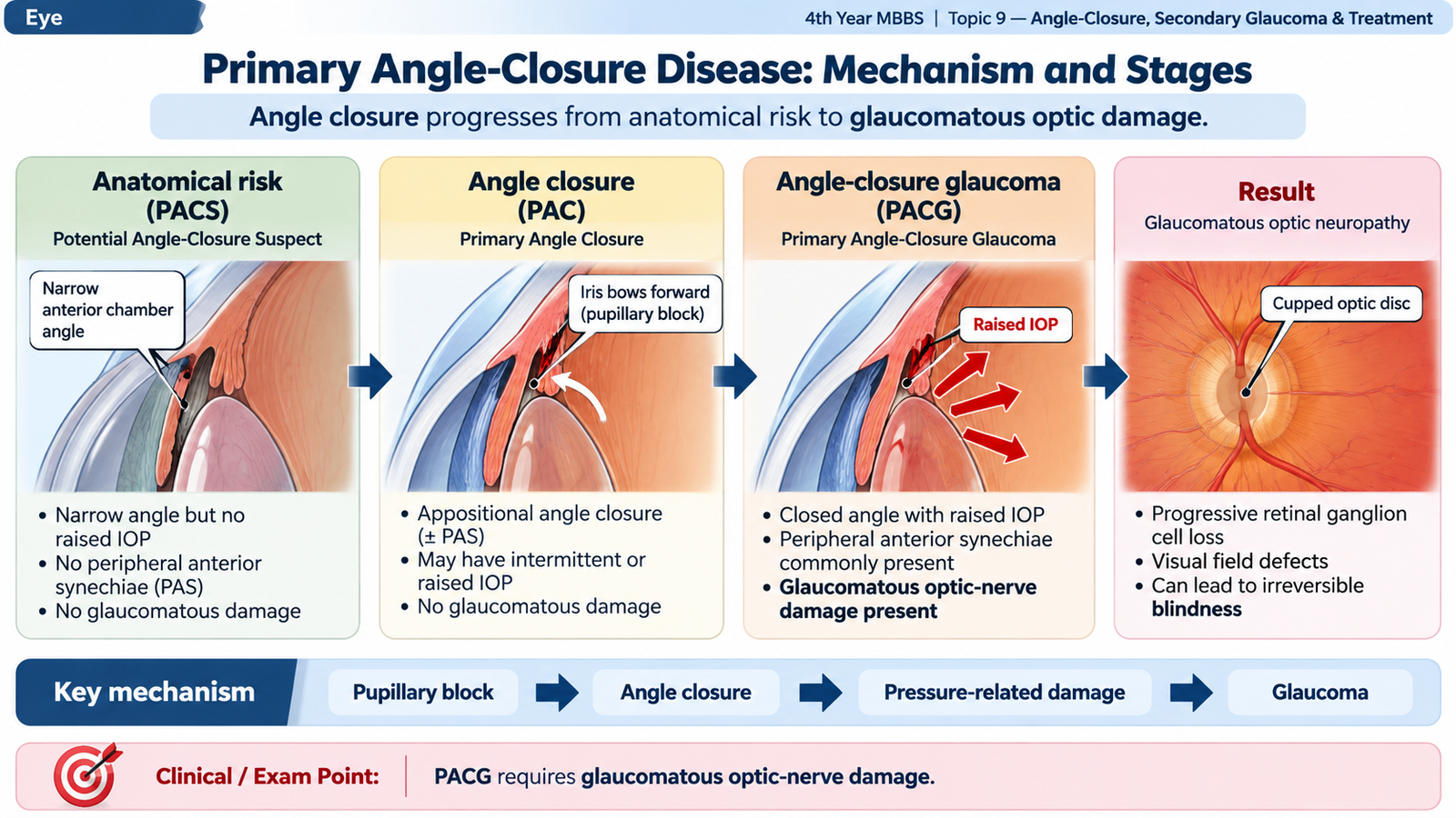
Primary angle-closure disease occurs in an eye in which the anterior chamber angle is anatomically narrow and can become obstructed by the peripheral iris. The anterior chamber angle contains the trabecular meshwork, which is the main conventional route for aqueous humor drainage. When the iris covers this drainage area, aqueous outflow falls and intraocular pressure may rise.
Relevant aqueous physiology
Aqueous humor is produced by the ciliary processes in the posterior chamber. It passes through the pupil into the anterior chamber and then drains mainly through the trabecular meshwork and Schlemm’s canal. Therefore, the trabecular meshwork must remain accessible for normal drainage.
How pupillary block closes the angle
The common mechanism of primary angle closure is relative pupillary block. Aqueous encounters increased resistance while passing between the posterior surface of the iris and the anterior lens. Pressure therefore becomes relatively higher behind the iris. This pushes the peripheral iris forward, producing iris bombé. The bowed peripheral iris then contacts and obstructs the trabecular meshwork.
An anatomically shallow anterior chamber, a relatively thick or anteriorly positioned lens and a short hypermetropic eye can bring the iris and lens closer together and increase the tendency to angle closure. Aging may increase this tendency because the crystalline lens becomes thicker. Plateau iris configuration is another mechanism in which the peripheral iris remains crowded against the angle because of its anatomical configuration, even after pupillary block has been relieved.
Primary angle-closure spectrum
Modern terminology separates anatomical angle closure from established glaucomatous optic-nerve damage. This distinction is important because an eye may have a dangerously narrow or closed angle before glaucoma has actually damaged the optic nerve.
Peripheral anterior synechiae (PAS) are permanent adhesions between the peripheral iris and the angle structures. Repeated or prolonged iridotrabecular contact can therefore convert reversible appositional closure into permanent structural closure.
Clinical course terminology
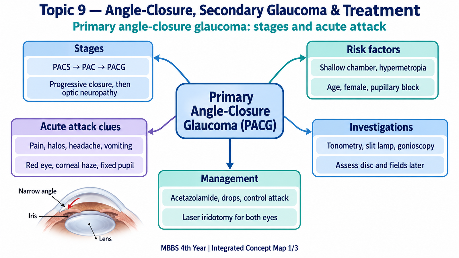
Students may also encounter traditional descriptions such as latent or predisposed disease, intermittent or subacute attacks, acute angle closure, chronic angle closure and end-stage or absolute glaucoma. These terms describe the clinical course. The modern PACS → PAC → PACG spectrum more clearly identifies whether structural angle damage and glaucomatous optic neuropathy are present.
B. Acute Angle Closure: Etiology, Recognition, Investigations and Management
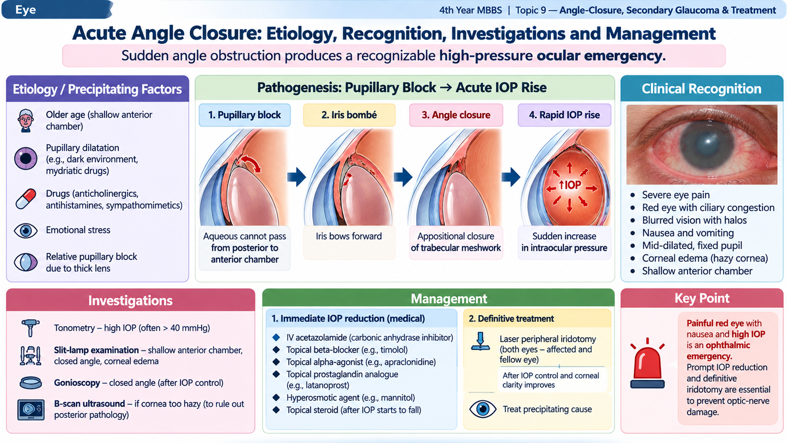
Acute angle closure occurs when the anterior chamber angle closes suddenly and intraocular pressure rises rapidly. The sudden pressure rise produces severe ocular symptoms and can damage the optic nerve if treatment is delayed. It is therefore a sight-threatening ophthalmic emergency.
Etiology and precipitating factors
The usual primary mechanism is pupillary block in an anatomically predisposed eye. Anything that increases contact between the iris and lens or crowds the peripheral iris toward the angle may precipitate an attack.
- Narrow anterior chamber angle and shallow anterior chamber.
- Hypermetropic or relatively short eye, in which anterior segment structures are more crowded.
- Increasing lens thickness with age, which reduces available anterior segment space.
- Family or anatomical predisposition to narrow angles.
- Pupillary dilatation, for example in darkness or after certain anticholinergic or sympathomimetic drugs, may trigger closure in a susceptible eye.
- Plateau iris configuration may produce persistent or recurrent angle crowding by a non-pupillary-block component.
Why the symptoms occur
Corneal endothelial function is disturbed by the very high pressure, so the cornea becomes edematous and loses transparency. This explains the sudden blurring and colored halos around lights. Severe ocular pain may be accompanied by headache, nausea and vomiting. These systemic symptoms can occasionally distract attention from the eye.
Clinical presentation
- Sudden severe ocular or periocular pain
- Red eye
- Rapid reduction of vision
- Colored halos around lights
- Headache
- Nausea and vomiting
- Reduced visual acuity
- Conjunctival and ciliary congestion
- Hazy or edematous cornea
- Shallow anterior chamber
- Mid-dilated, sluggish or fixed pupil
- Markedly raised intraocular pressure
- Closed anterior chamber angle
Interpreting the ophthalmic examination
Corneal edema: The high intraocular pressure interferes with normal corneal endothelial fluid control. The cornea becomes hazy, producing blurred vision and halos.
Shallow anterior chamber: The peripheral iris has moved forward and is obstructing the angle. Comparing anterior chamber depth with the fellow eye can be helpful.
Mid-dilated pupil: High pressure causes iris sphincter ischemia and dysfunction. The pupil therefore becomes poorly reactive and typically remains in a mid-dilated position.
High intraocular pressure: The trabecular drainage pathway is suddenly obstructed, so aqueous continues to be produced but cannot drain normally.
Fellow eye: In primary disease, the fellow eye often has a similar narrow anatomical configuration and must not be ignored.
Investigations
Acute angle closure is mainly a clinical diagnosis supported by ophthalmic examination. Investigations should confirm raised pressure, demonstrate the angle problem and assess whether glaucomatous damage has occurred.
- Visual acuity: documents functional visual reduction and provides a baseline for recovery.
- Slit-lamp examination: shows corneal edema, shallow anterior chamber, pupil abnormality and associated inflammation.
- Tonometry: confirms elevated intraocular pressure and helps assess response to treatment.
- Gonioscopy: is the key examination for confirming angle closure. During a severe acute attack, corneal edema may make gonioscopy difficult, so definitive angle assessment may become easier after initial pressure reduction and corneal clearing.
- Optic-disc and fundus examination: should be performed when the media become sufficiently clear to assess glaucomatous or retinal pathology.
- Visual-field assessment: becomes important after the acute episode when established glaucomatous damage is suspected.
Emergency management
Treatment has two objectives. The first is to lower intraocular pressure rapidly and reduce ocular inflammation. The second is to relieve the mechanism causing angle closure so that the attack does not recur.
1. Rapid reduction of intraocular pressure
A combination of aqueous-suppressing drugs is commonly required because a single drug may not lower the pressure sufficiently during a severe attack.
- Topical beta-blocker: reduces aqueous humor production.
- Topical alpha-2 agonist: reduces aqueous production and also increases uveoscleral outflow.
- Carbonic anhydrase inhibitor: topical or systemic treatment reduces aqueous production; systemic acetazolamide is useful when rapid pressure reduction is required.
- Hyperosmotic agent: may be used when the pressure remains dangerously high despite initial treatment. It reduces ocular volume by drawing water osmotically from the eye.
2. Reduce inflammation and symptoms
Topical corticosteroid reduces the inflammatory response produced by the acute attack. Analgesic and antiemetic treatment may be required because pain, nausea and vomiting can be severe.
3. Pilocarpine when appropriate
Pilocarpine constricts the pupil and contracts the ciliary muscle, helping pull the peripheral iris away from the trabecular meshwork in pupillary-block disease. When the intraocular pressure is extremely high, however, the iris sphincter may be ischemic and initially unresponsive. Pilocarpine is therefore more useful after pressure has begun to fall rather than as the only first treatment.
4. Definitive relief of pupillary block
Laser peripheral iridotomy creates a small opening in the peripheral iris. Aqueous can then pass directly from the posterior chamber to the anterior chamber, equalizing pressure across the iris. This flattens iris bombé and removes the pupillary-block component of angle closure.
Iridotomy is performed once the eye is sufficiently stabilized and the cornea is clear enough for laser treatment. The fellow eye is also carefully assessed because it commonly shares the same narrow-angle anatomy and may require prophylactic iridotomy.
Laser peripheral iridoplasty may be considered when appositional closure persists because of plateau iris or when opening the angle requires contraction of the peripheral iris. Lens extraction becomes particularly important when a large or cataractous lens contributes substantially to anterior segment crowding.
C. Neovascular Glaucoma: Ischemia-Driven Secondary Glaucoma
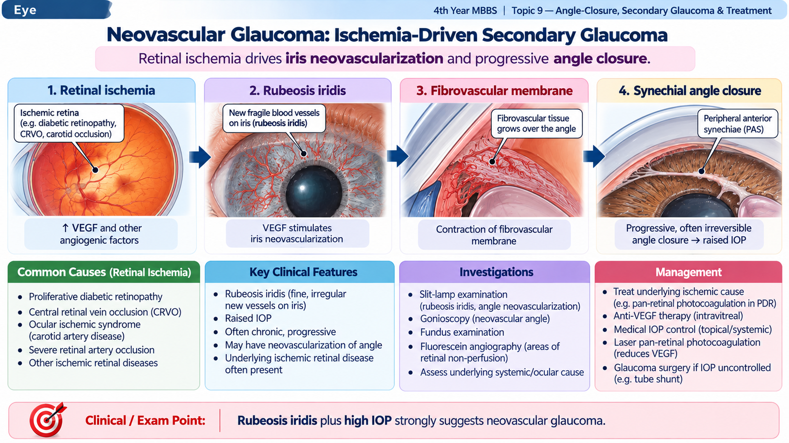
Neovascular glaucoma is a severe secondary glaucoma caused by retinal or ocular ischemia. Ischemic retina releases angiogenic mediators, particularly vascular endothelial growth factor (VEGF), which stimulate abnormal new vessels on the iris and within the anterior chamber angle. These vessels are accompanied by fibrovascular tissue that eventually obstructs and contracts across the aqueous drainage pathway.
Etiology
The important initiating problem is extensive retinal ischemia. Common clinically important causes include:
- Proliferative diabetic retinopathy
- Severe ischemic central retinal vein occlusion
- Ocular ischemic syndrome and other severe retinal ischemic disorders
Pathogenesis
The angle can therefore be relatively open early in the disease even though abnormal vessels are already present. Later, contraction of the fibrovascular membrane pulls the peripheral iris forward and produces permanent synechial angle closure. This explains why early recognition of iris neovascularization is important.
Clinical features and visual recognition
The typical patient already has severe retinal vascular disease and develops worsening vision, ocular pain and redness. Examination should deliberately look for abnormal vessels rather than assuming that every painful high-pressure eye has primary angle closure.
- Reduced vision: may result from both the underlying retinal ischemic disease and the raised intraocular pressure.
- Ocular pain and redness: become prominent as pressure rises.
- Rubeosis iridis: fine abnormal new vessels are seen on the iris, particularly around the pupillary margin and iris surface.
- Angle neovascularization: gonioscopy may show new vessels crossing structures where normal angle vessels should not run.
- Raised intraocular pressure: results from obstruction of trabecular outflow and later synechial angle closure.
- Corneal edema: may appear when pressure becomes markedly elevated.
- Peripheral anterior synechiae: indicate contraction and structural closure of the angle in advanced disease.
Investigations
Investigation has two purposes: confirm the glaucoma mechanism and identify the ischemic retinal disease that is driving neovascularization.
- Visual acuity: documents the degree of functional loss.
- Slit-lamp examination: identifies iris neovascularization, inflammation and corneal edema.
- Tonometry: measures the pressure elevation.
- Gonioscopy: detects angle neovascularization and shows whether the angle remains open or has developed synechial closure.
- Dilated fundus examination: searches for proliferative diabetic retinopathy, retinal vein occlusion or another ischemic retinal disorder.
- Fundus fluorescein angiography: may help demonstrate and define retinal non-perfusion when clinically required and when the fundus can be adequately visualized.
Management
Simply lowering the intraocular pressure is not enough. Neovascular glaucoma is driven by retinal ischemia, so effective management must address both the high pressure and the ischemic stimulus for new-vessel formation.
Control the ischemic drive
Panretinal photocoagulation (PRP) treats ischemic retina and reduces the continuing angiogenic stimulus when adequate retinal visualization is possible. Intravitreal anti-VEGF therapy can produce rapid regression of anterior-segment neovascularization and is particularly useful as an adjunct while definitive retinal treatment is arranged. Its effect on the ischemic stimulus is not a substitute for treating the underlying ischemic retina.
Lower intraocular pressure
Aqueous-suppressing drugs such as topical beta-blockers, alpha-2 agonists and carbonic anhydrase inhibitors are commonly useful. Systemic carbonic anhydrase inhibition may be required for a severe pressure rise. Miotics are generally not favored in an inflamed neovascular eye and do not correct the fibrovascular mechanism.
Control inflammation and pain
Topical anti-inflammatory treatment and appropriate pain control may be required because neovascular glaucoma is frequently painful and may have significant anterior-segment inflammation.
Surgery when pressure remains uncontrolled
Severe or refractory neovascular glaucoma frequently requires specialist glaucoma surgery. A glaucoma drainage device is an important option when medical therapy cannot maintain adequate pressure control. Cyclodestructive treatment may be considered in refractory disease, particularly when visual potential is poor and the major goal is pressure and pain control.
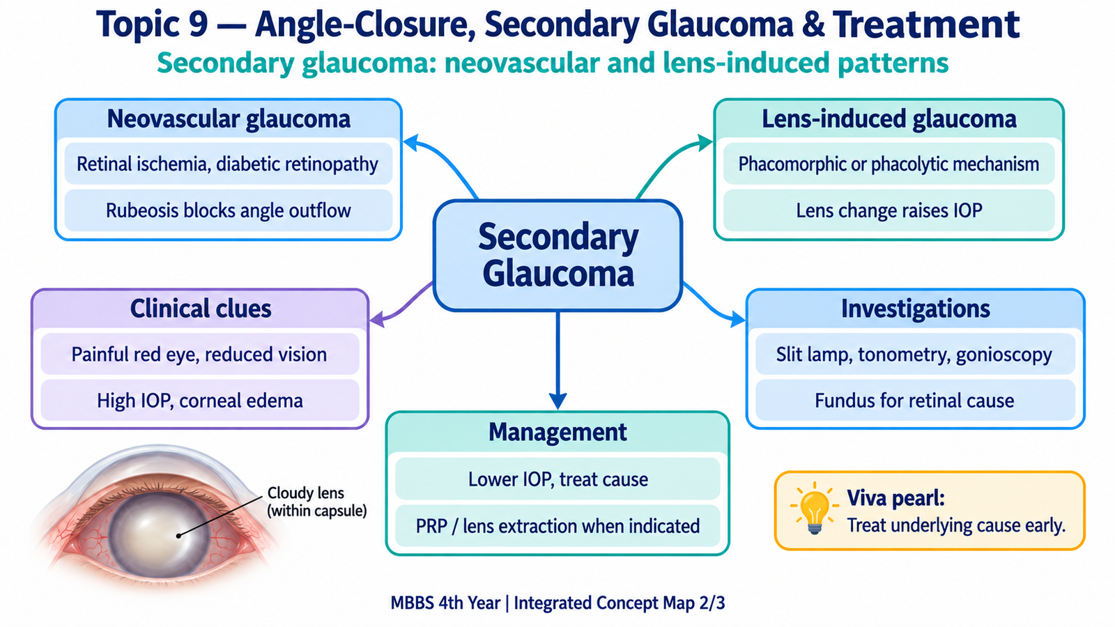
D. Lens-Induced Glaucoma: Phacomorphic, Phacolytic and Related Forms
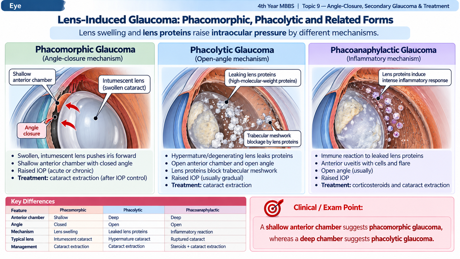
Lens-induced glaucoma refers to secondary elevation of intraocular pressure caused by abnormalities of the crystalline lens or escaped lens material. The lens can raise pressure by two major mechanisms: it may become enlarged and mechanically close the anterior chamber angle, or lens proteins and lens material may obstruct the trabecular meshwork and produce inflammation.
Phacomorphic glaucoma
Phacomorphic glaucoma is a secondary angle-closure glaucoma produced by a swollen or intumescent cataractous lens. As the lens enlarges, it pushes the iris-lens diaphragm forward and reduces anterior chamber depth. The peripheral iris then obstructs the trabecular meshwork.
The patient commonly presents with a painful red eye and marked reduction of vision in an eye with an advanced cataract. Examination shows corneal edema, a shallow anterior chamber, a swollen cataractous lens and raised intraocular pressure. Gonioscopy, when possible, demonstrates angle closure.
Phacolytic glaucoma
Phacolytic glaucoma develops in an eye with a hypermature cataract. Lens proteins leak through the lens capsule and enter the anterior chamber. These proteins, together with macrophages that ingest lens material, obstruct the trabecular meshwork. Unlike phacomorphic glaucoma, the main problem is trabecular blockage rather than mechanical angle crowding.
The eye is painful and red, vision is poor because of the cataract, and intraocular pressure is elevated. The anterior chamber is usually relatively deep because the lens is not closing the angle mechanically. Cells, flare and proteinaceous material may be present in the anterior chamber.
Other lens-related mechanisms
Lens-particle glaucoma occurs when lens material enters the anterior chamber after disruption of the lens capsule, for example following ocular trauma or ocular surgery. The particles mechanically obstruct trabecular drainage and may also provoke inflammation.
Lens-protein-related inflammatory glaucoma may occur when exposure to lens proteins produces significant intraocular inflammation. In these forms, raised pressure results from inflammatory material and impaired trabecular function.
Clinical evaluation and investigations
The diagnosis depends heavily on recognizing the abnormal lens and deciding whether the angle is mechanically closed or the trabecular meshwork is being blocked by lens material.
- Visual acuity: is usually markedly reduced because of the underlying cataract as well as the raised pressure.
- Slit-lamp examination: identifies an intumescent or hypermature cataract, anterior chamber depth, inflammation and lens particles or proteinaceous material.
- Tonometry: confirms the pressure elevation.
- Gonioscopy: distinguishes the closed angle of phacomorphic glaucoma from the usually open angle of phacolytic glaucoma.
- Fundus examination: should be attempted when possible. If a dense cataract prevents visualization and posterior segment information is clinically required, ocular ultrasonography may be used to assess structures behind the opaque lens.
- History of trauma or recent ocular surgery: strongly supports a lens-particle mechanism when retained lens material is present.
Management
Treatment initially controls the pressure and inflammation, but the abnormal lens remains the underlying cause. Therefore, definitive management requires removal of the causative lens or retained lens material once the eye is adequately stabilized.
Initial control
- Aqueous-suppressing antiglaucoma drugs lower intraocular pressure.
- Systemic carbonic anhydrase inhibition or a hyperosmotic agent may be required when the pressure is severely elevated.
- Topical corticosteroid is useful when significant lens-induced inflammation is present.
- Miotics are not routinely useful in lens-induced glaucoma and may aggravate anterior segment crowding in a phacomorphic eye.
Definitive treatment
Phacomorphic glaucoma: after initial pressure and inflammatory control, cataract extraction removes the bulky lens responsible for angle crowding.
Phacolytic glaucoma: cataract extraction removes the source of leaking lens proteins and is therefore definitive treatment.
Lens-particle glaucoma: retained lens material may need surgical removal if it is causing persistent inflammation or pressure elevation.
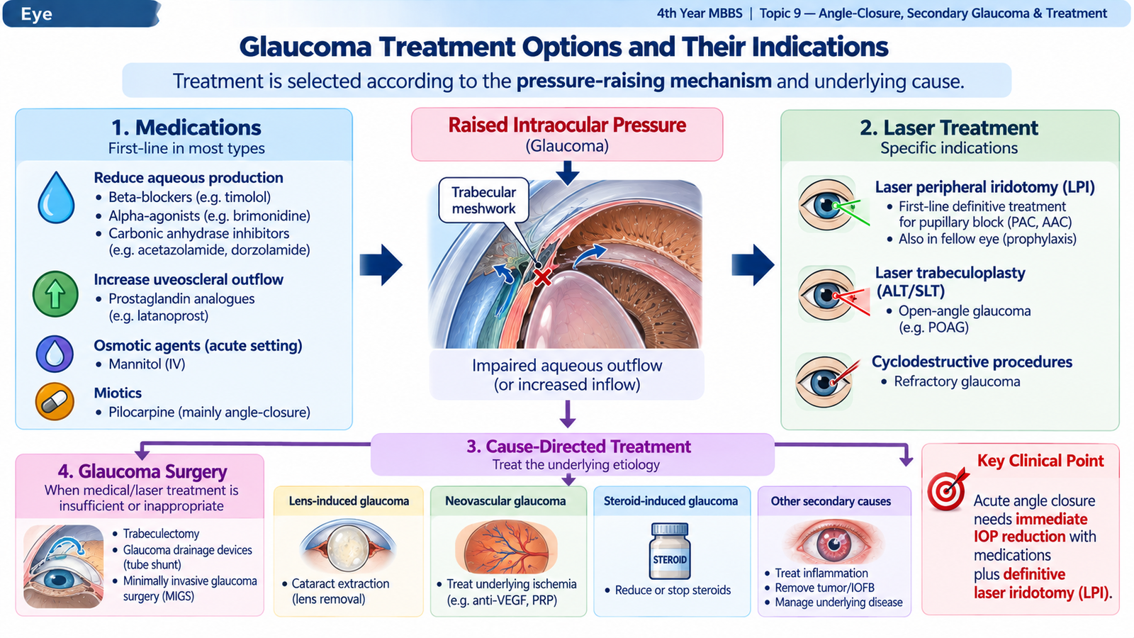
E. Glaucoma Treatment Options and Their Indications
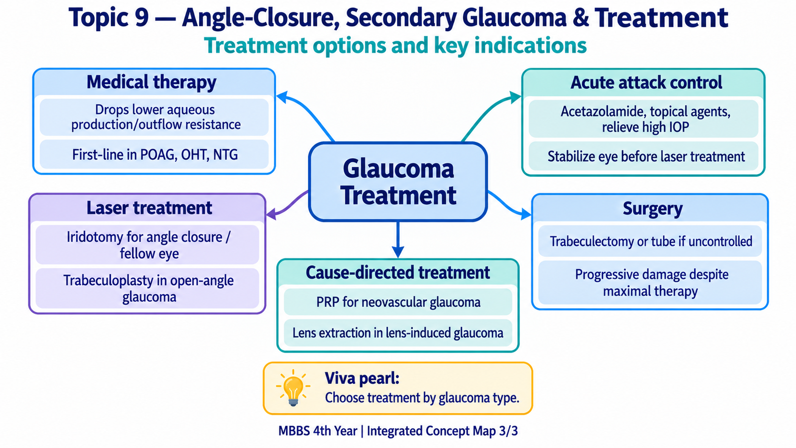
Glaucoma treatment aims to preserve vision by reducing intraocular pressure to a level at which further optic-nerve damage is unlikely. The best treatment depends on the mechanism of glaucoma, severity of disease, required pressure reduction, ocular anatomy, contraindications to medication and whether a reversible cause can be corrected. Treatment may therefore involve medication, laser treatment, correction of the underlying ocular cause, conventional glaucoma surgery or cyclodestructive procedures.
Medical treatment
Antiglaucoma drugs work mainly by either reducing aqueous humor production or increasing aqueous outflow. Chronic open-angle disease can often be treated initially with topical therapy, whereas an acute pressure crisis usually requires rapidly acting combinations.
| Treatment class | Main mechanism | Important indication / role | Important caution |
|---|---|---|---|
| Prostaglandin analogues | Increase mainly uveoscleral aqueous outflow | Common long-term treatment for open-angle glaucoma and ocular hypertension | Can cause conjunctival hyperemia, iris pigmentation and eyelash growth; not relied upon for an acute angle-closure emergency |
| Topical beta-blockers | Reduce aqueous production | Chronic therapy or part of rapid multi-drug pressure reduction | Use cautiously or avoid in significant asthma, bradycardia or heart block |
| Alpha-2 agonists | Reduce aqueous production and increase uveoscleral outflow | Adjunctive chronic therapy and rapid IOP reduction in acute situations | May cause ocular allergy, dry mouth, fatigue or systemic hypotensive effects |
| Carbonic anhydrase inhibitors | Reduce aqueous production by the ciliary epithelium | Topical drugs for chronic adjunctive therapy; systemic acetazolamide for rapid short-term reduction in severe IOP elevation | Systemic therapy requires attention to renal function, electrolytes and metabolic adverse effects |
| Miotics such as pilocarpine | Ciliary muscle contraction increases trabecular outflow and miosis pulls peripheral iris away from the angle | Most important in pupillary-block angle closure after initial IOP reduction | Not useful as sole initial therapy when iris ischemia prevents pupillary response; not routinely used in phacomorphic glaucoma |
| Hyperosmotic agents | Create an osmotic gradient that reduces ocular volume | Short-term emergency treatment when IOP is dangerously high or insufficiently responsive to initial therapy | Systemic fluid and cardiovascular or renal status must be considered |
Laser treatment
Laser peripheral iridotomy: indicated when pupillary block is responsible for primary angle closure. It is the definitive treatment after stabilization of an acute pupillary-block attack and is also used in appropriately selected fellow eyes or other eyes at significant risk of pupillary-block closure.
Laser trabeculoplasty: improves aqueous outflow through the trabecular meshwork and is used in open-angle glaucoma when the drainage angle is accessible. Selective laser trabeculoplasty may be used as initial treatment or as an adjunct when additional pressure lowering or reduced medication burden is desired.
Laser peripheral iridoplasty: contracts the peripheral iris and can help widen an angle that remains appositionally narrow, particularly in plateau iris configuration or when the pupillary-block component has already been addressed.
Panretinal photocoagulation: is not primarily a pressure-lowering laser procedure. In neovascular glaucoma it treats the ischemic retinal drive responsible for VEGF production and therefore addresses the underlying cause of neovascularization.
Lens and cause-directed treatment
Secondary glaucoma cannot always be treated adequately by lowering IOP alone. If a swollen cataract causes phacomorphic glaucoma or lens proteins produce phacolytic glaucoma, the definitive treatment is removal of the causative lens after initial stabilization. Similarly, neovascular glaucoma requires treatment of the ischemic retina as well as pressure control.
Glaucoma surgery
Surgery is considered when adequate pressure control cannot be achieved with appropriate tolerated medication and laser, when glaucomatous damage continues to progress, or when the particular form of glaucoma is unlikely to respond adequately to less invasive treatment.
Trabeculectomy: creates an alternative pathway for aqueous drainage from the anterior chamber to a subconjunctival filtering bleb. It is an important operation for glaucoma requiring substantial pressure lowering when medical or laser therapy is inadequate.
Glaucoma drainage devices: divert aqueous through a tube to an external plate. They are particularly useful in refractory glaucomas and many secondary glaucomas, including difficult neovascular glaucoma.
Lens extraction: is specifically indicated in lens-induced glaucoma and may also help selected angle-closure eyes in which a large lens is a major contributor to anterior segment crowding.
Cyclodestructive procedures: reduce aqueous production by treating part of the ciliary body. They are generally reserved for selected refractory glaucomas, especially when conventional pressure-control methods have failed or visual potential is poor.
Integrated Mechanism Flow
Although the initiating causes differ, the major glaucomas in this topic converge on one central problem: inadequate aqueous outflow and a harmful rise in intraocular pressure.
Pupillary block, retinal ischemia or lens abnormality
Angle closure or trabecular blockage
Aqueous production exceeds drainage
Pain, corneal edema and possible optic-nerve injury
Lower IOP rapidly and correct the cause
Important Comparison
The quickest way to separate the major secondary and angle-closure glaucomas in this topic is to combine the lens or iris appearance with anterior chamber depth and gonioscopic angle findings.
| Condition | Main mechanism | Anterior segment / angle | High-yield clue | Cause-directed treatment |
|---|---|---|---|---|
| Acute primary angle closure | Usually relative pupillary block | Shallow chamber; closed angle | Severe pain, halos, corneal edema, mid-dilated pupil | Laser peripheral iridotomy after initial IOP control |
| Neovascular glaucoma | Retinal ischemia → VEGF → fibrovascular angle membrane | May be open early; later synechially closed | Rubeosis iridis and angle neovascularization | Treat retinal ischemia with PRP ± anti-VEGF plus IOP control |
| Phacomorphic glaucoma | Swollen cataractous lens mechanically crowds angle | Shallow chamber; closed angle | Intumescent cataract with painful high-pressure eye | Cataract extraction after initial stabilization |
| Phacolytic glaucoma | Lens proteins and macrophages obstruct trabecular meshwork | Usually relatively deep chamber; open angle | Hypermature cataract with inflammatory material and high IOP | Cataract extraction after pressure and inflammation control |