
This chapter follows the KMU learning outcomes in a logical sequence. First understand how infection enters and spreads inside the eye, then connect this with the examination findings, investigations and emergency treatment. Revise the final high-yield points only after the main concepts are clear.
Topic 11 — Endophthalmitis & Panophthalmitis
A sight-threatening group of intraocular infections in which rapid recognition, appropriate investigation and urgent treatment are essential for preserving vision and, in severe panophthalmitis, the eye itself.
1. Topic Introduction
Endophthalmitis is severe inflammation, usually caused by infection, involving the internal cavities and tissues of the eye. It commonly develops after intraocular surgery, penetrating trauma or spread of microorganisms through the bloodstream. Because the vitreous cavity allows infection and inflammatory material to accumulate rapidly, retinal function may deteriorate and vision can be lost within a short period. Panophthalmitis is a more extensive and destructive process in which infection involves essentially all coats of the eye and may extend into surrounding tissues. In this chapter, you will learn the major causes, characteristic clinical findings, important investigations and management principles of both conditions, with particular attention to recognizing the signs that require immediate ophthalmic treatment.
A. Core Concept and Ocular Localization
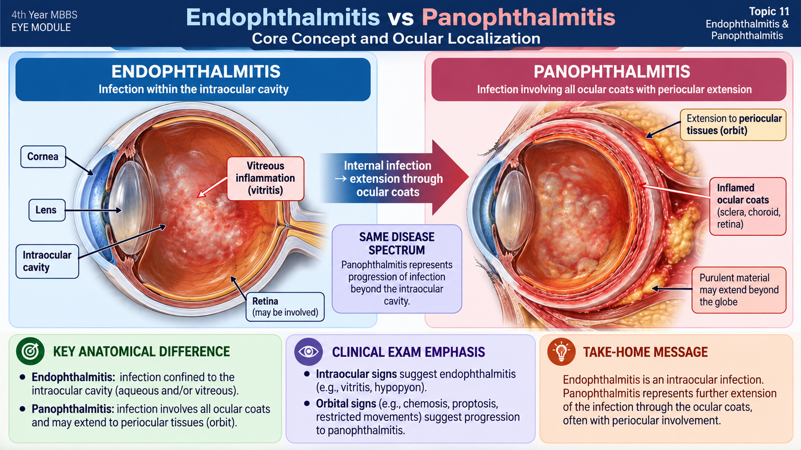
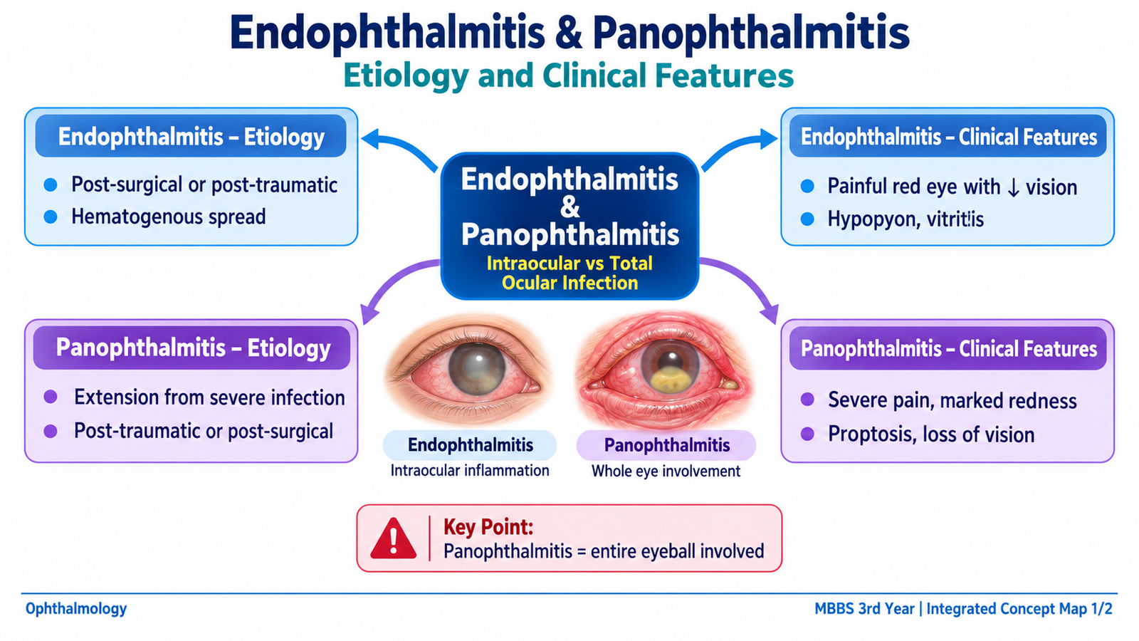
Endophthalmitis and panophthalmitis represent different degrees of severe ocular infection. Understanding exactly where the infection is located makes the clinical findings easier to understand.
Endophthalmitis
Endophthalmitis is an inflammatory process involving the internal structures of the eye, particularly the aqueous and vitreous cavities. In clinical practice, the important forms are usually infectious. The infection produces intense inflammation within the closed globe, so inflammatory cells, fibrin and microorganisms may accumulate in the anterior chamber and vitreous. The vitreous is especially important because it is relatively avascular. Once microorganisms enter it, local host defence is limited and infection may spread rapidly. Inflammatory mediators and microbial toxins can then damage the retina and other delicate intraocular tissues, explaining the rapid fall in vision.
Panophthalmitis
Panophthalmitis is a much more extensive suppurative infection in which the inflammatory process involves the intraocular contents as well as the ocular coats, including the sclera. It may also produce marked inflammation of adjacent orbital tissues. Therefore, endophthalmitis mainly presents as a severe intraocular infection, whereas panophthalmitis produces both intraocular destruction and striking external or orbital inflammatory signs.
B. Endophthalmitis: Etiology and Disease Development
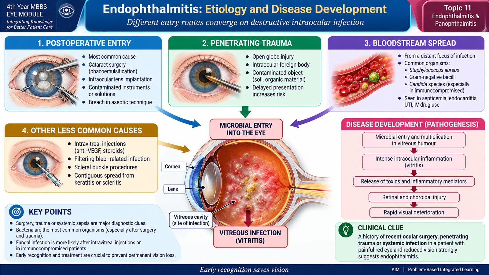
Endophthalmitis develops when microorganisms gain access to the internal eye. The source of entry is clinically important because it helps predict the likely organism and guides the investigation and management plan. The two major routes are exogenous entry from outside the eye and endogenous spread through the bloodstream.
Exogenous Endophthalmitis
In exogenous disease, microorganisms enter directly from the external environment. This is the more common mechanism in ophthalmic practice.
- Postoperative endophthalmitis: develops after intraocular surgery, particularly when organisms gain access to the eye during or after the procedure.
- Post-traumatic endophthalmitis: follows penetrating or open-globe injury, particularly when the wound is contaminated or an intraocular foreign body is present.
- Procedure-related infection: may rarely follow other intraocular procedures or injections.
- Direct extension: severe infection involving structures such as the cornea may occasionally extend deeper into the eye.
Acute postoperative cases are commonly bacterial. Coagulase-negative staphylococci and other Gram-positive organisms are important causes. Penetrating trauma, especially with soil contamination, may introduce particularly aggressive organisms such as Bacillus species.
Endogenous Endophthalmitis
In endogenous endophthalmitis, microorganisms reach the eye through the bloodstream during bacteremia or fungemia. They cross the blood-ocular barriers and establish infection within the eye. This form should therefore raise concern about an infection elsewhere in the body. Predisposing settings include severe systemic infection, impaired immunity, intravenous access associated with bloodstream infection and other conditions in which microorganisms circulate in the blood. Bacteria and fungi may both be responsible. Candida is an important fungal cause of endogenous intraocular infection.
How the Disease Produces Ocular Damage
The eye is a small closed space containing highly specialized tissues. Therefore, even a limited increase in inflammatory material can interfere with the optical pathway, while microbial toxins and inflammatory mediators may directly injure the retina. This combination explains why endophthalmitis can cause rapid and permanent visual loss.
C. Endophthalmitis: Clinical Features and Ophthalmic Examination
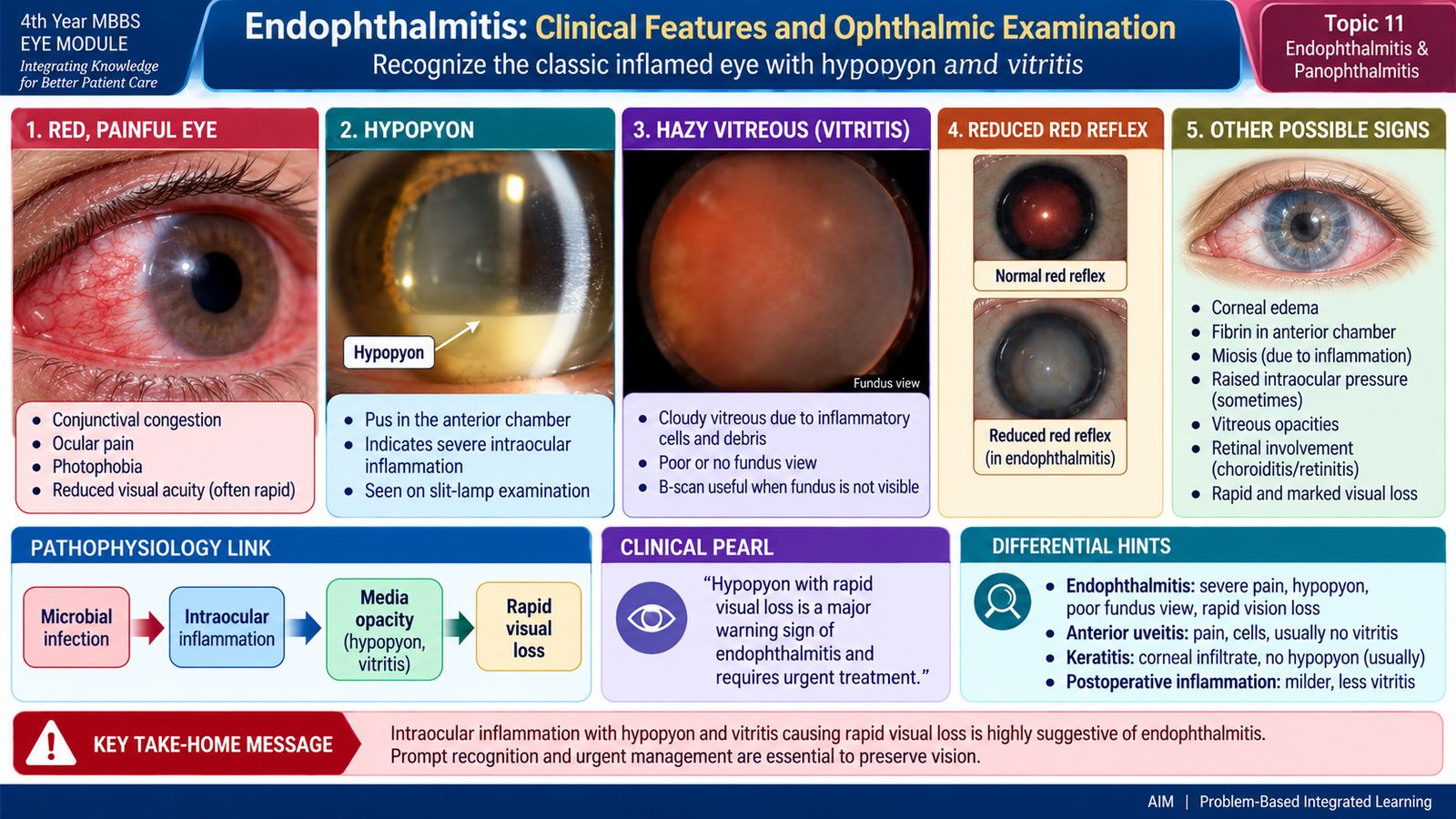
Endophthalmitis should be suspected when a patient develops a combination of decreased vision, ocular inflammation and characteristic anterior- or posterior-segment findings, especially after recent eye surgery, penetrating injury or in association with systemic infection. The presentation may be dramatic in acute bacterial infection but more gradual in some chronic or fungal cases.
Symptoms
- Rapid reduction of vision is one of the most important symptoms.
- Ocular pain is common in acute disease because of intense intraocular inflammation.
- Redness develops from marked ocular vascular congestion.
- Photophobia may occur because inflammation involves the anterior uveal tissues.
- Some patients report increasing discomfort or visual haze after a recent procedure or injury.
Pain is important but is not absolutely required. A relatively painless presentation does not exclude endophthalmitis, particularly in less acute forms.
Visual Acuity
Visual acuity is usually reduced, sometimes profoundly. The loss occurs because corneal edema, inflammatory material in the anterior chamber and especially vitreous inflammation obstruct the visual axis. Retinal dysfunction may further reduce vision.
Anterior Segment Findings
- Lid swelling may be present.
- Conjunctival and ciliary congestion produce the characteristic red eye.
- Corneal edema may make the cornea appear hazy.
- The anterior chamber may contain numerous inflammatory cells and fibrin.
- Hypopyon may develop as white inflammatory cells settle inferiorly in the anterior chamber.
Posterior Segment Findings
Vitritis means inflammatory cells and exudative material within the vitreous. It reduces vitreous transparency, producing a hazy or absent red reflex and making the fundus increasingly difficult to visualize.
- Reduced or absent red reflex.
- Hazy media because of vitreous inflammatory material.
- Poorly visible or completely obscured fundus in severe cases.
- Retinal involvement may contribute to profound loss of vision.
A relative afferent pupillary defect may appear in severe disease when retinal or optic-nerve function has been significantly compromised. Intraocular pressure may vary and should be interpreted in the overall clinical setting rather than used alone to diagnose the condition.
D. Endophthalmitis: Investigations and Diagnostic Approach
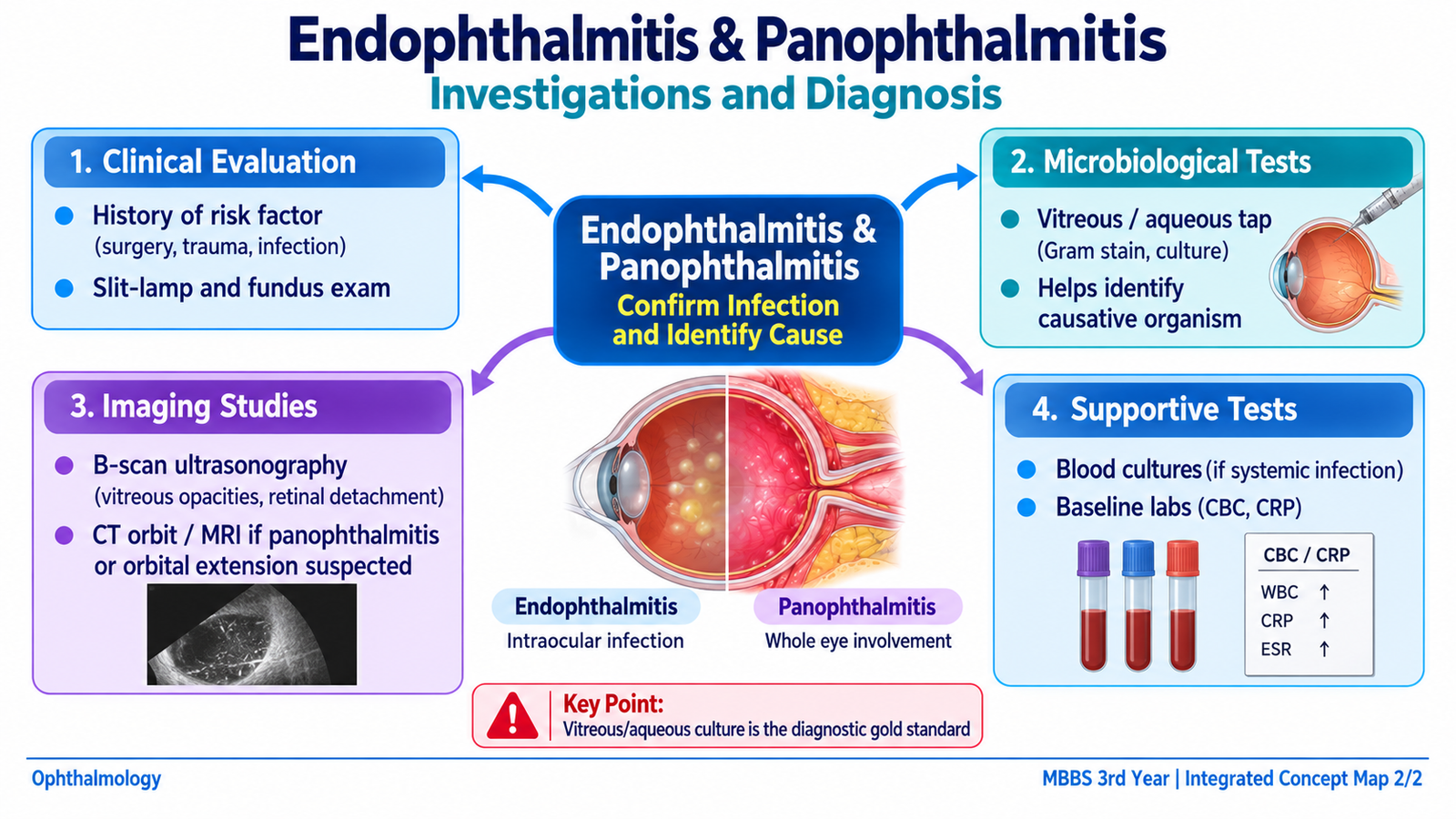
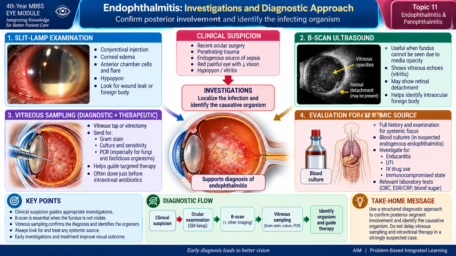
Endophthalmitis is primarily a clinical emergency. Investigations are used to confirm the extent of ocular involvement, identify the causative organism where possible and guide antimicrobial treatment. Investigation must not produce an unnecessary delay in emergency therapy.
1. Slit-Lamp Examination
Why it is used: to examine the anterior segment carefully. Important findings: corneal edema, anterior chamber cells and flare, fibrin and hypopyon. What they mean: they demonstrate severe intraocular inflammation and, together with the history and posterior-segment findings, support the diagnosis.
2. Fundus Examination
Why it is used: to assess the vitreous and retina when the media remain sufficiently clear. Important finding: vitreous haze or inflammatory debris may obscure retinal details. What it means: increasing loss of the fundus view reflects significant vitreous inflammation. A completely obscured fundus does not mean that the posterior segment can be ignored; it makes ultrasonography particularly useful.
3. B-Scan Ocular Ultrasonography
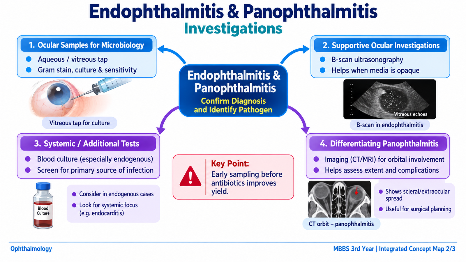
Why it is used: when the fundus cannot be visualized because of dense vitreous opacity. Important findings: mobile vitreous echoes, membranes or other evidence of vitreous inflammatory material may be detected. What it means: the scan confirms significant posterior-segment involvement and can also identify associated structural complications, such as retinal detachment, that may influence treatment planning.
4. Aqueous and Vitreous Sampling
Samples of intraocular fluid may be obtained for microbiological examination. Vitreous material is particularly valuable because the major infection is frequently located within the posterior segment.
- Direct microscopy may demonstrate bacteria or fungi.
- Gram staining assists in bacterial assessment.
- Appropriate fungal examination may be performed when fungal infection is suspected.
- Culture helps identify the organism.
- Antimicrobial susceptibility results may allow subsequent therapy to be refined.
5. Investigation for an Endogenous Source
When endogenous endophthalmitis is suspected, the eye may be only one manifestation of systemic infection. Blood cultures and clinical evaluation for the primary infective focus are therefore important. The purpose is to identify and treat the source of bacteremia or fungemia in addition to treating the eye.
Trauma-Related Assessment
After penetrating trauma, appropriate ocular or orbital imaging may be required when a retained intraocular foreign body is suspected. This is important because retained contaminated material increases the risk of persistent infection and may require surgical management.
E. Endophthalmitis: Management
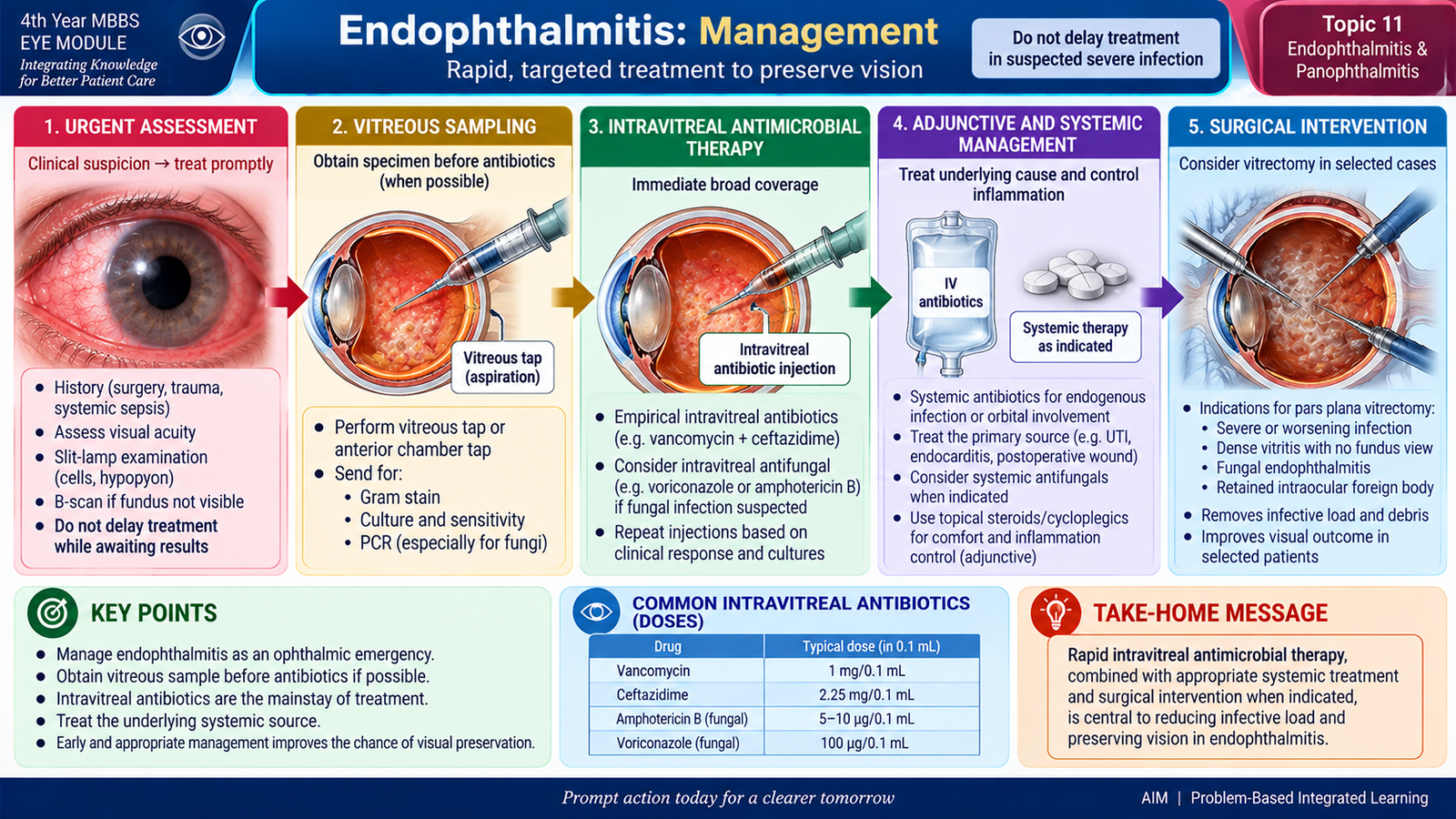
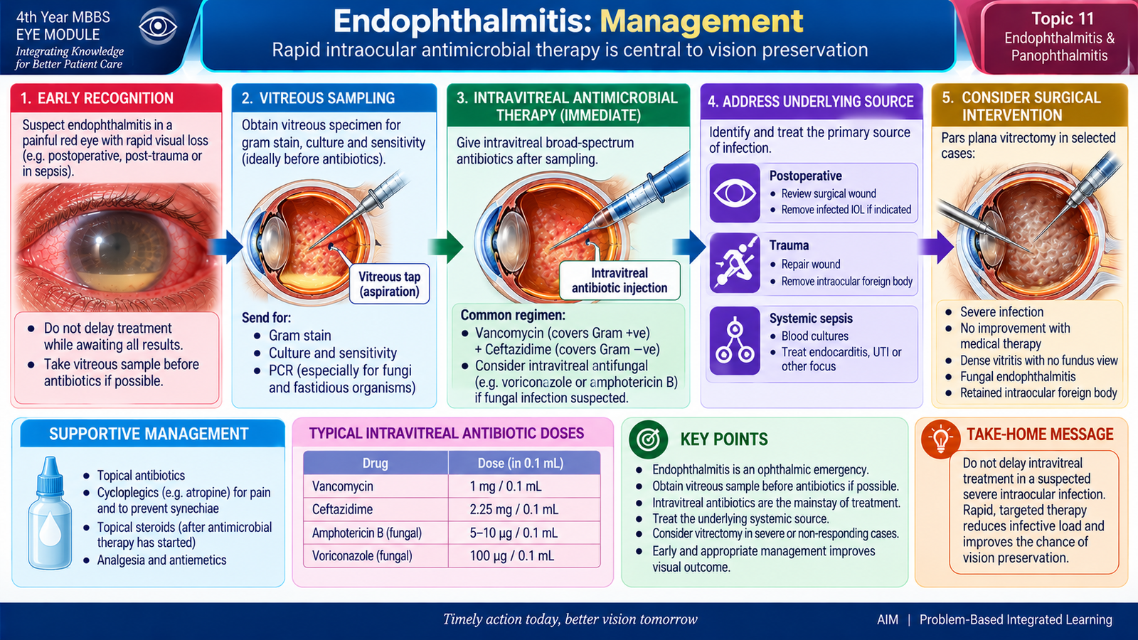
Endophthalmitis is an ophthalmic emergency. The central management principle is to deliver effective antimicrobial treatment directly to the infected intraocular tissues as rapidly as possible while obtaining appropriate specimens and addressing the underlying source of infection.
Immediate Priorities
- Urgent ophthalmology assessment.
- Document visual acuity and key ocular findings.
- Obtain appropriate aqueous or vitreous samples when indicated.
- Begin intraocular antimicrobial treatment promptly.
- Assess whether surgery is required.
Intravitreal Antimicrobial Therapy
Intravitreal treatment places the antimicrobial directly into the vitreous cavity, producing an effective drug concentration at the main site of infection. This is the cornerstone of treatment for severe bacterial endophthalmitis. Initial therapy is usually broad enough to cover both major Gram-positive and Gram-negative bacterial groups. Commonly used intravitreal agents include vancomycin for Gram-positive coverage and ceftazidime for Gram-negative coverage. Therapy can later be modified according to microbiological results and the clinical response. When fungal endophthalmitis is suspected or confirmed, appropriate intraocular antifungal treatment is required rather than relying on antibacterial agents.
Supporting Ophthalmic Treatment
Topical therapy may be used to control associated anterior-segment inflammation and infection, but topical medication alone cannot adequately treat infection within the vitreous cavity. Cycloplegic treatment may be used when significant anterior uveal inflammation is present. Corticosteroid therapy, when considered by the ophthalmologist, is used only as an adjunct after adequate antimicrobial treatment has been established.
Role of Systemic Antimicrobials
Systemic treatment is especially important in endogenous endophthalmitis because the bloodstream is the route by which microorganisms reached the eye. The systemic infective focus must also be found and treated. Systemic antimicrobials may also be required in selected severe trauma-related infections, particularly when there is an open-globe injury or infection extends beyond the intraocular cavity. They do not replace appropriate intravitreal therapy for established severe intraocular infection.
Pars Plana Vitrectomy
Vitrectomy removes infected and inflammatory vitreous material from the eye. This can reduce the microbial and inflammatory load and improve access to the posterior segment. It is particularly considered when disease is severe, vision is profoundly reduced, dense vitreous inflammation prevents useful visualization, structural complications are present or the infection does not respond adequately to initial treatment. The exact decision depends on the ophthalmic assessment and clinical severity.
F. Panophthalmitis: Etiology and Disease Development
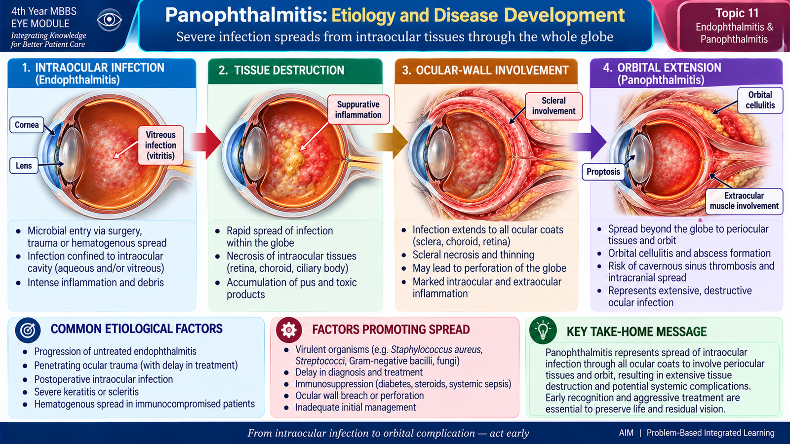
Panophthalmitis represents a destructive progression of ocular infection. The infection is no longer limited mainly to the aqueous and vitreous cavities; it involves the ocular coats and can produce severe inflammation in surrounding tissues. For this reason, the affected eye often has much more dramatic external signs than uncomplicated endophthalmitis.
Etiology
Panophthalmitis may develop when a severe ocular infection progresses despite the natural barriers of the globe or when a highly virulent infection produces rapid tissue destruction.
- Severe or untreated endophthalmitis may extend through the ocular tissues.
- Penetrating ocular trauma, particularly contaminated injury, can introduce aggressive organisms into several ocular structures.
- Postoperative infection may occasionally progress to widespread ocular involvement.
- Endogenous infection may occur during severe bacteremia or fungemia.
Pathological Progression
The major conceptual difference is therefore extension. Endophthalmitis is predominantly an internal ocular infection, while panophthalmitis indicates that the infection has become much more extensive and structurally destructive.
G. Panophthalmitis: Clinical Features and Investigations
Panophthalmitis usually presents as a severely painful, intensely inflamed eye with profound visual impairment and prominent external swelling. The extension of inflammation beyond the internal eye explains the orbital-type signs that distinguish it from less extensive endophthalmitis.
Clinical Features
- Severe ocular pain due to extensive inflammation.
- Profound reduction or loss of vision because the retina and other essential ocular tissues are severely damaged.
- Marked conjunctival congestion and chemosis.
- Pronounced eyelid edema.
- Corneal edema or opacity may be present.
- Anterior chamber inflammation with hypopyon may be visible.
- Proptosis may occur because inflammation extends into surrounding orbital tissues.
- Restricted and painful ocular movements may develop because the periocular tissues are inflamed.
- The globe may be extremely tender.
- Systemic fever or toxicity may accompany endogenous or widely spreading infection.
Investigations
The investigation strategy has two aims: confirm the extent of ocular destruction and identify the infecting organism and its source.
Ophthalmic Examination
Visual acuity, anterior-segment findings, pupil response and any available posterior-segment view are documented. Particular attention is paid to proptosis, ocular movement and scleral or periocular involvement because these indicate that infection is no longer confined to the inner eye.
B-Scan Ultrasonography
When the posterior segment cannot be seen, B-scan ultrasonography can demonstrate dense vitreous inflammatory material and associated structural abnormalities. Its usefulness depends on the integrity and clinical condition of the globe.
Orbital Imaging
When extension into the orbit is suspected, appropriate orbital imaging can define the extent of surrounding tissue involvement. In traumatic cases, imaging can also help assess for a retained foreign body where clinically appropriate.
Microbiological Investigation
Intraocular or other appropriate specimens are examined and cultured to identify the organism and guide antimicrobial therapy. In endogenous disease, blood cultures and evaluation of the systemic infective source are also important.
H. Panophthalmitis: Management and Vision-Threatening Priorities
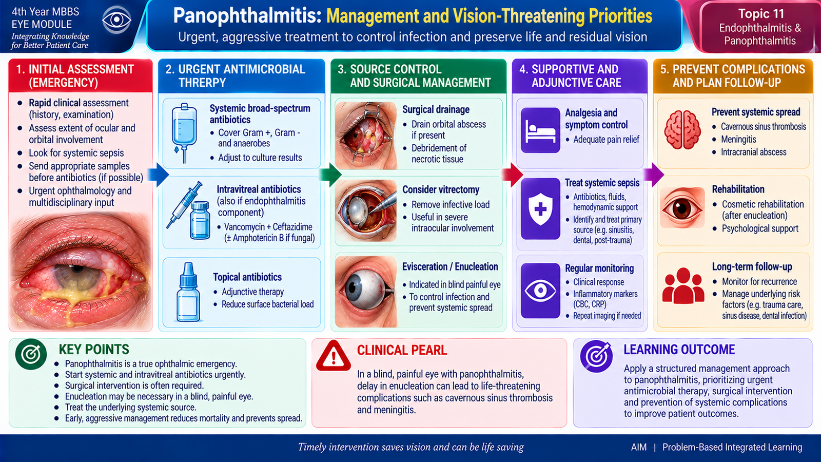
Panophthalmitis requires immediate specialist treatment because both vision and preservation of the globe may be threatened. Management is more aggressive than for infection confined mainly to the intraocular cavity because the disease may involve the ocular wall, surrounding tissues and sometimes a systemic infective source.
Immediate Management Principles
- Urgent ophthalmology management, often in a hospital setting.
- Rapid assessment of remaining visual function and the structural state of the globe.
- Obtain appropriate microbiological samples whenever this can be done without delaying essential treatment.
- Begin broad antimicrobial treatment promptly.
Antimicrobial Therapy
Because the infection involves the intraocular tissues, intravitreal antimicrobial therapy may be required. Because panophthalmitis may also involve the ocular coats and surrounding tissues, systemic antimicrobial therapy has an important role, particularly when there is extraocular extension or an endogenous source. Treatment is adjusted according to the suspected organism, microbiological results and clinical response. Fungal infection requires appropriate antifungal therapy rather than antibacterial treatment alone.
Surgical Management
When useful visual potential remains and the globe is structurally salvageable, vitrectomy may be considered to remove infected vitreous and reduce the intraocular infective load. In advanced disease, the eye may become irreversibly blind, severely painful and structurally destroyed. If infection remains uncontrolled despite appropriate therapy, globe-sacrificing surgery may become necessary to eradicate the infective focus. The exact procedure depends on the extent of ocular-wall involvement and specialist assessment.
Treat the Systemic Source
If panophthalmitis has arisen from bloodstream infection, treating the eye alone is inadequate. The primary source of bacteremia or fungemia must also be identified and treated to control the systemic disease and prevent further metastatic infection.
3. Integrated Mechanism Flow
Intervention point: Early recognition and rapid antimicrobial treatment aim to stop the sequence before irreversible retinal damage and widespread ocular destruction occur.
4. Important Comparison — Endophthalmitis vs Panophthalmitis
| Feature | Endophthalmitis | Panophthalmitis |
|---|---|---|
| Main extent | Predominantly intraocular cavities and tissues | All ocular coats with possible periocular extension |
| Vision | Usually markedly reduced | Usually profoundly reduced or lost |
| Anterior eye | Redness, cells, fibrin, hypopyon | Severe inflammation with marked external involvement |
| Vitreous | Vitritis is characteristic | Severe intraocular suppuration is part of widespread disease |
| Proptosis / motility | Usually not dominant features | Proptosis and restricted movements may be prominent |
| Investigation emphasis | B-scan and intraocular microbiological sampling | Same principles plus assessment of extraocular extension |
| Management | Urgent intravitreal antimicrobial therapy ± vitrectomy | Aggressive intravitreal/systemic therapy ± major surgery |
⭐ 5. AIM High-Yield Review
Endophthalmitis & Panophthalmitis
Use these videos after completing the learning material to reinforce etiology, clinical recognition, investigations and management.
▶️ Video 1 — Endophthalmitis
Covers the major causes and types of endophthalmitis, clinical features, hypopyon and vitritis, investigations including vitreous sampling and B-scan ultrasonography, and principles of urgent treatment.
▶️ Video 2 — Panophthalmitis
Reinforces panophthalmitis as extensive suppurative inflammation involving the whole eye and helps differentiate it from infection confined mainly to the intraocular contents.