
Topic 13 — Optic Neuritis, Optic Neuropathies & Papilledema
1. Topic Introduction
The optic nerve carries visual information from the retina to the brain. Disease affecting this pathway may therefore reduce visual acuity, colour vision and contrast sensitivity and may produce characteristic visual-field and pupillary abnormalities. In this chapter, you will first learn how to recognize optic-nerve dysfunction. You will then study optic neuritis and its important forms, followed by hereditary, nutritional and toxic optic neuropathies. The final part deals with papilledema, which is optic-disc swelling caused specifically by raised intracranial pressure. Particular attention is given to distinguishing papilledema from other causes of disc swelling, because this distinction has important implications for both vision and the patient’s general neurological safety.
A. Optic Nerve Function and Clinical Localization
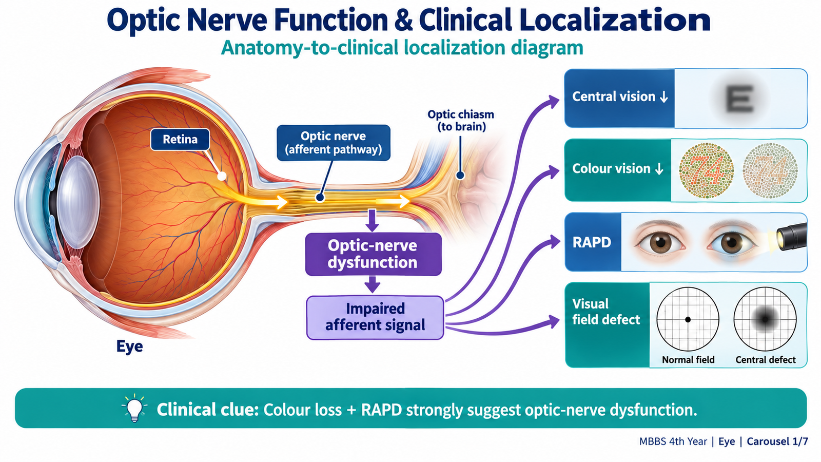
Before studying individual optic neuropathies, it is useful to understand what happens when the optic nerve is damaged. The optic nerve contains axons of retinal ganglion cells. These axons carry information responsible for central vision, colour discrimination, visual fields and the afferent limb of the pupillary light reflex. Because several visual functions travel together through the nerve, optic-nerve disease produces a recognizable combination of abnormalities rather than isolated reduction of visual acuity.
Core Clinical Pattern
An optic neuropathy should be suspected when visual loss is accompanied by disproportionate loss of colour vision, a characteristic visual-field defect or an abnormal afferent pupillary response. The fundus may show a swollen optic disc, a pale optic disc or even a completely normal disc, depending on the site and stage of disease.
- Visual acuity: may be reduced because fibres responsible for central vision are affected.
- Colour vision: red and other colours may appear faded or washed out. Dyschromatopsia is often an early clue to optic-nerve dysfunction.
- Visual field: central or centrocecal defects are common when the papillomacular fibres are involved.
- Relative afferent pupillary defect (RAPD): occurs when one optic nerve conducts the afferent light signal less effectively than the other. It is therefore especially helpful in unilateral or markedly asymmetric disease.
- Optic disc: it may initially appear normal when the lesion lies behind the globe, become swollen in an anterior process, or become pale after permanent axonal loss.
Why the Optic Disc May Look Normal
Only the short anterior part of the optic nerve is directly visible during fundoscopy. Inflammation behind the globe may therefore cause marked visual dysfunction while the optic disc still appears normal. This is the basis of the classic description of retrobulbar optic neuritis: the patient cannot see, while the examiner initially cannot see the lesion.

B. Optic Neuritis: Classification, Etiology and Mechanism
Optic neuritis is inflammation of the optic nerve that produces acute or subacute impairment of optic-nerve function. Demyelination is a common mechanism, particularly in young adults, but optic neuritis may also occur with other immune-mediated or infectious conditions. Classification is useful because the appearance of the optic disc and the overall clinical pattern help determine the likely cause and the urgency of further investigation.
Classification
Optic neuritis can be classified according to the location and ophthalmoscopic appearance of inflammation.
- Retrobulbar neuritis: inflammation is mainly behind the visible optic-nerve head. The optic disc may initially look normal despite definite visual dysfunction.
- Papillitis: inflammation involves the anterior optic nerve and produces visible optic-disc swelling.
- Neuroretinitis: optic-disc swelling is associated with a characteristic macular star caused by leakage around the disc. Infectious or inflammatory causes are important in this pattern.
Clinically, another useful distinction is between typical demyelinating optic neuritis and atypical optic neuritis. Typical disease commonly presents as a painful, unilateral optic neuropathy in a young adult and often improves substantially. Atypical features such as very severe visual loss, marked bilateral involvement, recurrent attacks, unusual disc appearances or prominent systemic disease should prompt investigation for alternative inflammatory or infectious causes.
Etiology
Demyelination is the most important cause in typical optic neuritis. It may occur as an isolated episode or in association with a central nervous system demyelinating disorder. Other forms of optic neuritis occur when immune-mediated inflammation or infection directly affects the optic nerve.
- Demyelinating disease: including optic neuritis associated with multiple sclerosis.
- Other immune-mediated demyelinating disorders: including neuromyelitis optica spectrum disorder and MOG-antibody-associated disease.
- Systemic inflammatory disease: such as sarcoidosis or other autoimmune inflammatory disorders.
- Infections: selected viral, bacterial or granulomatous infections may involve the optic nerve.
- Post-infectious inflammation: inflammation may occasionally follow a systemic infection.
How Inflammation Produces Visual Loss
Myelin allows rapid conduction along optic-nerve fibres. When inflammation damages myelin, visual signals become slow or fail to travel normally. This explains the rapid reduction in visual function. If inflammation also damages axons themselves, some visual deficit may persist and the optic disc may later become pale because of optic atrophy.
C. Optic Neuritis: Clinical Features, Investigations and Management
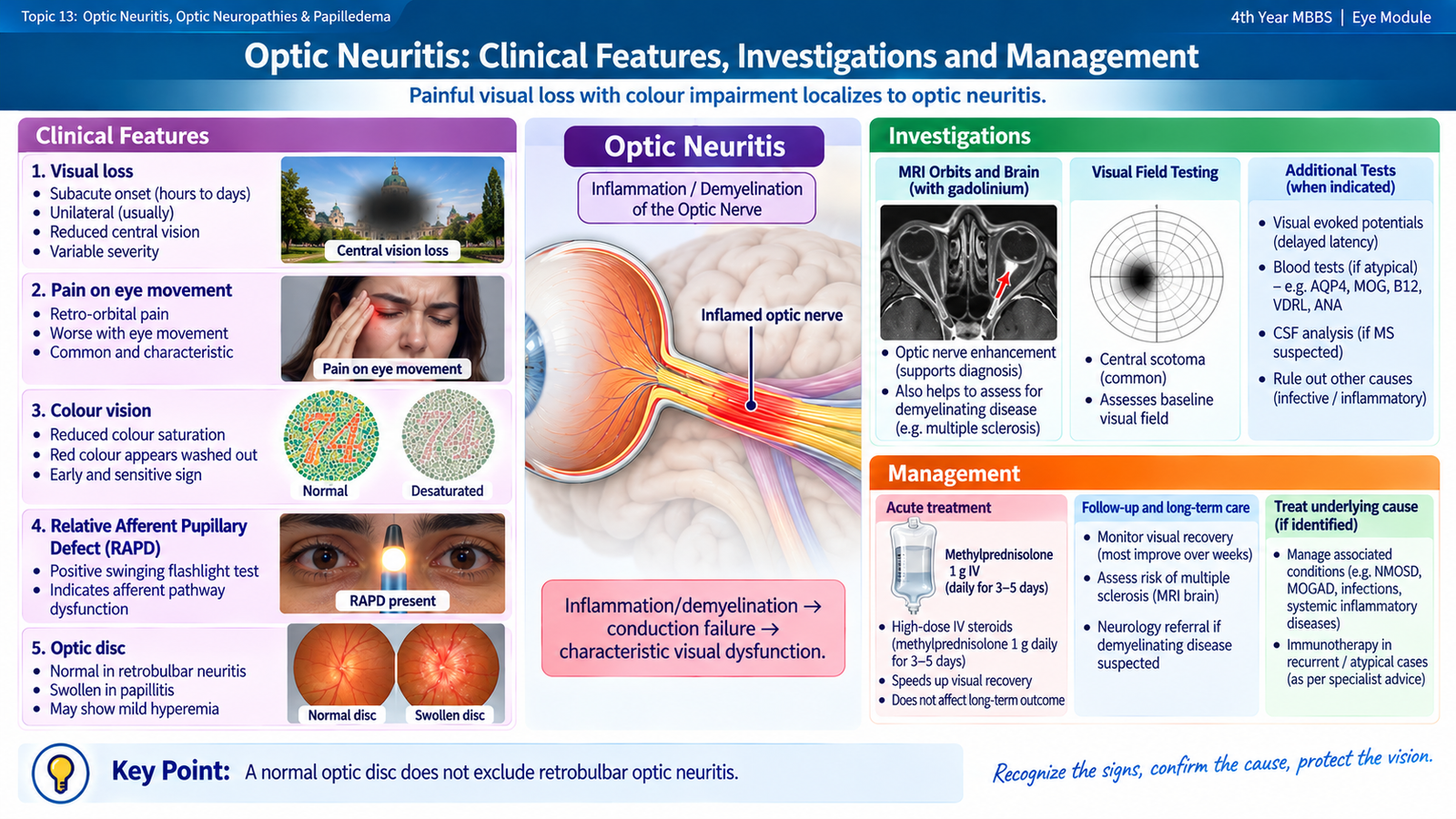
Typical optic neuritis is recognized from the combination of visual symptoms, optic-nerve examination findings and appropriate imaging. The whole pattern is more important than any single finding. A young adult with rapidly developing monocular visual loss, pain on moving the eye, reduced colour perception and an RAPD has a classic optic-nerve pattern even when the optic disc appears normal.
Clinical Features
- Onset: acute or subacute visual loss developing over hours to several days.
- Laterality: typical demyelinating optic neuritis is commonly unilateral in adults.
- Pain: pain around the eye, particularly during eye movement, is common. Movement stretches tissues around the inflamed retrobulbar optic nerve and may provoke discomfort.
- Reduced visual acuity: severity varies from mild blurring to marked visual loss.
- Dyschromatopsia: colours, especially red, appear less vivid because optic-nerve conduction affecting colour pathways is impaired.
- Reduced contrast sensitivity: objects may appear less distinct even when the acuity reduction is modest.
- Visual-field defect: a central or centrocecal scotoma is characteristic, although other field defects can occur.
Ophthalmic Examination
Visual acuity establishes the degree of functional loss. Colour testing often demonstrates disproportionate colour desaturation. In unilateral or asymmetric disease, the swinging-flashlight test may show an RAPD, because the affected optic nerve transmits a weaker afferent light signal.
The optic disc depends on the anatomical site of inflammation:
- Retrobulbar neuritis: the disc initially appears normal.
- Papillitis: the optic disc is swollen and its margins become blurred.
- After axonal loss: optic-disc pallor may develop.
Investigations
Investigation aims to confirm optic-nerve dysfunction, identify the inflammatory pattern and look for an associated neurological or systemic disorder. Tests should be chosen according to the clinical presentation rather than performed indiscriminately.
- Visual-field testing: demonstrates and documents the field defect, commonly a central scotoma.
- Colour-vision testing: provides objective evidence of dyschromatopsia and helps monitor recovery.
- MRI of the orbits and brain: may demonstrate enhancement or abnormal signal in the inflamed optic nerve. Brain imaging also helps identify demyelinating lesions that may indicate an associated central nervous system disorder.
- Optical coherence tomography (OCT): documents the retinal nerve-fibre layer and ganglion-cell complex. Swelling may be present in anterior disease, while thinning may appear later after axonal loss.
- Visual evoked potential (VEP): may show delayed visual conduction, classically reflected by increased latency in demyelination.
- Targeted laboratory investigations: are particularly important when the presentation is atypical or when infectious, inflammatory or other immune-mediated disease is suspected.
Why MRI matters: it does more than show optic-nerve inflammation. In a patient with suspected demyelinating optic neuritis, associated brain lesions can influence the likelihood that the episode is part of a wider neurological demyelinating disorder and therefore influence neurological follow-up.
Management
Management depends on whether the attack is a typical demyelinating episode or part of an atypical inflammatory or infectious disorder. The diagnosis should therefore be established before treatment is generalized to every patient.
- Ophthalmology or neuro-ophthalmology assessment: confirms the diagnosis and identifies atypical features.
- Neurological assessment: is important when demyelinating disease is suspected.
- Systemic corticosteroid therapy: may be used under specialist supervision in appropriate acute demyelinating optic neuritis. In typical disease, corticosteroids can accelerate visual recovery, although the final degree of recovery is often good even without treatment.
- Treat the underlying cause: infectious or systemic inflammatory optic neuritis requires treatment directed at that disorder rather than treatment of the optic nerve alone.
- Long-term disease management: patients with multiple sclerosis or another immune-mediated demyelinating disorder may require neurological disease-specific therapy.
- Follow-up: visual acuity, colour vision, visual fields and structural changes may be monitored to document recovery or persistent axonal damage.
D. Hereditary Optic Neuropathies
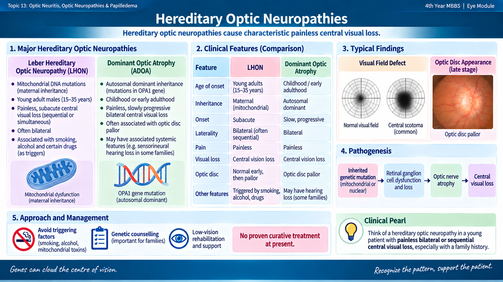
Hereditary optic neuropathies are genetically determined disorders in which retinal ganglion cells and their optic-nerve axons progressively lose function. Unlike typical optic neuritis, visual loss is usually painless and commonly affects both eyes. The two important undergraduate patterns are Leber hereditary optic neuropathy and dominant optic atrophy.
Leber Hereditary Optic Neuropathy
Leber hereditary optic neuropathy (LHON) results from pathogenic mutations of mitochondrial DNA. Because mitochondrial DNA is inherited from the mother, transmission is maternal, although not every carrier develops visual loss. Young adult males are affected more commonly clinically.
The disease preferentially damages retinal ganglion cells and fibres of the papillomacular bundle. This explains why loss of central vision, colour impairment and central or centrocecal scotomas dominate the clinical picture.
Clinical pattern
- Painless, subacute central visual loss.
- One eye may be affected first, with the second eye becoming involved later.
- Central or centrocecal visual-field defect.
- Impaired colour vision.
- The acute fundus may show peripapillary nerve-fibre swelling and characteristic small peripapillary vascular abnormalities.
- Established disease eventually produces optic-disc pallor due to axonal loss.
Dominant Optic Atrophy
Dominant optic atrophy is usually an autosomal dominant optic neuropathy, commonly related to abnormal mitochondrial function. It often begins in childhood or early life and progresses slowly. Visual loss is usually bilateral and relatively symmetrical.
- Gradual, painless reduction of vision in both eyes.
- Dyschromatopsia.
- Central or centrocecal field defects.
- Characteristic temporal optic-disc pallor may develop because fibres in the papillomacular bundle are prominently affected.
Investigations
The diagnosis is based on recognizing the clinical pattern and confirming hereditary disease when appropriate.
- Family history: may suggest mitochondrial or autosomal dominant inheritance, although an apparently negative history does not completely exclude hereditary disease.
- Visual acuity, colour vision and visual fields: document the functional pattern.
- Fundus examination and OCT: demonstrate optic-nerve structural changes and later retinal nerve-fibre or ganglion-cell loss.
- Genetic testing: can confirm the suspected hereditary diagnosis and assist family counselling.
- Additional investigation: is considered when the presentation is atypical or another treatable optic neuropathy must be excluded.
Management
Established optic-nerve axonal loss is difficult to reverse, so management focuses on correct diagnosis, reducing avoidable mitochondrial stress, supporting remaining vision and counselling the patient and family.
- Neuro-ophthalmic assessment and confirmation of diagnosis.
- Genetic counselling for the patient and family.
- Avoidance of smoking and excessive alcohol, particularly in mitochondrial optic neuropathy.
- Correction of any coexisting nutritional deficiency.
- Low-vision rehabilitation when permanent central visual loss limits daily activities.
- Disease-specific treatment may be considered by specialist services in selected hereditary optic neuropathies.
E. Nutritional and Toxic Optic Neuropathies
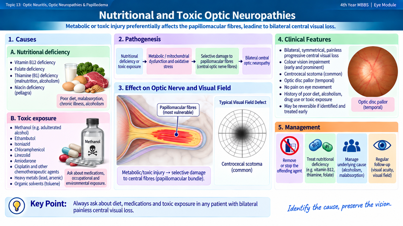
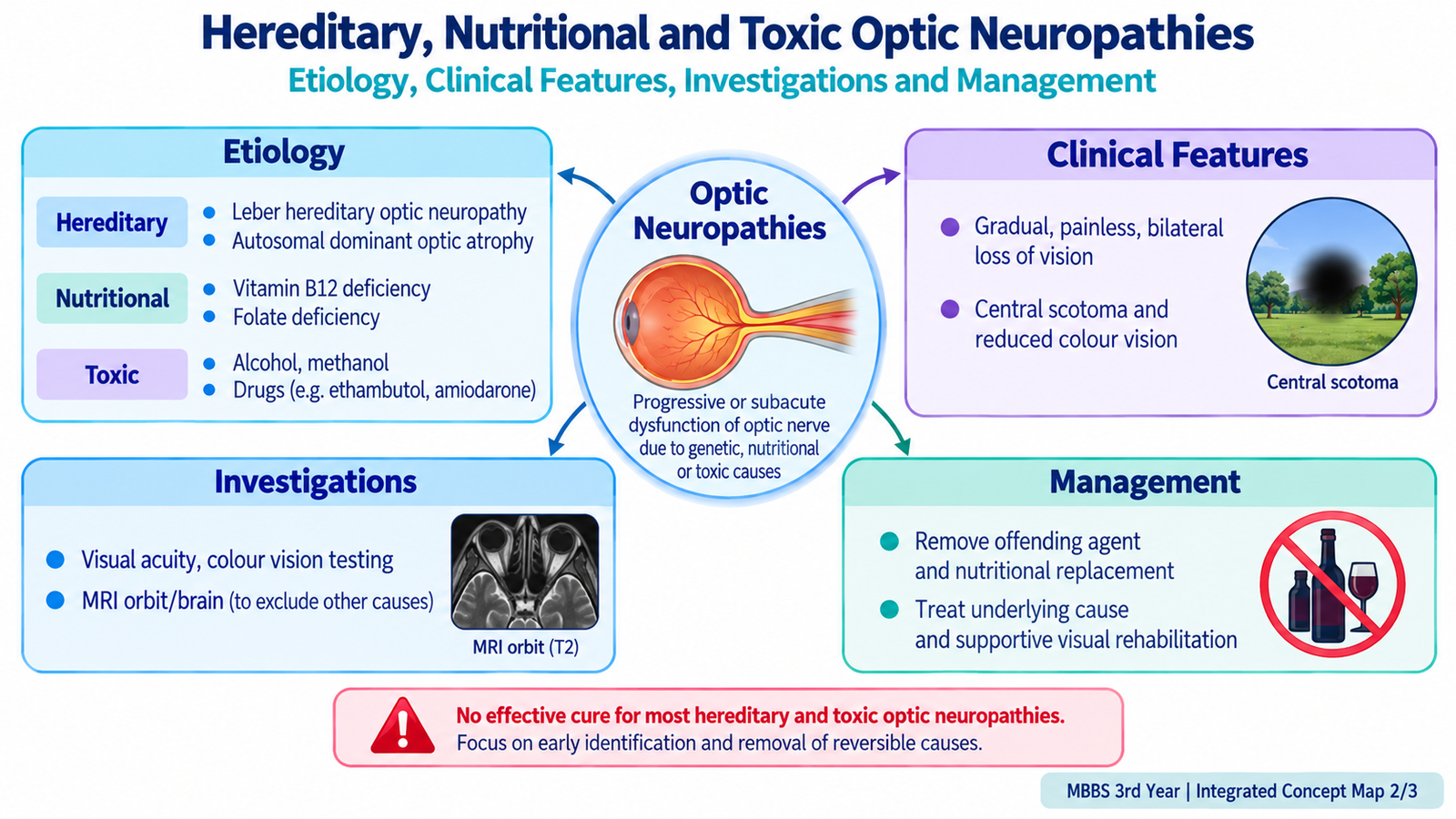
Nutritional and toxic optic neuropathies are acquired disorders in which metabolic deficiency or exposure to a toxic substance damages retinal ganglion cells and optic-nerve fibres. They often affect the papillomacular bundle, so the typical pattern is painless, bilateral and relatively symmetrical central visual dysfunction. Recognizing this pattern is important because early removal of the toxin or correction of the deficiency may prevent further permanent damage.
Nutritional Optic Neuropathy
Normal optic-nerve function has a high metabolic requirement. Deficiency of nutrients required for mitochondrial energy production and neuronal metabolism can therefore impair retinal ganglion-cell and axonal function.
Important causes and risk settings
- Vitamin B12 deficiency.
- Folate or thiamine deficiency.
- Copper deficiency in appropriate clinical settings.
- Severe dietary inadequacy or malnutrition.
- Chronic alcohol misuse, particularly when associated with poor nutrition.
- Malabsorption or previous gastrointestinal surgery when these produce nutritional deficiency.
Clinical features
- Gradual, painless bilateral reduction of central vision.
- Loss of colour discrimination.
- Central or centrocecal scotomas.
- Optic discs may appear relatively normal early.
- Temporal or diffuse optic pallor may develop after persistent axonal loss.
Toxic Optic Neuropathy
Toxic optic neuropathy occurs when a drug or chemical interferes with cellular metabolism or directly injures retinal ganglion cells and optic-nerve axons. The pattern is often similar to nutritional optic neuropathy because the papillomacular fibres are particularly vulnerable.
Important causes
- Ethambutol: an important drug-associated cause of bilateral optic neuropathy.
- Methanol: may produce profound optic-nerve injury and is a medical emergency.
- Other medications or toxic exposures may cause optic neuropathy and should be considered from the history when the pattern is compatible.
Ethambutol toxicity classically produces reduced central vision, dyschromatopsia and central or centrocecal field defects. Methanol exposure may cause severe bilateral visual loss together with systemic toxicity. Because continued exposure may worsen irreversible axonal injury, the exposure history is central to diagnosis.
Investigations
Investigation begins with careful history because the diagnosis often depends on identifying a nutritional risk or toxic exposure that explains a bilateral optic-nerve pattern.
- Visual acuity and colour vision: document the functional deficit.
- Visual fields: commonly demonstrate central or centrocecal scotomas.
- Fundus examination: may be normal initially and later show optic pallor.
- OCT: can document loss of retinal ganglion cells and retinal nerve-fibre tissue.
- Nutritional investigations: are selected according to the history and may include assessment of vitamin B12 and other suspected deficiencies.
- Medication and exposure review: should identify potentially toxic agents and the duration of exposure.
- Additional tests: are used when another inflammatory, hereditary or compressive optic neuropathy must be excluded.
Management
Management is aimed at removing the damaging factor before additional axons are lost.
- Nutritional disease: identify and replace the deficient nutrient and correct the underlying dietary or absorptive problem.
- Suspected drug toxicity: promptly involve the prescribing team so that the responsible drug can be stopped or replaced when clinically appropriate.
- Methanol exposure: requires immediate emergency medical and toxicology management because both systemic and visual injury can progress rapidly.
- Follow-up: visual acuity, colour vision and fields are monitored for recovery or progression.
- Visual rehabilitation: may be needed when irreversible optic atrophy remains.
F. Papilledema and Optic Disc Swelling: Core Distinction
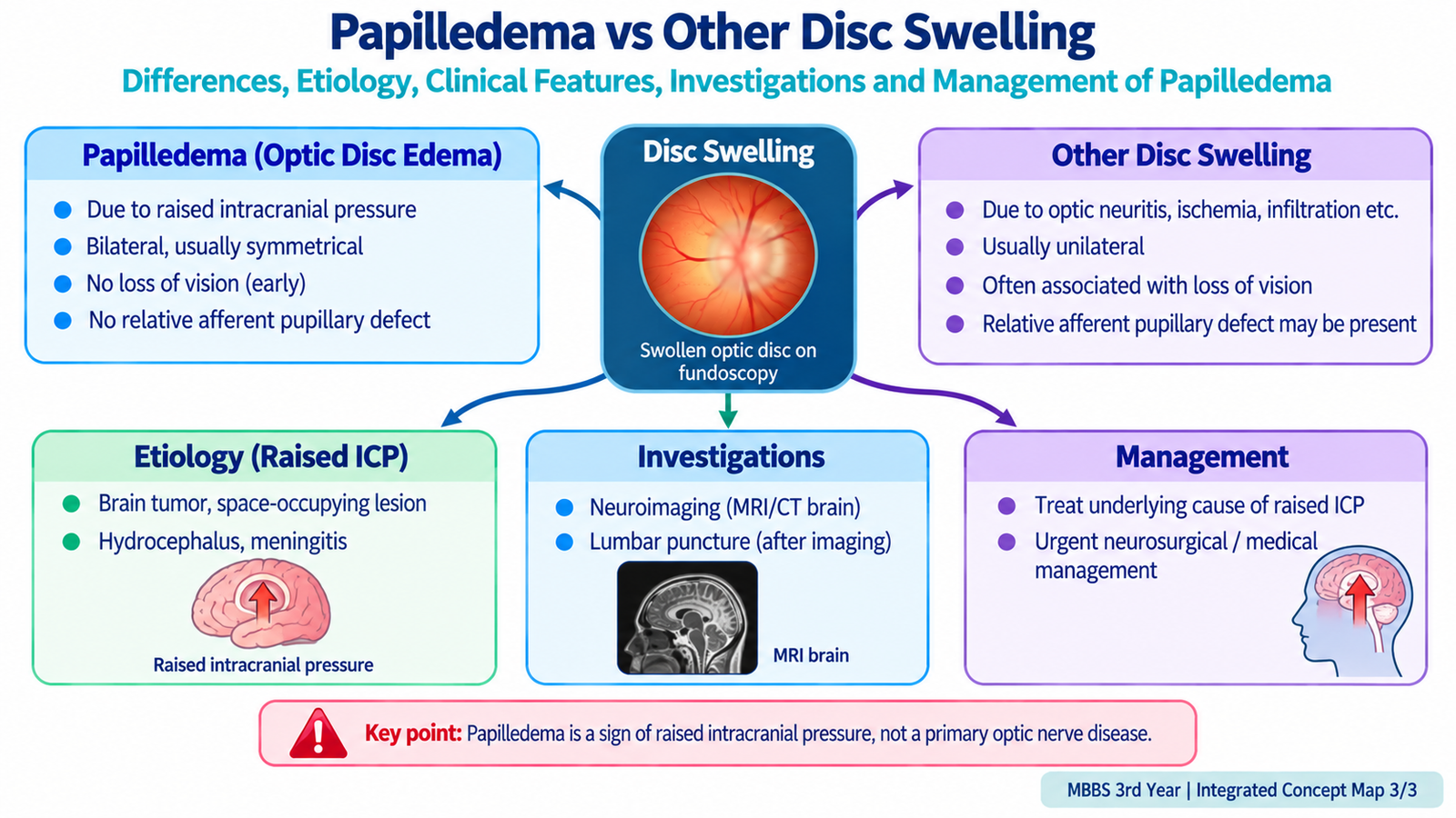
The terms papilledema and optic-disc swelling must not be used as synonyms. Optic-disc swelling is a descriptive examination finding: the optic nerve head is elevated and its margins appear blurred. Many local ocular and optic-nerve disorders can produce this appearance. Papilledema is a specific diagnosis in which the disc swelling occurs because intracranial pressure is raised.
Optic Disc Swelling
Disc swelling may result from inflammation, ischemia, raised intracranial pressure, infiltration or other local or systemic disease. It may be unilateral or bilateral. Visual function may be affected early when the optic nerve itself is injured.
Examples include:
- papillitis;
- ischemic optic neuropathy;
- neuroretinitis;
- infiltrative optic-nerve disease;
- severe vascular or retinal disease producing disc edema.
Papilledema
Papilledema usually affects both optic discs because raised cerebrospinal fluid pressure is transmitted through the subarachnoid space surrounding both optic nerves. In the early stage, central visual acuity may remain relatively normal because the retinal ganglion-cell axons are compressed and swollen but have not yet undergone major permanent loss.
This differs from optic neuritis, in which the optic nerve itself is inflamed and visual function—especially acuity, colour vision and the afferent pupillary response—may deteriorate early.

G. Papilledema: Etiology, Clinical Features, Investigations and Management
Papilledema is an ophthalmic sign of raised intracranial pressure and may therefore indicate serious neurological disease. The immediate clinical priorities are to recognize the disc appearance, identify symptoms suggesting increased intracranial pressure, protect visual function and determine the intracranial cause. Treatment directed only at the eye is not sufficient because the underlying disorder lies within the cranial compartment.
Etiology
Any disorder that increases intracranial pressure sufficiently can produce papilledema. The important mechanisms are an intracranial space-occupying process, impaired cerebrospinal fluid circulation or absorption, impaired cerebral venous drainage, or raised pressure without an identifiable structural lesion.
- Intracranial mass lesion: such as a tumour or other space-occupying process.
- Intracranial hemorrhage or marked cerebral swelling: may increase intracranial volume and pressure.
- Hydrocephalus: causes increased cerebrospinal fluid volume and pressure.
- Meningeal inflammation: may interfere with cerebrospinal fluid circulation or absorption.
- Cerebral venous sinus thrombosis: impairs venous drainage and may raise intracranial pressure.
- Idiopathic intracranial hypertension: produces raised intracranial pressure without an intracranial mass or hydrocephalus after appropriate evaluation.
Pathogenesis
The optic nerve is surrounded by meninges and a continuation of the intracranial subarachnoid space. Raised intracranial pressure is therefore transmitted along the optic-nerve sheath to the region of the optic nerve head. This interferes with normal axoplasmic transport within the nerve fibres and causes venous congestion. Axons become swollen, producing elevation and blurring of the optic disc.
Clinical Features
Symptoms may arise from raised intracranial pressure itself or from its effect on the optic nerves. Visual acuity is often preserved early, so a patient with significant papilledema may initially report little permanent visual loss.
- Headache: a common symptom of raised intracranial pressure.
- Nausea or vomiting: may accompany increased intracranial pressure.
- Transient visual obscurations: brief episodes in which vision dims or blacks out, often lasting only a short time.
- Pulsatile tinnitus: may occur particularly in idiopathic intracranial hypertension.
- Diplopia: may occur if raised intracranial pressure produces a sixth cranial nerve palsy.
- Progressive visual-field loss: develops when papilledema is persistent or severe.
- Permanent visual reduction: may occur late after chronic axonal injury and secondary optic atrophy.
Ophthalmic Examination
Visual acuity may be normal or only mildly reduced in early papilledema. This is important because preserved acuity does not make the disc swelling harmless. Colour vision is also usually relatively preserved early compared with optic neuritis.
When papilledema is approximately symmetrical, an RAPD is generally absent because both afferent pathways are affected similarly. A significant RAPD suggests asymmetrical optic-nerve dysfunction or another optic neuropathy.
Fundus appearance
- Elevation and swelling of the optic nerve head.
- Blurred optic-disc margins.
- Hyperemia of the disc.
- Reduced or obscured physiological cup.
- Venous congestion and tortuosity.
- Small peripapillary hemorrhages may be present.
- Retinal vessels may become partially obscured as they cross the swollen disc margin.
These findings occur because swollen nerve fibres expand the optic nerve head and obscure its normal boundaries. Venous congestion develops because raised tissue pressure interferes with venous outflow.
Visual field
An enlarged blind spot is a classic early visual-field finding. The swollen disc elevates the adjacent peripapillary retina, enlarging the physiological blind spot. With prolonged disease, more extensive peripheral field loss may develop as optic-nerve fibres become permanently damaged.
Investigations
The investigation of papilledema is directed first toward confirming that the disc swelling is genuine and then identifying why intracranial pressure is raised. Because an intracranial mass may be present, the order of investigations is important.
1. Assess visual function
- Visual acuity.
- Colour vision.
- Pupillary responses.
- Formal visual-field testing.
- Fundus examination and documentation of the optic discs.
Visual-field testing is particularly important because deterioration of the field may reveal threatened vision before severe central acuity loss develops.
2. Neuroimaging
Brain imaging is required to look for a mass lesion, hydrocephalus, structural disease or another cause of raised intracranial pressure. MRI is particularly useful when available, while venous imaging may be required when cerebral venous sinus thrombosis is suspected.
Why imaging comes before lumbar puncture: if a space-occupying lesion is causing raised intracranial pressure, removing cerebrospinal fluid without first identifying that lesion may be dangerous because it can create a pressure gradient within the cranial cavity.
3. Lumbar puncture when appropriate
After imaging has excluded a contraindicating intracranial mass or obstructive process, lumbar puncture may be used to measure cerebrospinal fluid opening pressure and analyze the fluid when clinically indicated. This is particularly important in establishing idiopathic intracranial hypertension and in evaluating suspected meningeal disease.
4. OCT and fundus documentation
OCT can quantify optic-nerve-head and retinal nerve-fibre-layer changes and is useful for follow-up. However, OCT shows the structural swelling; it does not by itself establish why intracranial pressure is raised.
5. General clinical assessment
Neurological examination and appropriate systemic assessment help identify the underlying cause. Blood pressure should also be measured because severe hypertensive retinopathy can produce optic-disc edema and must not be mistaken automatically for papilledema.
Management
Papilledema is a sign rather than the final disease diagnosis. Effective treatment therefore requires reduction of raised intracranial pressure and treatment of its cause while visual function is monitored closely.
- Urgent referral: new papilledema requires prompt ophthalmic and neurological assessment because the underlying cause may be life-threatening.
- Intracranial mass or hydrocephalus: requires appropriate neurological or neurosurgical management.
- Infectious or inflammatory intracranial disease: requires treatment directed at the specific cause.
- Cerebral venous sinus thrombosis: requires urgent cause-specific medical management.
- Idiopathic intracranial hypertension: management includes reduction of raised intracranial pressure, appropriate weight reduction when relevant, and medical therapy such as acetazolamide under specialist supervision.
- Visual monitoring: serial visual-field assessment and optic-disc evaluation are essential because central acuity alone may underestimate progressive optic-nerve damage.
- Threatened vision: progressive visual loss despite medical treatment may require urgent procedures that lower intracranial pressure or protect the optic nerve, such as cerebrospinal-fluid diversion or optic-nerve-sheath fenestration in selected patients.

3. Integrated Mechanism Flow
Raised intracranial pressure
Pressure reaches optic-nerve sheath
Axoplasmic stasis and venous congestion
Optic-disc swelling
Enlarged blind spot and visual symptoms
Chronic axonal loss and optic atrophy
4. Important Comparison — Papilledema versus Other Optic Disc Swelling
This distinction is clinically important because papilledema points toward raised intracranial pressure, whereas other forms of disc swelling may result from primary disease of the optic nerve or surrounding ocular structures.
| Feature | Papilledema | Other Optic Disc Swelling |
|---|---|---|
| Meaning | Disc edema specifically from raised intracranial pressure | Descriptive disc edema from various local or systemic causes |
| Laterality | Usually bilateral, sometimes asymmetric | May be unilateral or bilateral |
| Visual acuity early | Often relatively preserved | May be reduced early, depending on cause |
| Colour vision | Often preserved early | Frequently reduced when the optic nerve itself is diseased |
| RAPD | Usually absent when swelling is symmetrical | May be present in unilateral or asymmetric optic neuropathy |
| Associated symptoms | Headache, vomiting, transient visual obscurations, pulsatile tinnitus or diplopia may occur | Depend on cause; optic neuritis commonly produces painful visual loss |
| Early visual field | Enlarged blind spot is typical | Central or other nerve-specific defects may occur |
| Main clinical implication | Search urgently for the cause of raised intracranial pressure | Identify the specific ocular, optic-nerve or systemic cause |
⭐ 5. AIM High-Yield Review
Optic Neuritis, Optic Neuropathies & Papilledema
Use these videos after reading the learning material to reinforce clinical recognition, optic-disc findings and important neuro-ophthalmology concepts.