
📘 Topic 14 — Childhood Blindness, Leukocoria & Amblyopia
1. Topic Introduction
Childhood visual loss requires particular attention because a child’s visual system is still developing. Some ocular diseases directly damage the eye, while others prevent a clear image from reaching the retina and thereby interfere with normal development of the visual cortex. Leukocoria, meaning a white pupillary reflex, is especially important because it may be the first visible sign of serious disease such as retinoblastoma or congenital cataract. This chapter explains how to recognize and investigate leukocoria, then develops the major conditions responsible for childhood visual impairment: retinoblastoma, congenital cataract, congenital glaucoma and amblyopia. The final section brings these conditions together into a practical approach to childhood blindness and its management.
A. Leukocoria — Importance, Causes & Differentiation
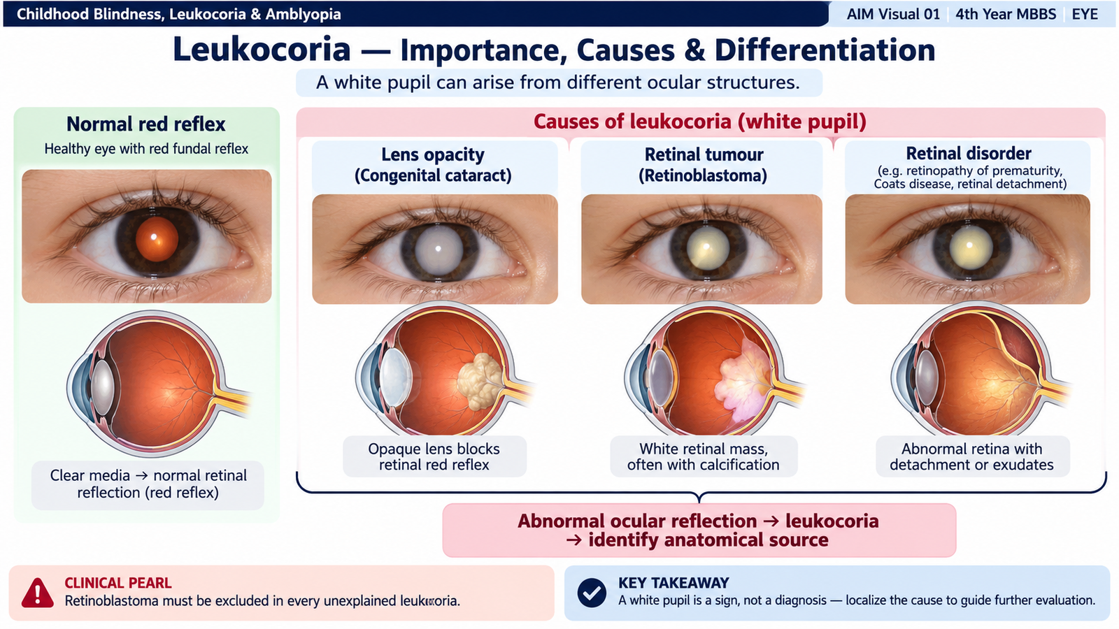
Leukocoria means an abnormal white appearance of the pupil instead of the normal red reflex. It is not a diagnosis by itself. It is a visible sign produced when light entering the eye is reflected back from an abnormal lens, vitreous or retina rather than from a normally perfused fundus. In a child, leukocoria must always be taken seriously. A harmless explanation should never be assumed until important ocular diseases have been excluded. The main clinical concern is that leukocoria may represent a potentially fatal intraocular malignancy such as retinoblastoma or a treatable cause of severe visual deprivation such as congenital cataract.
Why is a white pupil important?
- It may be the presenting sign of a sight-threatening ocular disorder.
- Retinoblastoma must be excluded because delay may threaten both the eye and the child’s life.
- Congenital cataract may cause irreversible deprivation amblyopia if a clear visual axis is not restored early.
- Some retinal disorders causing leukocoria can progress to retinal detachment or permanent retinal damage.
- Leukocoria detected by parents or in photographs still requires proper ophthalmic assessment.
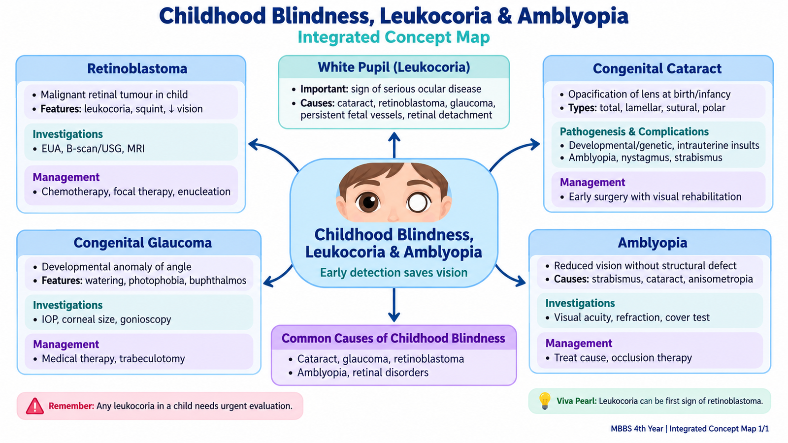
Major causes of leukocoria
The site of pathology provides a useful way to understand the differential diagnosis. An opacity in the lens produces leukocoria in congenital cataract, while a white retinal mass, retinal exudation, fibrovascular tissue or retinal detachment may produce a similar appearance from the posterior segment.
| Condition | Main site | Helpful distinguishing clue |
|---|---|---|
| Retinoblastoma | Retina | White retinal tumour; intraocular calcification may be demonstrated on imaging. |
| Congenital cataract | Lens | Visible lens opacity with absent or abnormal red reflex. |
| Persistent fetal vasculature | Retrolental region | Usually unilateral; affected eye may be small with retrolental fibrovascular tissue. |
| Coats disease | Retina | Retinal telangiectasia with marked exudation, typically unilateral; calcification is not characteristic. |
| Retinopathy of prematurity | Retina | History of prematurity; advanced disease may produce retinal detachment and leukocoria. |
| Ocular toxocariasis | Retina/vitreous | Unilateral inflammatory retinal granuloma or vitreous inflammation may mimic a white intraocular lesion. |
B. Evaluation & Investigations of a Child with Leukocoria
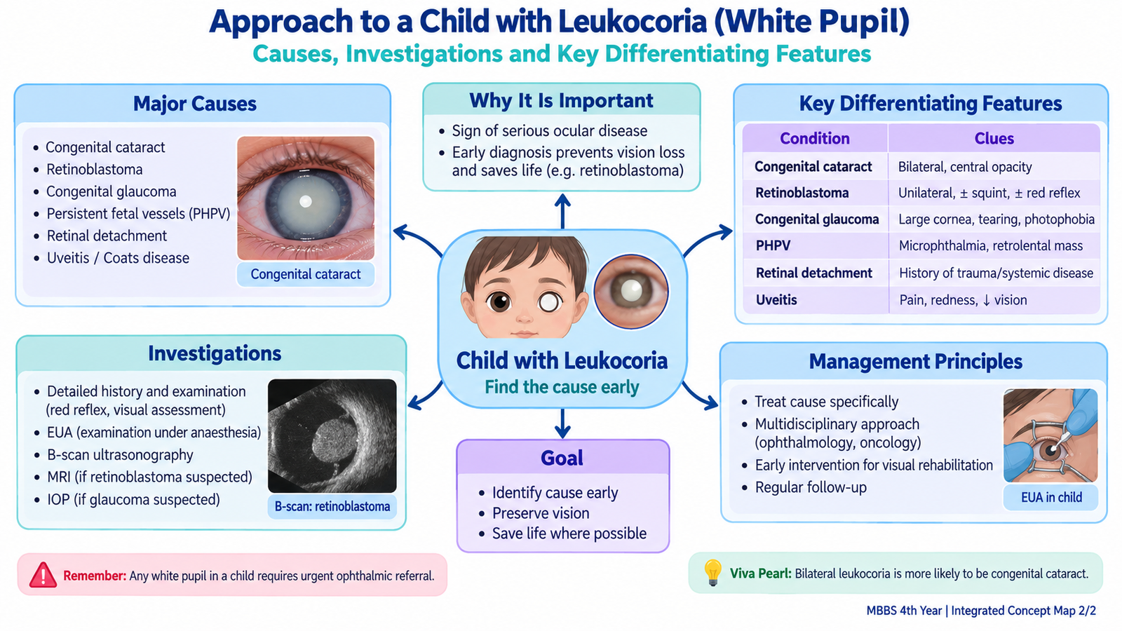
The investigation of leukocoria should answer three questions: Is the white reflex genuine? Where is the abnormality located? Could it represent retinoblastoma or another urgent sight-threatening disorder? Evaluation therefore begins with careful ophthalmic examination and then uses imaging when the posterior segment cannot be adequately assessed or when a tumour is suspected.
History
History helps establish the onset, laterality and likely cause. Important points include whether the white pupil is constant or intermittent, whether one or both eyes are affected, and whether strabismus or poor vision has been noticed.
- Age when the abnormal reflex was first noticed.
- Unilateral or bilateral involvement.
- Associated squint, reduced visual behaviour, redness or pain.
- History of prematurity when retinopathy of prematurity is possible.
- Family history of retinoblastoma or childhood eye disease.
- Previous ocular inflammation or trauma when relevant.
Ophthalmic examination
The red reflex is a rapid screening observation. A normal transparent ocular media allows light to reach the retina and produces a reddish-orange reflection. An opacity or abnormal mass may interrupt this reflex and produce asymmetry, darkness or whiteness.
- Visual behaviour or visual acuity: assesses the functional effect of the lesion.
- External inspection: looks for asymmetry in globe size or associated abnormalities.
- Pupillary examination: assesses shape and reactions.
- Red-reflex examination: identifies asymmetry or an abnormal white reflex.
- Anterior-segment examination: determines whether the lesion lies in the cornea, anterior chamber or lens.
- Dilated fundus examination: is essential when the visual axis permits examination of the retina.
Important investigations
Investigation should be directed by the suspected location. If a lens opacity clearly explains the leukocoria, the child requires assessment of its visual significance and associated ocular abnormalities. If a posterior segment lesion is suspected, ocular imaging becomes more important.
- Examination under anaesthesia: may be required in an uncooperative young child to allow detailed examination of both eyes, particularly when retinoblastoma is suspected.
- Ocular ultrasonography: is especially useful when the fundus is not visible. In retinoblastoma it may demonstrate an intraocular mass with highly reflective calcification.
- MRI of the orbits and brain: is useful when retinoblastoma is suspected because it assesses optic-nerve, orbital and intracranial involvement without exposing the child to ionizing radiation.
- Cycloplegic refraction: is useful after serious structural causes have been assessed, particularly when reduced vision or amblyopia is suspected.

C. Retinoblastoma
Retinoblastoma is a malignant tumour arising from the developing retina in early childhood. It is one of the most important diagnoses behind leukocoria because delayed recognition can lead not only to irreversible visual loss but also to extraocular tumour spread and death.
Etiology and genetic basis
Retinoblastoma develops when both functional copies of the RB1 tumour-suppressor gene are lost in a susceptible retinal cell. The RB protein normally helps regulate progression through the cell cycle. Loss of this control allows abnormal retinal cells to proliferate.
In the heritable form, a pathogenic RB1 alteration is already present in the germline, so fewer additional events are needed for tumour formation. These children are therefore more likely to develop bilateral or multifocal tumours. Sporadic disease is more often unilateral and unifocal.
Clinical features
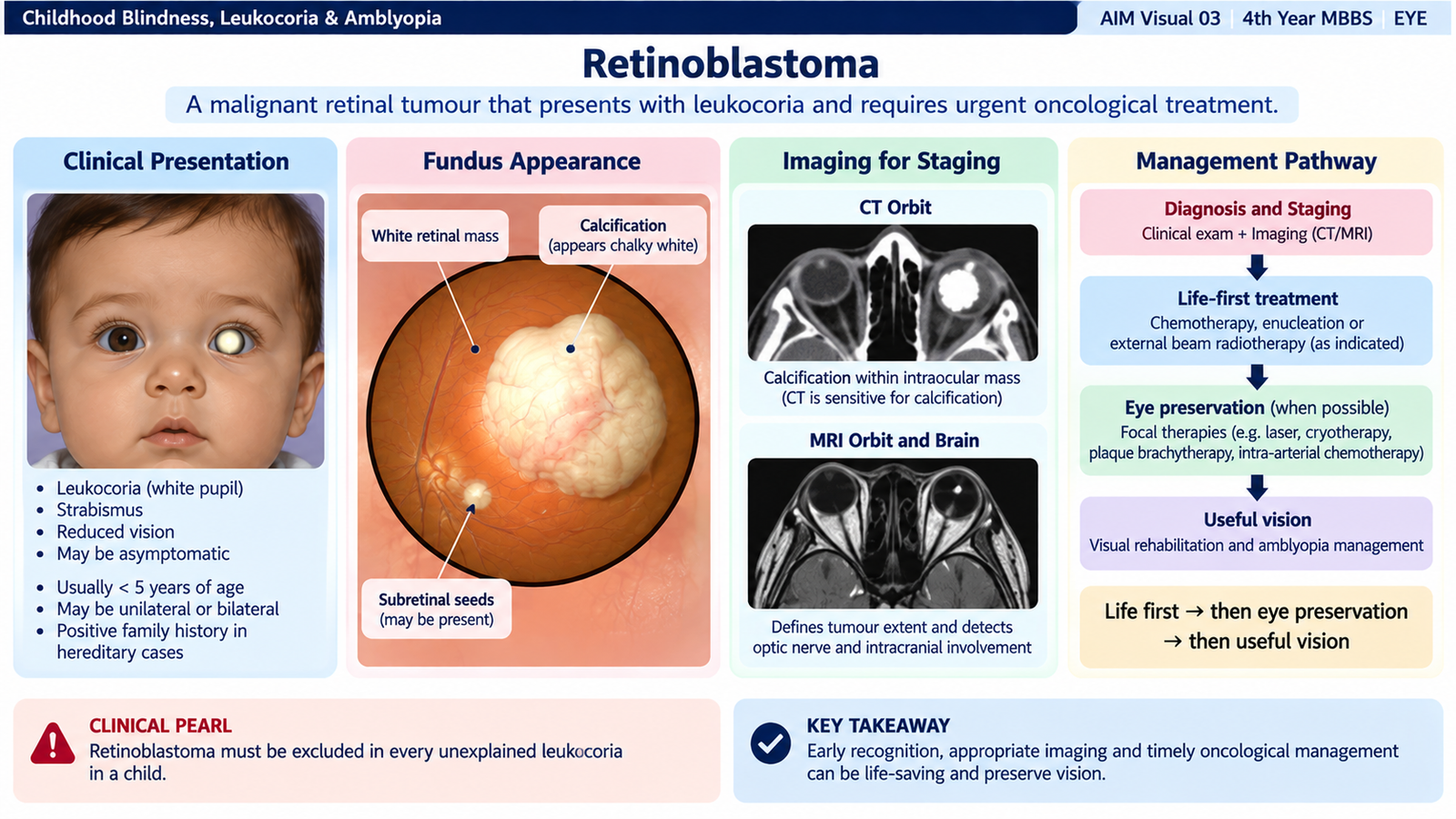
The presentation depends on tumour size and location. Leukocoria is the classic presenting sign because the pale tumour reflects light back through the pupil instead of producing the normal red reflex.
- Leukocoria — the most characteristic presenting sign.
- Strabismus — may occur when macular or central vision is impaired.
- Reduced visual behaviour or poor fixation.
- Red or painful eye in advanced disease.
- Secondary glaucoma may occur when advanced intraocular disease raises intraocular pressure.
- Proptosis may occur in very advanced extraocular disease.
Fundus appearance
Ophthalmoscopy typically reveals a creamy-white retinal mass. Tumour calcification is common and is an important diagnostic clue. Tumour growth may extend into the vitreous or subretinal space and may be accompanied by retinal detachment.
Investigations
Diagnosis is based mainly on ophthalmic examination and imaging. The aim is to confirm the ocular tumour, examine both eyes, assess the extent of disease and identify features that influence treatment.
- Dilated fundus examination: identifies the number, size and location of retinal tumours.
- Examination under anaesthesia: allows detailed assessment in small children.
- Ocular ultrasonography: demonstrates the mass and characteristic calcification.
- MRI of brain and orbits: evaluates optic-nerve, orbital and intracranial extension.
- Genetic assessment: is important in suspected hereditary disease and may guide examination of relatives and future surveillance.
CT can demonstrate calcification but is not preferred when MRI and ultrasonography can provide the required information, particularly because children with hereditary RB1 abnormalities have an increased vulnerability to radiation-associated second malignancies.
Management principles
Treatment has three priorities in descending order: save life, save the eye, and preserve useful vision whenever possible. Management depends on tumour extent, laterality, location and the visual potential of the affected eye.
- Enucleation: removal of the eye is used particularly for a large advanced unilateral tumour when useful vision cannot realistically be preserved or when the eye poses a major oncological risk.
- Systemic chemotherapy: may reduce tumour size and allow additional local treatment.
- Intra-arterial chemotherapy: delivers chemotherapy directly to the ophthalmic circulation in selected eyes and is used in specialist centres.
- Intravitreal chemotherapy: may be used for selected vitreous tumour seeds.
- Focal therapy: laser photocoagulation, thermotherapy or cryotherapy can destroy selected small tumours.
- Radiotherapy: has a limited selective role because of long-term adverse effects, particularly in hereditary disease.
D. Congenital Cataract
Definition
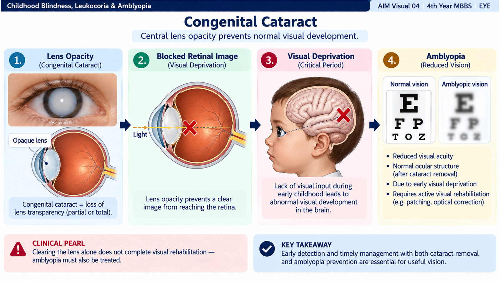
A congenital cataract is an opacity of the crystalline lens that is present at birth or becomes evident during early infancy as a result of abnormal lens development. The clinical importance of the cataract depends not only on its presence but also on its density, size, position and effect on the visual axis. A small peripheral opacity may have little effect on vision, whereas a dense central cataract prevents formation of a clear retinal image. In a young child this visual deprivation can permanently interfere with development of the visual cortex.
Types
Congenital cataracts are commonly described according to the part of the lens affected and their morphological appearance.
- Total cataract: the entire lens becomes opaque.
- Nuclear cataract: opacity predominantly involves the embryonic or fetal nucleus.
- Lamellar or zonular cataract: a particular layer or zone of the lens is opaque while other layers remain relatively clear.
- Anterior polar cataract: small opacity at the anterior pole of the lens.
- Posterior polar cataract: opacity at the posterior pole, close to the visual axis.
- Sutural cataract: opacity follows the Y-shaped lens sutures.
- Membranous cataract: severe disruption or absorption of lens material leaves a thin opaque membrane.
Pathogenesis and visual consequences
The normal lens remains transparent because its fibres and proteins are arranged in an orderly way and allow light to pass with minimal scattering. Disturbance of lens development alters this organization and produces an opacity.
This explains why congenital cataract is different from cataract in an elderly adult. In the child, restoring a transparent optical pathway is only one part of treatment. The developing brain must also learn to use the affected eye.
Clinical features
- Abnormal or absent red reflex.
- Leukocoria when the opacity is dense.
- Poor fixation or reduced visual behaviour.
- Strabismus, particularly with unilateral visual deprivation.
- Nystagmus may develop in severe bilateral deprivation that has been present during early visual development.
Complications
The most important complication is deprivation amblyopia. A dense cataract prevents patterned visual stimulation from reaching the retina, so the corresponding visual cortex fails to develop normal visual acuity.
- Severe amblyopia.
- Strabismus.
- Nystagmus in longstanding severe bilateral deprivation.
- Poor long-term vision despite technically successful surgery if treatment is delayed.
Management
Management is determined by whether the cataract is visually significant. A small opacity that does not obstruct the visual axis may be observed, while a dense central cataract that prevents normal visual development requires prompt specialist treatment.
- Assessment of visual significance: determine density, position, laterality and effect on fixation and red reflex.
- Surgical removal: required for a visually significant cataract that obstructs the visual axis.
- Optical rehabilitation: clear retinal focus must be restored using an intraocular lens where appropriate, spectacles or contact lenses depending on the child’s circumstances.
- Amblyopia treatment: particularly important in unilateral or asymmetrical cataract.
- Long-term follow-up: monitors refractive change, visual development and complications such as secondary glaucoma or renewed visual-axis opacification.

E. Congenital Glaucoma
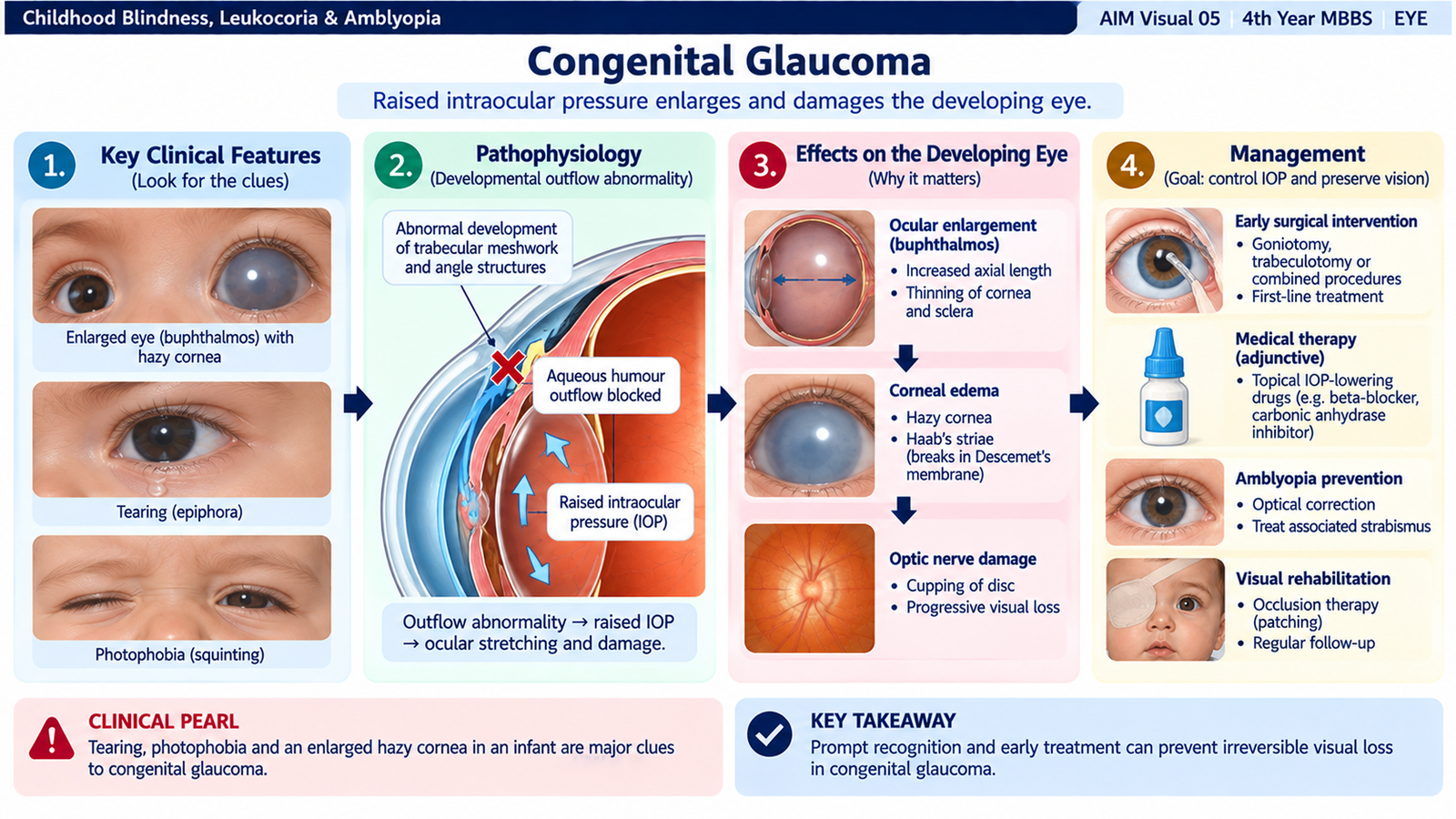
Congenital glaucoma is glaucoma occurring because the aqueous-outflow pathway of the developing eye is abnormal. In primary congenital glaucoma, abnormal development of the anterior chamber angle and trabecular outflow region reduces drainage of aqueous humour. Intraocular pressure rises and damages the developing eye.
Etiology and pathogenesis
The main mechanism is developmental abnormality of the aqueous outflow angle, often termed trabeculodysgenesis. Aqueous humour continues to be produced, but it cannot leave the eye normally through the trabecular pathway.
The sclera and cornea of an infant are more elastic than those of an adult. Therefore, persistent raised intraocular pressure can enlarge the entire globe. This produces buphthalmos, literally an abnormally enlarged eye.
Clinical features
The classic symptom complex consists of epiphora, photophobia and blepharospasm. These symptoms are mainly related to corneal epithelial edema caused by raised intraocular pressure.
- Epiphora: excessive watering.
- Photophobia: discomfort in bright light.
- Blepharospasm: forceful eyelid closure.
- Corneal haze or edema: due to raised intraocular pressure.
- Enlarged cornea and globe: caused by stretching of the developing ocular coats.
- Buphthalmos: enlarged eye in longstanding raised pressure.
- Reduced visual behaviour if corneal opacity or optic-nerve damage becomes significant.
Important examination findings
Increased pressure stretches the cornea and may produce breaks in Descemet membrane. These curvilinear breaks are called Haab striae. Continued pressure also damages retinal ganglion-cell axons at the optic nerve and produces glaucomatous optic-disc cupping.
- Raised intraocular pressure.
- Increased corneal diameter.
- Corneal edema or haze.
- Haab striae.
- Optic-disc cupping.
- Asymmetrical enlargement when one eye is more severely affected.
Investigations
Evaluation aims to confirm elevated intraocular pressure, document enlargement of the eye, assess the drainage angle and determine whether optic-nerve injury has occurred.
- Tonometry: confirms raised intraocular pressure.
- Measurement of corneal diameter: documents enlargement caused by chronic pressure.
- Corneal examination: assesses edema and Haab striae.
- Gonioscopy: evaluates the anterior chamber angle.
- Optic-disc examination: assesses glaucomatous cupping.
- Examination under anaesthesia: may be necessary for complete assessment in a young child.
Management
Primary congenital glaucoma is mainly a surgical disease because the basic problem is an abnormal aqueous-outflow pathway. Medical therapy may lower pressure temporarily, but it does not correct the developmental obstruction.
- Goniotomy: opens the abnormal trabecular tissue from within the anterior chamber when the cornea permits adequate visualization.
- Trabeculotomy: opens the trabecular outflow pathway from an external approach.
- Trabeculectomy or combined procedures: may be required in selected or resistant cases.
- Glaucoma drainage procedures: may be used when conventional surgery is unsuccessful.
- Medical pressure reduction: may serve as an adjunct while definitive surgical treatment is planned.
- Visual rehabilitation: refractive correction and amblyopia treatment may also be required.
F. Amblyopia
Definition
Amblyopia is reduced best-corrected visual acuity caused by abnormal visual development during childhood, without an ocular structural lesion sufficient to explain the degree of visual loss. It is therefore primarily a developmental disorder of visual processing rather than a disease that can simply be seen on fundus examination. Normal visual development requires each eye to provide the brain with a clear and appropriately aligned image. If one eye sends a blurred, blocked or persistently misaligned image during the sensitive period of visual development, the brain reduces its use of that image. Continued suppression results in reduced visual function in the affected eye.
Etiology and types
The type of amblyopia reflects the type of abnormal visual input responsible for cortical suppression.
- Strabismic amblyopia: persistent ocular misalignment causes the brain to suppress the image from the deviating eye to avoid confusion or diplopia.
- Anisometropic amblyopia: unequal refractive errors produce one clear and one chronically blurred retinal image, so the brain preferentially uses the clearer eye.
- Isoametropic or ametropic amblyopia: a high uncorrected refractive error in both eyes produces bilateral blurred retinal images.
- Deprivation amblyopia: an obstruction such as a dense congenital cataract or severe ptosis prevents normal visual stimulation.
- Meridional amblyopia: uncorrected astigmatism causes persistent blur in a particular orientation during visual development.
Pathogenesis
Deprivation amblyopia is particularly important because a severely obstructed visual axis may provide almost no useful patterned input. This is why early management of congenital cataract is essential even though the retina and optic nerve may initially be structurally normal.
Clinical features
- Reduced visual acuity in one or both eyes.
- Difference in visual acuity between the two eyes.
- Strabismus may coexist.
- Abnormal fixation preference may be noticed in young children.
- Ocular examination may be structurally normal in pure refractive or strabismic amblyopia.
- Vision does not become normal simply by looking through a pinhole if established amblyopia is present.
Investigations and assessment
Amblyopia is diagnosed only after measuring vision carefully and excluding structural ocular or neurological causes of poor vision. The examination must therefore identify both the reduced acuity and the reason abnormal visual development occurred.
- Age-appropriate visual acuity or fixation assessment.
- Cycloplegic refraction: identifies anisometropia, high bilateral refractive error or astigmatism.
- Cover testing and ocular alignment assessment: detects strabismus.
- Red reflex and anterior-segment examination: exclude visual-axis obstruction such as cataract.
- Dilated fundus examination: excludes retinal or optic-nerve pathology.
Management
Treatment aims to provide the brain with a clear image from the amblyopic eye and encourage active use of that eye. Management therefore starts with the underlying cause and is followed by specific amblyopia therapy when required.
- Correct refractive error: appropriate spectacles or contact lenses provide the clearest retinal image possible.
- Remove visual deprivation: treat a visually significant cataract or other obstruction promptly.
- Occlusion therapy: covering the better-seeing eye encourages use of the amblyopic eye.
- Pharmacological penalization: atropine may be used in selected children to reduce the advantage of the better eye.
- Treat associated strabismus: according to the underlying ocular alignment problem while continuing visual rehabilitation.
- Monitor vision: treatment is adjusted according to response and to avoid excessive reduction of vision in the better eye.

G. Common Causes of Childhood Blindness & Management Plan
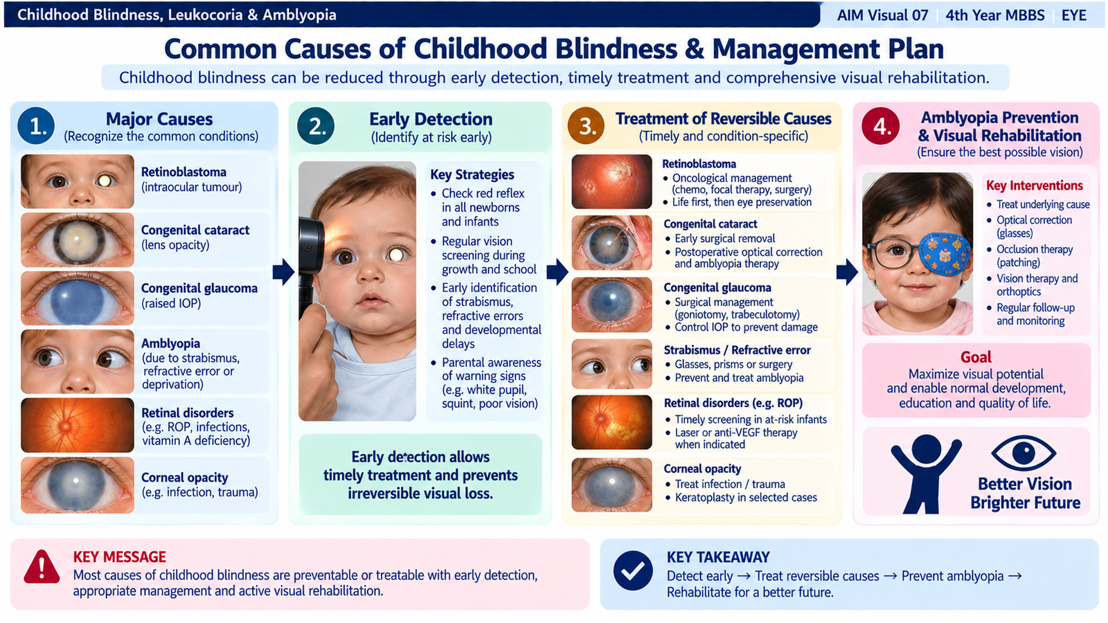
Childhood blindness may result from disease affecting almost any part of the visual pathway, from the cornea and lens to the retina, optic nerve or visual cortex. For undergraduate clinical practice, the most useful approach is to identify conditions in which vision can be preserved or restored by timely prevention, diagnosis, treatment or visual rehabilitation.
Important causes
- Congenital and developmental cataract.
- Congenital and childhood glaucoma.
- Retinopathy of prematurity.
- Retinoblastoma and other serious retinal disorders.
- Retinal dystrophies and congenital retinal abnormalities.
- Corneal opacity or scarring following nutritional deficiency, infection or ocular injury.
- Uncorrected refractive error.
- Amblyopia due to strabismus, refractive error or visual deprivation.
- Optic-nerve and central visual pathway disorders.
Management plan
Management is most effective when organized according to the stage at which visual loss can be prevented. Some disorders must be prevented before they develop, some require rapid treatment to preserve the eye, and others require rehabilitation when irreversible damage has already occurred.
Recognize abnormal red reflex, leukocoria, strabismus, poor visual behaviour, abnormal globe size and other warning signs.
Urgently assess suspected retinoblastoma, congenital glaucoma and other sight- or life-threatening ocular disease.
Clear significant cataract, control glaucoma, treat relevant retinal disease and correct refractive error.
Recognize and treat amblyopia whenever unequal, blurred or deprived visual input has occurred.
Prevent ocular injury, address nutritional and infectious causes of corneal damage and ensure appropriate assessment of infants at risk of retinopathy of prematurity.
Use appropriate optical aids, low-vision rehabilitation and educational support when normal vision cannot be restored.
Condition-based management overview
| Cause | Main management principle |
|---|---|
| Congenital cataract | Prompt treatment of visually significant cataract + optical rehabilitation + amblyopia therapy. |
| Congenital glaucoma | Early pressure control, mainly by surgery, with long-term visual follow-up. |
| Retinoblastoma | Urgent specialist oncological management: life first, then globe and vision preservation. |
| Refractive error / amblyopia | Accurate refraction, optical correction and amblyopia treatment where needed. |
| Retinopathy of prematurity | Identify at-risk premature infants, perform appropriate retinal surveillance and treat sight-threatening disease. |
| Irreversible retinal / optic-nerve disease | Maximize residual vision and provide low-vision and educational rehabilitation. |
3. Integrated Mechanism Flow
⭐ AIM High-Yield Review
- Leukocoria is a sign, not a diagnosis; retinoblastoma must always be excluded.
- Congenital cataract produces leukocoria from the lens; retinoblastoma produces it from a retinal tumour.
- Ultrasonographic calcification strongly supports retinoblastoma in the appropriate clinical setting.
- MRI is important in suspected retinoblastoma for evaluating optic-nerve, orbital and intracranial extension.
- Retinoblastoma treatment priorities are life → eye → useful vision.
- The major visual danger of dense congenital cataract is deprivation amblyopia.
- Successful congenital cataract care requires surgery when indicated, optical correction and amblyopia treatment.
- Congenital glaucoma classically produces epiphora, photophobia and blepharospasm.
- Buphthalmos occurs because raised intraocular pressure stretches the relatively elastic infant eye.
- Haab striae are breaks in Descemet membrane caused by corneal stretching in congenital glaucoma.
- Definitive treatment of primary congenital glaucoma is mainly surgical.
- Amblyopia is reduced vision caused by abnormal visual development, not by an ocular lesion sufficient to explain the loss.
- Major amblyopia mechanisms are strabismus, unequal refractive error, bilateral blur and visual deprivation.
- Childhood blindness is best reduced through early detection, treatment of reversible causes, prevention of amblyopia and rehabilitation of irreversible loss.
- Any child with unexplained leukocoria requires prompt ophthalmic evaluation.
Childhood Blindness, Leukocoria & Amblyopia
Watch these focused videos after completing the learning material to reinforce the major clinical concepts.
Retinoblastoma & Leukocoria — High-Yield Review
Reinforces leukocoria, retinoblastoma presentation, RB1 concepts, differential diagnosis and management principles.
Pathogenesis & Management of Pediatric Cataracts
Covers pediatric cataract types, presentation, investigation, surgical principles and postoperative visual rehabilitation.
Diagnosis & Management of Congenital Glaucoma
Reinforces congenital glaucoma pathophysiology, clinical recognition, investigations and medical and surgical management.
Amblyopia — Development of the Amblyopic Eye
A concise explanation of how abnormal visual input during childhood interferes with visual cortical development.