
📖 AIM Learning Material
Topic 1 — Musculoskeletal Foundations, Metabolic Bone Diseases and Bone-Mineral Homeostasis
A clear first-learning chapter on bone structure, metabolic bone diseases, bone-mineral medicines, musculoskeletal pain, childhood bone disorders, ergonomics, prevention, disability and rehabilitation.
Topic Introduction
Bone is a living tissue that is continuously renewed by osteoclasts and osteoblasts. Its strength depends on the amount of bone, the arrangement of cortical and trabecular architecture, and proper mineralization with calcium and phosphate. This chapter explains the main metabolic bone disorders: osteoporosis, osteomalacia, rickets and Paget disease. It then connects these disorders with their clinical features, investigations, treatment and prevention. You will also learn how calcium, vitamin D and antiresorptive drugs act, how NSAIDs are used for musculoskeletal pain, why childhood bone disorders produce characteristic deformities, and how ergonomics and rehabilitation reduce disability. The aim is to understand each condition through simple cause-and-effect links rather than isolated facts.
A. Bone Structure, Osteoporosis, Osteomalacia and Rickets
To understand metabolic bone disease, first remember that cortical bone provides dense outer strength, while trabecular bone forms an internal lattice with a large remodeling surface. Osteoblasts form osteoid, osteoclasts resorb mineralized bone, and osteocytes sense mechanical strain. RANKL promotes osteoclast formation by activating RANK, while osteoprotegerin limits this signal. Normal mineralization requires adequate calcium, phosphate and vitamin D. Mechanical loading helps maintain bone and muscle; prolonged immobility reduces both.
Osteopenia and osteoporosis
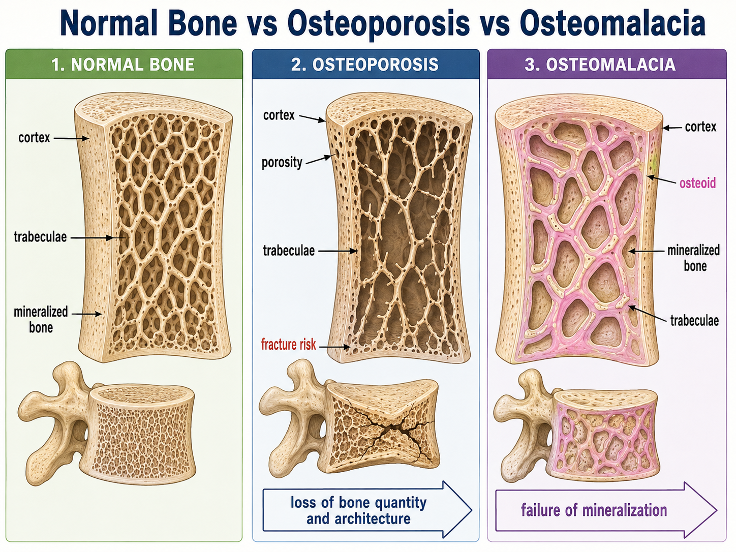
Osteopenia indicates bone mass below the expected healthy range but less severe than osteoporosis. Osteoporosis is a systemic skeletal disorder characterized by reduced bone mass, deterioration of bone microarchitecture and increased susceptibility to fragility fracture. The remaining bone is usually mineralized normally; the principal problem is that too little structurally competent bone remains. Bone strength depends on both quantity and quality. A person may therefore fracture not only because bone mineral content is reduced, but also because trabecular connectivity, cortical thickness, microdamage repair and overall geometry have deteriorated.
Classification
- Primary osteoporosis: includes postmenopausal bone loss and age-related osteoporosis.
- Secondary osteoporosis: results from diseases, nutritional problems, prolonged immobility or medicines such as glucocorticoids.
- Localized osteoporosis: may occur around an immobilized or disused limb.
Major risk factors
- Advancing age
- Female sex and menopause
- Low peak bone mass
- Previous fragility fracture
- Family tendency
- Low calcium or vitamin D intake
- Physical inactivity
- Smoking and harmful alcohol use
- Low body weight or malnutrition
- Recurrent falls
- Long-term glucocorticoid exposure
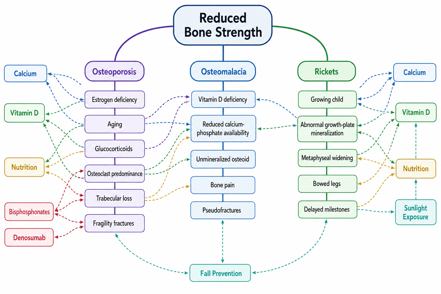
Pathogenesis of postmenopausal osteoporosis
Age-related and glucocorticoid-associated bone loss
With aging, osteoblast replication and function decline, calcium and vitamin D adequacy may decrease, and reduced activity lowers mechanical stimulation. Glucocorticoids suppress osteoblast function, reduce calcium absorption, increase calcium loss and may promote muscle weakness, thereby combining skeletal loss with greater fall risk.
Morphology
- Thinning of cortical bone with increased porosity.
- Thinning and loss of trabecular plates.
- Loss of trabecular connectivity.
- Microfractures and vertebral compression.
- Normal mineralization of the reduced bone matrix.
Osteomalacia and rickets
Osteomalacia is defective mineralization of newly formed osteoid in adults after epiphyseal closure. Rickets is defective mineralization of growing bone and growth-plate cartilage in children. In both, the body produces osteoid, but mineral deposition is inadequate.
Common causes
- Vitamin D deficiency due to poor intake, inadequate sunlight exposure or malabsorption.
- Impaired vitamin D activation in chronic kidney or liver disease.
- Phosphate deficiency or renal phosphate loss.
- Medicines or disorders interfering with vitamin D metabolism.
Mechanism
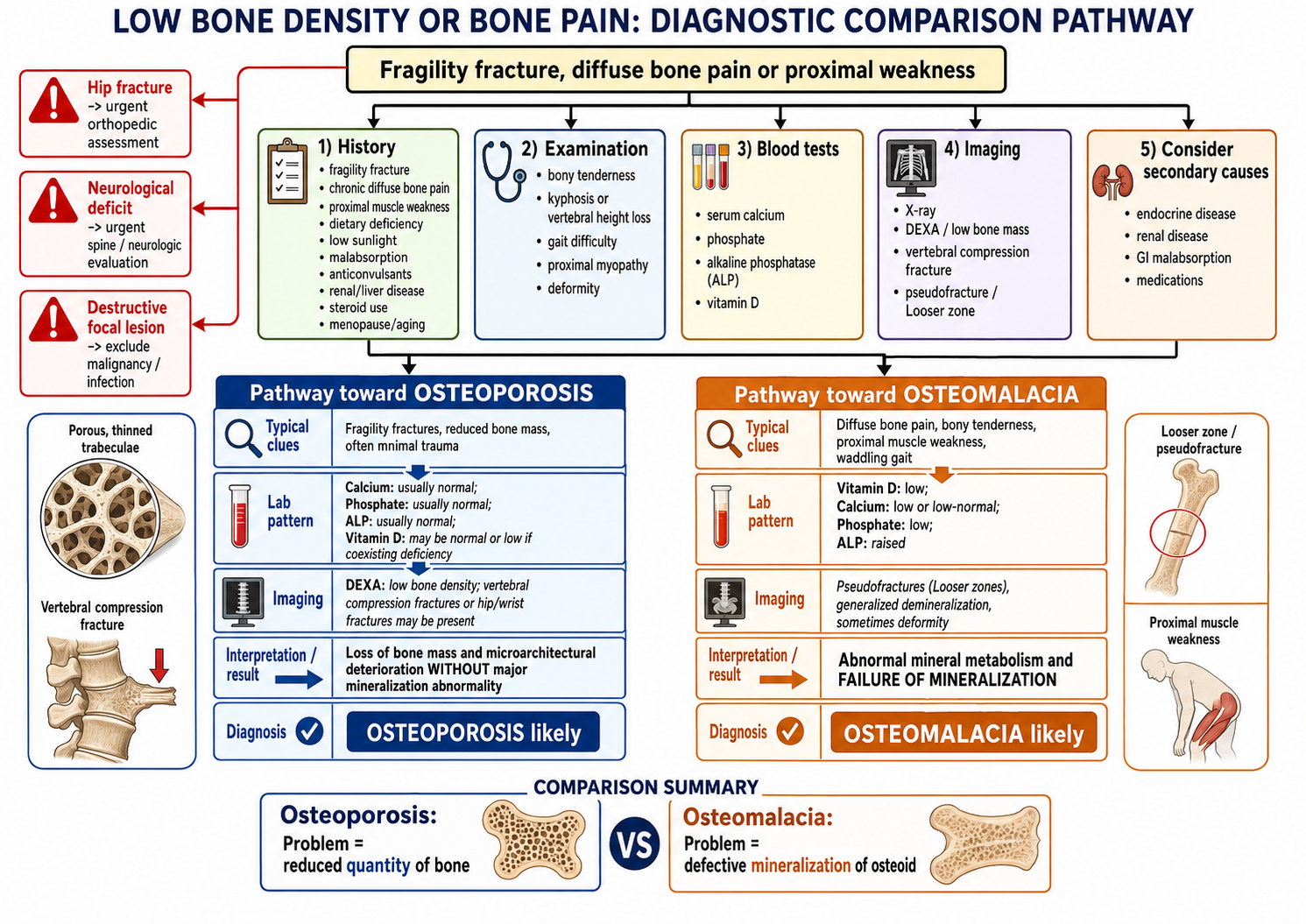
Clinical distinction
| Feature | Osteoporosis | Osteomalacia | Rickets |
|---|---|---|---|
| Main defect | Reduced amount and quality of bone | Defective mineralization of adult osteoid | Defective mineralization of growing bone and growth plates |
| Typical pain | Often silent until fracture | Diffuse bone pain and proximal weakness | Bone tenderness, delayed milestones and deformity |
| Structural clue | Fragility fracture, vertebral compression | Pseudofracture or reduced mineralization | Widened, cupped and frayed metaphyses |
| Laboratory tendency | Often no characteristic mineral abnormality | Low phosphate and raised alkaline phosphatase may occur | Similar mineral disturbance with raised alkaline phosphatase |
B. Paget Disease: Excessive and Disorganized Bone Remodeling
Paget disease of bone
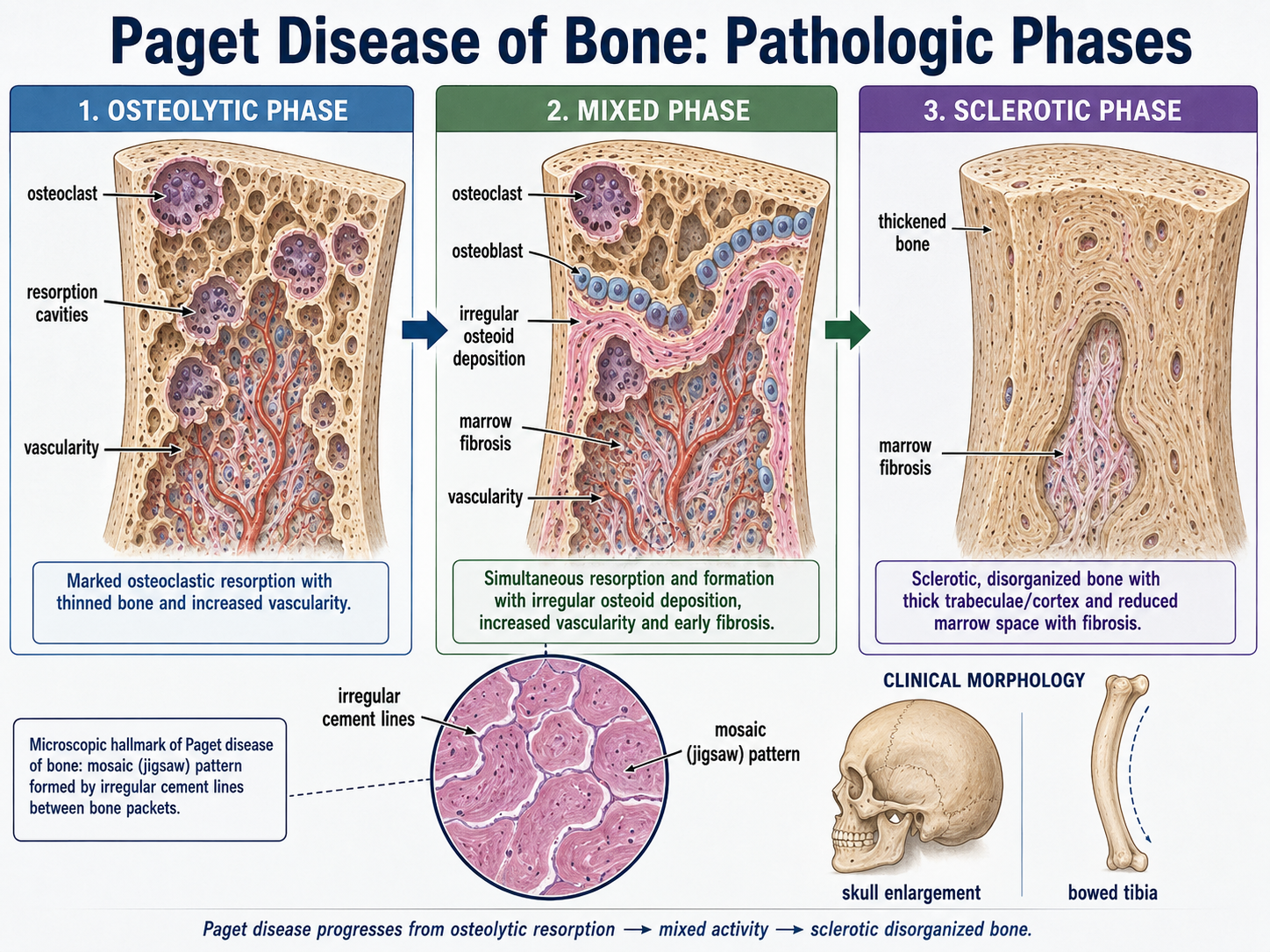
Paget disease, also called osteitis deformans, is a localized disorder of bone remodeling in which excessive osteoclastic resorption is followed by excessive but disorganized osteoblastic bone formation. The resulting bone may become enlarged and dense on imaging, yet its architecture is mechanically unsound.
Pathogenetic phases
- Osteolytic phase: marked osteoclastic resorption creates large resorption spaces.
- Mixed phase: osteoclasts and osteoblasts are both highly active.
- Sclerotic or burnt-out phase: dense but disorganized bone is deposited, with reduced cellular activity later.
Morphology
- Bone enlargement with cortical and trabecular thickening.
- Disorganized arrangement of lamellar bone.
- Irregular cement lines producing a mosaic or jigsaw pattern microscopically.
- Marrow replacement by loose connective tissue and prominent vessels during active disease.
- Bone deformity and microfractures despite increased density.
Clinical and diagnostic clues
- Many patients are asymptomatic and diagnosed incidentally.
- Bone pain, deformity or pathological fracture may occur.
- Skull involvement can produce enlargement, headache, hearing impairment or cranial nerve compression.
- Long-bone involvement may produce bowing.
- Serum alkaline phosphatase may be increased due to high osteoblastic activity, while calcium and phosphate may remain relatively preserved.
- Radiographs may show a mixture of lytic and sclerotic changes with bone expansion.
Complications
- Pathological fracture and secondary osteoarthritis due to altered mechanics.
- Nerve compression, including hearing impairment when the skull is involved.
- High-output cardiac strain in extensive active disease because of increased skeletal vascularity.
- Rare malignant transformation to an aggressive bone sarcoma.
C. Drugs Affecting Bone and Mineral Homeostasis
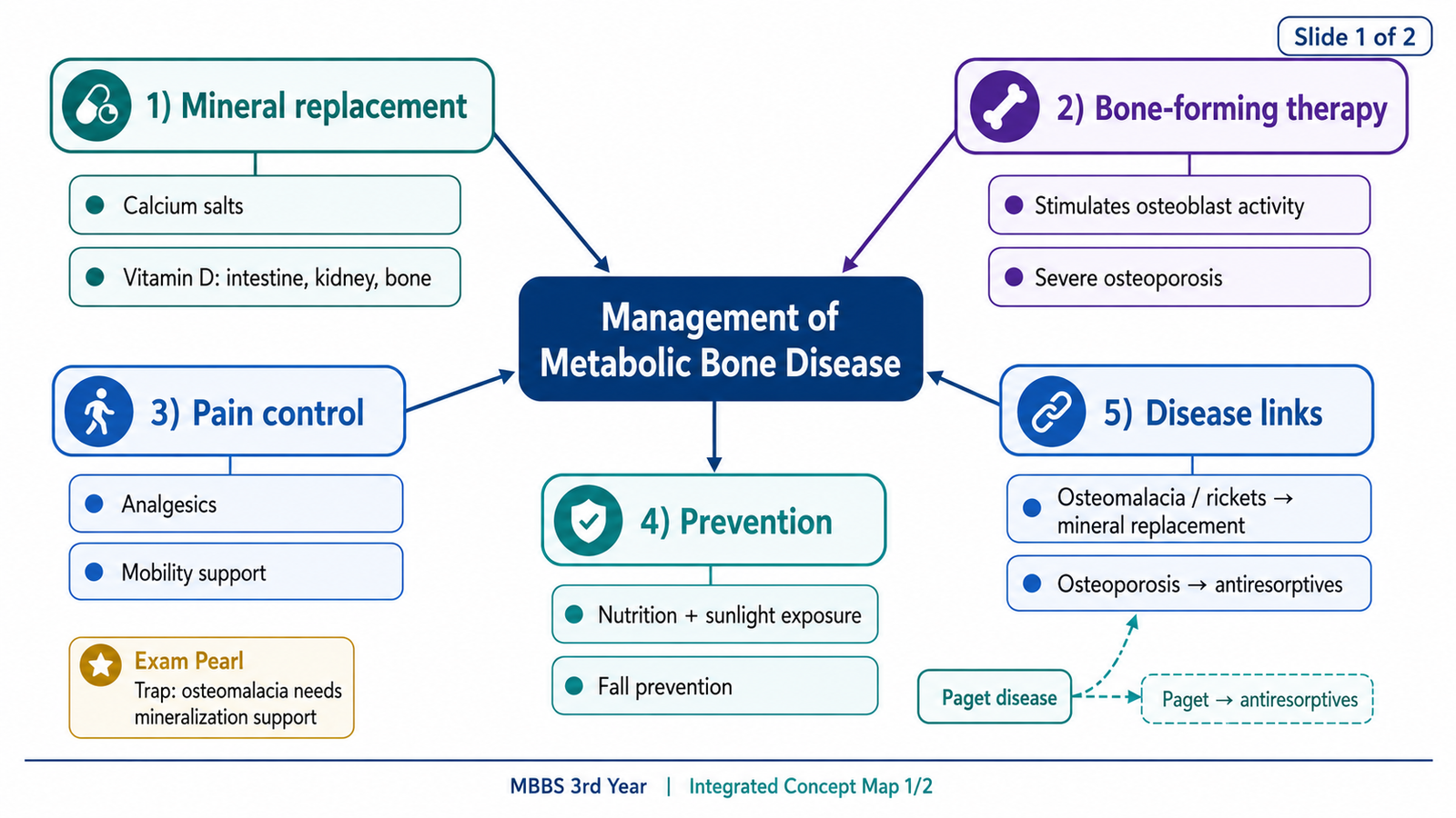
Drug treatment is selected according to the main abnormality. Replacement therapy supplies calcium or vitamin D when mineral availability is inadequate. Antiresorptive drugs reduce excessive osteoclast activity, while bone-forming drugs increase osteoblast-dependent formation in selected patients.
Classification of drugs used in metabolic bone disorders
| Therapeutic role | Drug groups | Main effect |
|---|---|---|
| Replace deficient mineral | Calcium salts | Supply calcium for physiological and skeletal requirements |
| Improve mineral absorption | Vitamin D preparations and active analogues | Increase calcium and phosphate availability |
| Suppress resorption | Bisphosphonates, denosumab, calcitonin, raloxifene | Reduce osteoclast-mediated bone loss |
| Stimulate formation | Parathyroid-hormone-related anabolic therapy and other bone-forming agents | Increase osteoblast-dependent bone formation when used appropriately |
Calcium preparations
- Calcium carbonate
- Calcium citrate
- Calcium gluconate
- Calcium chloride
- Other oral calcium salts formulated for supplementation
Clinical uses of calcium salts
- Prevention or correction of dietary calcium deficiency.
- Adjunctive support in osteoporosis when intake is inadequate.
- Management of states associated with hypocalcemia.
- Support of mineralization in rickets or osteomalacia when calcium deficiency contributes.
- Selected emergency use of intravenous calcium in serious symptomatic hypocalcemia or specific toxicological and cardiac circumstances under monitoring.
Important cautions with calcium
Excessive supplementation can cause gastrointestinal discomfort, constipation, hypercalcemia or contribute to stone formation in susceptible individuals. Oral calcium may reduce absorption of certain medicines by binding them in the gastrointestinal tract; timing may therefore need separation.
Vitamin D preparations
- Cholecalciferol, representing vitamin D3.
- Ergocalciferol, representing vitamin D2.
- Calcifediol, the 25-hydroxylated form.
- Calcitriol, the active 1,25-dihydroxylated form.
- Active or partially activated analogues used when activation is impaired.
Actions of vitamin D
Increases absorption of calcium and phosphate by promoting synthesis of transport proteins and related absorptive mechanisms.
Supports renal conservation of calcium and contributes to coordinated mineral balance, although its renal effects interact with parathyroid hormone and the prevailing mineral state.
Provides adequate calcium and phosphate for mineralization. In regulatory settings, active vitamin D can also facilitate bone resorption indirectly by influencing osteoblast-lineage signaling to osteoclast precursors.
Clinical uses of vitamin D
- Prevention and treatment of nutritional vitamin D deficiency.
- Treatment of rickets and osteomalacia caused by deficient vitamin D supply or action.
- Adjunctive use in osteoporosis when vitamin D status or intake is inadequate.
- Use of active preparations when renal activation is impaired or when a rapid active form is specifically required.
- Management of selected hypocalcemic states under appropriate monitoring.
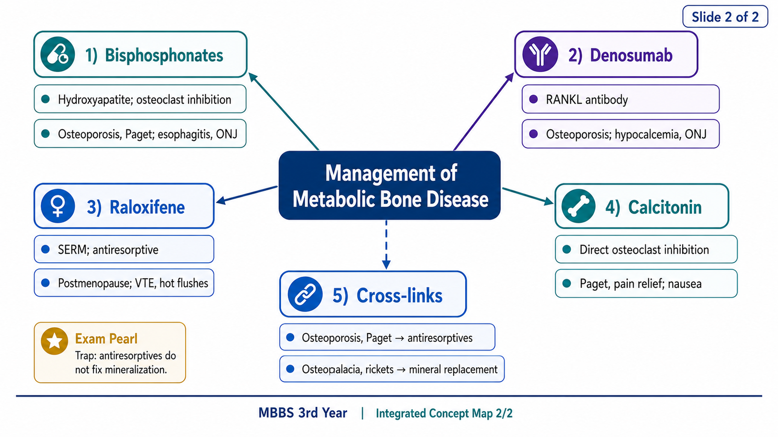
Bisphosphonates
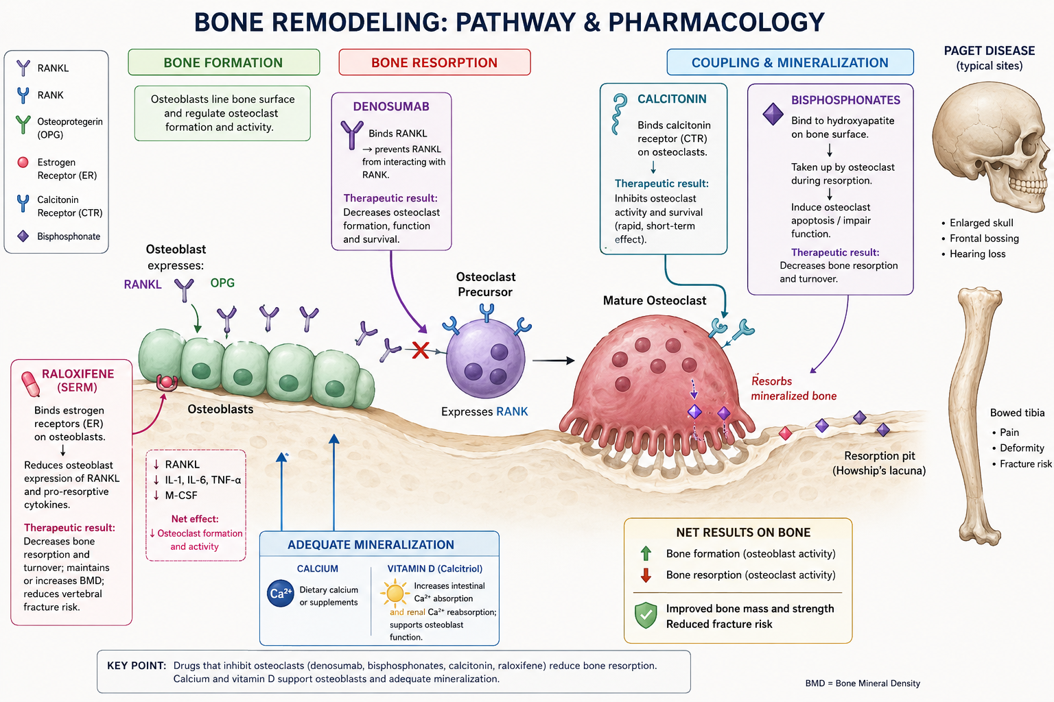
Bisphosphonates are analogues of pyrophosphate with strong affinity for hydroxyapatite. They become concentrated at sites of active bone remodeling and are taken up by osteoclasts during resorption.
Nitrogen-containing bisphosphonates inhibit an enzyme in the mevalonate pathway, impairing protein prenylation required for osteoclast cytoskeletal organization, vesicular transport and survival. Non-nitrogen compounds form toxic intracellular metabolites within osteoclasts.
Clinical uses of bisphosphonates
- Postmenopausal and other forms of osteoporosis.
- Glucocorticoid-associated osteoporosis.
- Paget disease when suppression of excessive remodeling is required.
- Reduction of skeletal complications in selected conditions with excessive bone resorption.
- Management of certain hypercalcemic states.
Adverse effects and precautions
- Upper gastrointestinal irritation and esophagitis with oral preparations.
- Acute-phase symptoms after some intravenous preparations.
- Hypocalcemia, particularly when vitamin D or calcium deficiency is uncorrected.
- Renal caution with some preparations.
- Rare osteonecrosis of the jaw, especially with potent or prolonged therapy in high-risk settings.
- Rare atypical femoral fractures with prolonged suppression of remodeling.
Calcitonin
Calcitonin binds receptors on osteoclasts and rapidly reduces their resorptive activity. It can therefore lower calcium released from bone and reduce excessive turnover.
It may be used in selected cases of Paget disease, hypercalcemia and for short-term relief of pain associated with an acute vertebral compression fracture. Its anti-osteoporotic effect is generally weaker than that of major first-line antiresorptive agents. Adverse effects include nausea, flushing and local nasal symptoms with intranasal use. Hypersensitivity may occur. Its effect can diminish with continuing exposure because of reduced responsiveness.
Raloxifene
Raloxifene is a selective estrogen receptor modulator. It behaves as an estrogen agonist in bone, reducing osteoclast-driven resorption, while acting as an antagonist in breast tissue and without providing full estrogenic stimulation in all reproductive tissues.
Important adverse effects include hot flushes, leg cramps and increased risk of venous thromboembolism. It should be avoided in patients with active or significant previous thromboembolic disease.
Denosumab
Denosumab is a monoclonal antibody that binds RANKL. By preventing RANKL from activating RANK on osteoclast precursors and mature osteoclasts, it reduces osteoclast formation, activity and survival.
Important concerns include hypocalcemia, infection or skin reactions in some patients, and rare jaw osteonecrosis or atypical femoral fracture. Bone loss may accelerate after discontinuation if subsequent management is not planned, because its antiresorptive effect is reversible.
Classification of drugs used to treat osteoporosis
- Antiresorptive agents: bisphosphonates, denosumab, raloxifene, calcitonin and selected hormonal approaches.
- Bone-forming agents: intermittent parathyroid-hormone-related anabolic therapy and other agents that promote formation.
- Supportive replacement: calcium and vitamin D when dietary intake or status is inadequate.
D. NSAIDs in Musculoskeletal Disorders
Clinical applications of NSAIDs
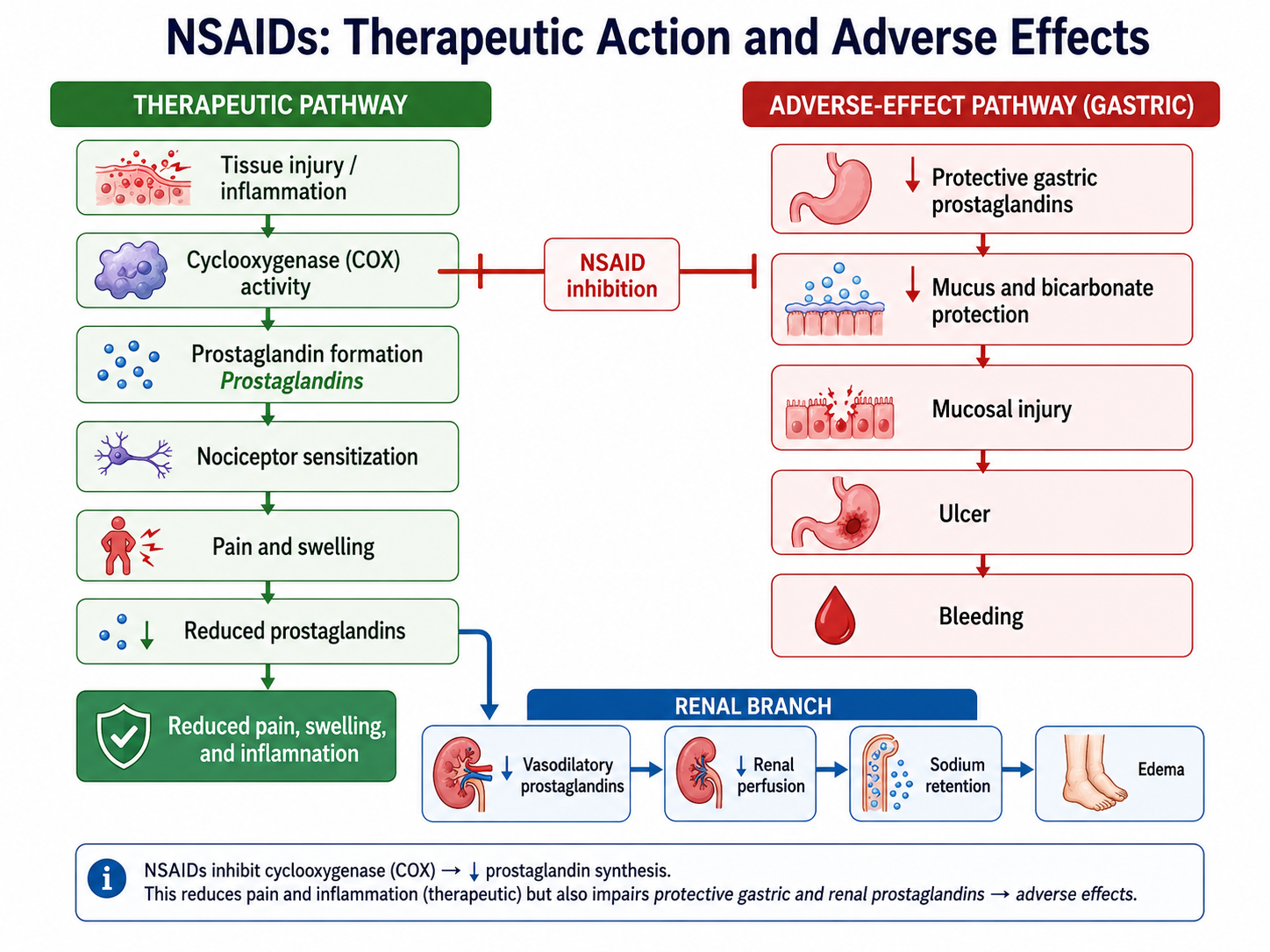
Non-steroidal anti-inflammatory drugs are used in musculoskeletal disorders because they reduce pain, inflammatory swelling and stiffness. They are useful when prostaglandin-mediated inflammation contributes to symptoms, but they do not correct the underlying loss of bone mass or mineralization defect.
Relevant musculoskeletal uses
- Inflammatory joint pain and stiffness.
- Acute soft-tissue sprain, strain or overuse injury.
- Osteoarthritis-related pain when non-drug measures are insufficient.
- Short-term pain associated with musculoskeletal injury.
- Selected inflammatory spinal or periarticular conditions.
Drug-selection logic
The lowest effective exposure for the shortest appropriate period is generally preferred. Selection depends on age, gastrointestinal history, renal function, cardiovascular risk, concurrent medicines and whether a topical preparation can provide adequate local benefit with less systemic exposure.
Important adverse effects
- Gastrointestinal: dyspepsia, gastritis, ulceration and bleeding due to reduced protective prostaglandins.
- Renal: sodium retention, edema and reduced renal perfusion in susceptible patients.
- Cardiovascular: worsening hypertension, fluid retention and altered thrombotic balance with some agents.
- Respiratory or hypersensitivity: bronchospasm or allergic reactions in susceptible individuals.
- Platelet-related: impaired aggregation with non-selective cyclooxygenase inhibition.
Why NSAIDs do not treat osteoporosis
NSAIDs can reduce pain from associated osteoarthritis, soft-tissue inflammation or an acute fracture, but they do not rebuild lost trabeculae, correct vitamin D deficiency or block the major osteoclast pathways responsible for osteoporosis. Symptomatic relief must therefore not delay diagnosis and disease-specific management.
E. Clinical Recognition, Investigation and Management of Osteoporosis and Osteomalacia
Clinical presentation of osteoporosis
Osteoporosis is often clinically silent until a fracture occurs. A fracture resulting from a minor fall or routine activity is a major diagnostic clue. Common sites include vertebrae, hip and distal radius.
- Acute back pain from vertebral compression.
- Progressive height loss and thoracic kyphosis.
- Wrist fracture after a low-energy fall.
- Hip fracture with marked loss of mobility and independence.
- Chronic pain and functional limitation after repeated vertebral fractures.
Clinical presentation of osteomalacia
- Diffuse bone discomfort, often involving the pelvis, lower back, ribs or lower limbs.
- Proximal muscle weakness causing difficulty climbing stairs or rising from a chair.
- Waddling gait.
- Bone tenderness.
- Insufficiency fractures or pseudofractures.
Important causes and risk factors to seek in history
- Menopause, advanced age and previous fragility fracture.
- Dietary calcium and vitamin D intake.
- Sunlight exposure and clothing or lifestyle factors that reduce exposure.
- Long-term glucocorticoid or other bone-affecting medicine use.
- Reduced mobility, neurological disease or prolonged bed rest.
- Malabsorption, chronic kidney disease or chronic liver disease.
- Smoking, harmful alcohol use and recurrent falls.
- Features of endocrine or systemic illness causing secondary bone loss.
Investigations
- Bone mineral-density assessment where clinically indicated
- Radiographs for suspected fracture or deformity
- Assessment of vertebral compression when suggested by pain or height loss
- Serum calcium
- Serum phosphate
- Alkaline phosphatase
- Vitamin D status where appropriate
- Renal and liver function
- Relevant endocrine evaluation
- Malabsorption assessment when suspected
- Medication review
Differential diagnosis
| Clinical problem | Differentiating clue |
|---|---|
| Osteoporosis | Fragility fracture or vertebral compression with relatively preserved mineral biochemistry |
| Osteomalacia | Diffuse bone pain, proximal weakness and biochemical evidence of impaired mineralization |
| Osteoarthritis | Joint-related pain worsened by use, with local stiffness and structural joint changes |
| Inflammatory arthritis | Prolonged inflammatory stiffness, synovitis and systemic inflammatory features |
| Malignant or infective bone lesion | Focal persistent pain, constitutional symptoms, destructive lesion or inflammatory evidence |
| Myopathy | Predominant muscle weakness without the typical bone pain or fragility pattern |
Broad management plan for osteoporosis
- Confirm fracture status and identify secondary causes.
- Review medicines that increase bone loss, sedation or fall risk.
- Ensure adequate dietary calcium, vitamin D and overall nutrition.
- Encourage suitable weight-bearing, resistance and balance activity.
- Reduce smoking, harmful alcohol exposure and environmental fall hazards.
- Select appropriate antiresorptive or bone-forming treatment according to fracture risk, contraindications and patient factors.
- Manage pain without relying on unsafe long-term NSAID exposure.
- Arrange rehabilitation after fracture or functional decline.
Broad management plan for osteomalacia
- Identify the cause of defective mineralization.
- Correct vitamin D, calcium or phosphate deficiency as appropriate.
- Treat malabsorption, renal, hepatic or medication-related contributors.
- Monitor clinical recovery and relevant biochemical abnormalities.
- Provide safe mobility support and treat fractures when present.
F. Bone Pain, Nutritional Rickets and Important Childhood Skeletal Disorders
Nutritional rickets: causation
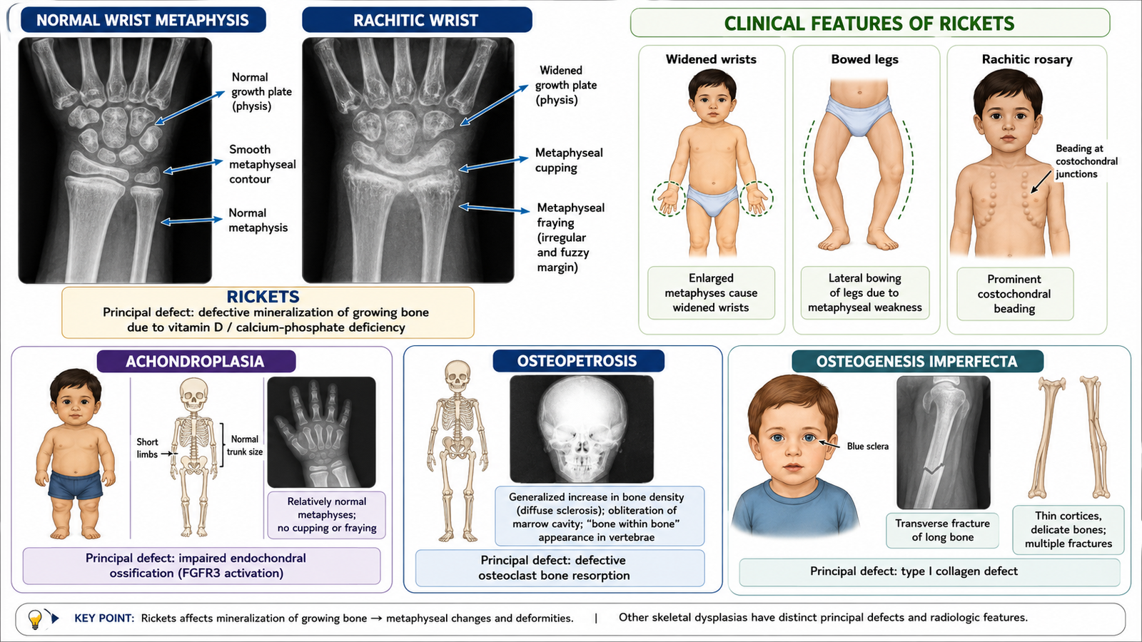
Nutritional rickets develops when a growing child has inadequate vitamin D action, inadequate calcium intake or both. Because the growth plate remains active, impaired mineralization affects not only osteoid but also the orderly mineralization and maturation of growth-plate cartilage.
Clinical presentation
- Delayed sitting, standing or walking.
- Bone pain or irritability.
- Widening of wrists and ankles.
- Bowing of weight-bearing legs or other lower-limb deformity.
- Prominent costochondral junctions.
- Frontal prominence or delayed closure of skull fontanelles in younger children.
- Muscle weakness and hypotonia.
- Growth impairment in more persistent disease.
Laboratory and radiological findings
- Raised alkaline phosphatase due to increased osteoblastic activity.
- Reduced phosphate is common in active deficiency states.
- Calcium may be low or maintained by secondary hormonal compensation.
- Radiographs show metaphyseal widening, cupping and fraying.
- Bone density may appear reduced, with delayed or irregular mineralization.
Prevention
- Adequate maternal and child nutrition.
- Age-appropriate dietary calcium and vitamin D.
- Safe sunlight exposure according to local circumstances and skin-health considerations.
- Recognition of high-risk infants, children with restricted diets and those with malabsorption or chronic disease.
- Early treatment of deficiency before deformity and growth disturbance become established.
- Caregiver education regarding diet, development and warning signs.
Growing pains
Growing pains are a benign pattern of recurrent limb discomfort in otherwise healthy children. They usually occur in both legs, often later in the day or at night, and are not accompanied by persistent swelling, morning stiffness, focal bony tenderness, systemic illness or progressive functional loss.
Important skeletal dysplasias and inherited bone disorders
| Condition | Core abnormality | Important clinical clues | Main distinction |
|---|---|---|---|
| Achondroplasia | Abnormal endochondral bone growth, especially affecting long bones | Disproportionate short limbs, relatively preserved trunk length, large head and characteristic facial profile | Bone mineralization is not the principal defect |
| Osteopetrosis | Failure of osteoclast-mediated bone resorption | Dense but brittle bones, fractures, reduced marrow space, anemia or cranial nerve compression | Radiodense bone may still fracture because architecture and remodeling are abnormal |
| Osteogenesis imperfecta | Inherited defect involving type I collagen | Recurrent fractures, bone deformity, blue sclerae, hearing impairment or dental abnormalities in some forms | Defective matrix quality rather than simple vitamin deficiency |
| Nutritional rickets | Defective growth-plate mineralization | Widened wrists, bowed legs, delayed milestones and metaphyseal cupping | Acquired mineralization disorder with characteristic biochemical changes |
G. Ergonomics, Epidemiology and Prevention of Musculoskeletal Disorders
Ergonomics
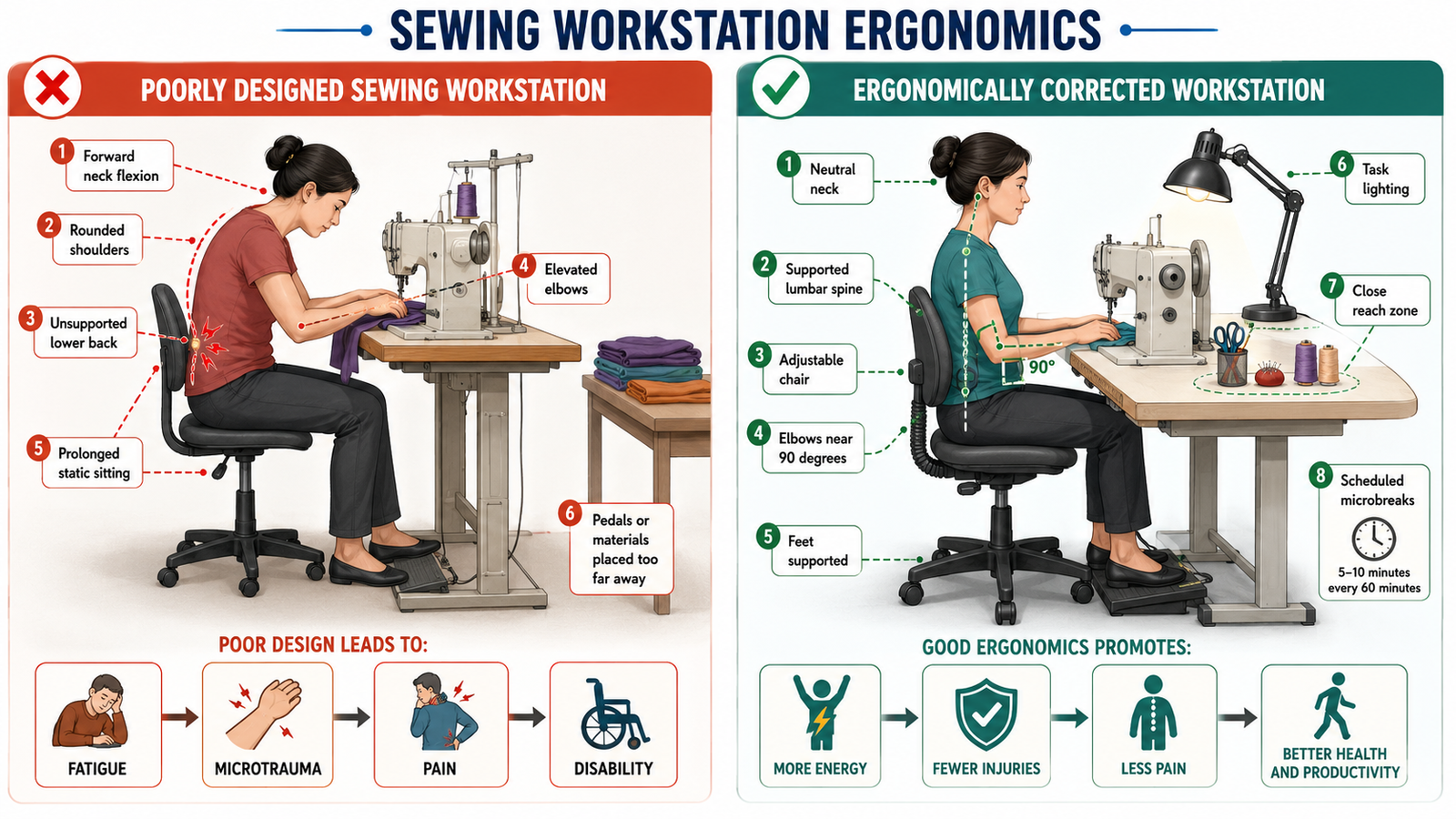
Ergonomics is the scientific adaptation of work, tools, tasks and the environment to human anatomical, physiological and psychological capabilities. Its practical purpose is to fit the work to the worker rather than forcing the worker to tolerate poorly designed work.
Principles of workplace ergonomics
- Maintain neutral joint and spinal positions where possible.
- Reduce excessive force, repetitive motion and prolonged static posture.
- Place frequently used items within comfortable reach.
- Adjust work height, seating, lighting and visual angle to the task and worker.
- Use mechanical assistance or team handling for heavy loads.
- Allow task variation, microbreaks and recovery time.
- Train workers in safe movement and early symptom reporting.
- Consider individual differences, disability and pregnancy.
Mechanism of occupational musculoskeletal injury
Epidemiology of musculoskeletal disorders
Musculoskeletal disorders affect children, working-age adults and older people, but the pattern differs by age, sex, occupation, nutrition, activity level and comorbidity. The burden includes pain, restricted mobility, reduced work capacity, dependence on caregivers and increased healthcare use.
- Age and sex
- Body weight and fitness
- Previous injury
- Bone health and nutrition
- Comorbid disease
- Repetition and force
- Awkward posture
- Vibration
- Manual handling
- Slippery or unsafe environments
- Poverty and food insecurity
- Access to safe recreation
- Healthcare access
- Education and workplace protection
- Availability of rehabilitation
Prevention and control
- Primordial prevention: promote healthy urban, school and workplace design before harmful patterns become established.
- Primary prevention: nutrition, physical activity, ergonomic redesign, worker education, safe lifting, fall prevention and avoidance of unnecessary bone-harming exposure.
- Secondary prevention: early recognition of pain, weakness, low-trauma fracture, vitamin deficiency or work-related symptoms, followed by timely assessment and treatment.
- Tertiary prevention: fracture care, rehabilitation, assistive devices, workplace modification and prevention of recurrent disability.
- Quaternary prevention: avoid unnecessary imaging, long-term analgesic exposure, excessive supplementation and inappropriate treatment of low-risk individuals.
Population prevention of osteoporosis, osteomalacia and rickets
- Improve awareness of calcium, vitamin D and balanced nutrition.
- Promote safe physical activity throughout life to improve peak bone mass and maintain strength.
- Identify high-risk older adults, postmenopausal women and patients exposed to long-term glucocorticoids.
- Recognize high-risk infants and children before deformity develops.
- Reduce falls through environmental modification, vision correction, medication review and balance training.
- Improve access to diagnosis, essential treatment and rehabilitation.
H. Disability, Rehabilitation and Essential Anatomical Reinforcement
Disability and public-health impact
Disability results when an impairment interacts with personal and environmental barriers to restrict activity or participation. A vertebral fracture, hip fracture, congenital skeletal disorder or chronic occupational injury may affect walking, self-care, communication, education, employment and social participation.
Public-health issues faced by persons with disability
- Limited physical access to buildings, transport and healthcare facilities.
- Financial barriers and loss of employment.
- Reduced educational and social participation.
- Dependence on caregivers and caregiver burden.
- Stigma, discrimination and reduced autonomy.
- Inadequate access to assistive devices and rehabilitation services.
- Greater risk of secondary complications from immobility and social isolation.
Types of rehabilitation
Clinical treatment, pain control, prevention of complications and restoration of physical function.
Strength, balance, mobility, gait training and safe use of assistive devices.
Return-to-work planning, job modification, retraining and ergonomic adaptation.
Support for family, community participation, independent living and reduction of stigma.
Adaptation of learning environments and support for children or adults whose condition affects education.
Support for adjustment, confidence, coping and participation after injury or chronic disability.
Measures required for rehabilitation
- Early multidisciplinary assessment of impairment, activity limitation and environmental barriers.
- Clear functional goals agreed with the patient and family.
- Pain management, nutrition and treatment of the underlying bone disorder.
- Progressive strength, balance and mobility training.
- Home-safety and fall-risk assessment.
- Appropriate walking aids, braces or other assistive technology.
- Workplace or school modification.
- Community follow-up and prevention of recurrent injury.
Important anatomical and physiological characteristics of the musculoskeletal system
The musculoskeletal system provides support, protects organs, permits movement and stores minerals. Bones act as levers, joints guide movement, muscles generate force, tendons transmit force and ligaments support stability. Function depends on coordinated anatomy, muscle strength, proprioception and continuous bone remodeling.
- Long bones transmit load through cortical shafts and distribute force near joints through trabecular metaphyseal and epiphyseal regions.
- Articular cartilage reduces friction and distributes compressive load but has limited intrinsic repair capacity.
- Synovial joints depend on capsules, ligaments, muscles and proprioception for stability.
- Skeletal muscle generates movement, stabilizes joints and protects bone by reducing falls.
- Bone remodels in response to mechanical stress, microdamage, mineral needs and hormonal signals.
- Immobilization causes muscle atrophy, reduced bone loading and accelerated functional decline.
Regional anatomy reinforcement: orbit
The orbit is a pyramidal bony cavity containing the eyeball, extraocular muscles, optic nerve, vessels, nerves, lacrimal structures and orbital fat. Its roof, floor, medial wall and lateral wall are formed by contributions from several skull bones. The optic canal carries the optic nerve and ophthalmic artery, while the superior and inferior orbital fissures transmit important neurovascular structures. The thin orbital floor and medial wall are clinically important in facial trauma. Fracture can cause extraocular muscle entrapment, diplopia, altered sensation or communication with adjacent sinuses.
Regional anatomy reinforcement: eyeball
The wall of the eyeball has three principal coats: an outer fibrous coat formed by sclera and cornea, a vascular coat containing choroid, ciliary body and iris, and an inner neural coat formed by the retina. The lens separates anterior aqueous compartments from the vitreous cavity. Extraocular muscles control gaze, while the optic nerve carries retinal output. Connective-tissue disorders can influence scleral appearance, as illustrated by blue sclerae in some patients with osteogenesis imperfecta.
Regional anatomy reinforcement: ear
The external ear collects sound, the middle ear transmits vibrations through the tympanic membrane and auditory ossicles, and the inner ear converts mechanical energy into neural signals through the cochlea. The vestibular apparatus supports balance. The facial and vestibulocochlear nerves have important anatomical relationships within the temporal bone. Bone-remodeling disorders affecting the skull may impair hearing through narrowing of neural pathways or alteration of ossicular and cochlear mechanics. Hearing impairment can also occur in osteogenesis imperfecta because of abnormal connective-tissue and ossicular properties.
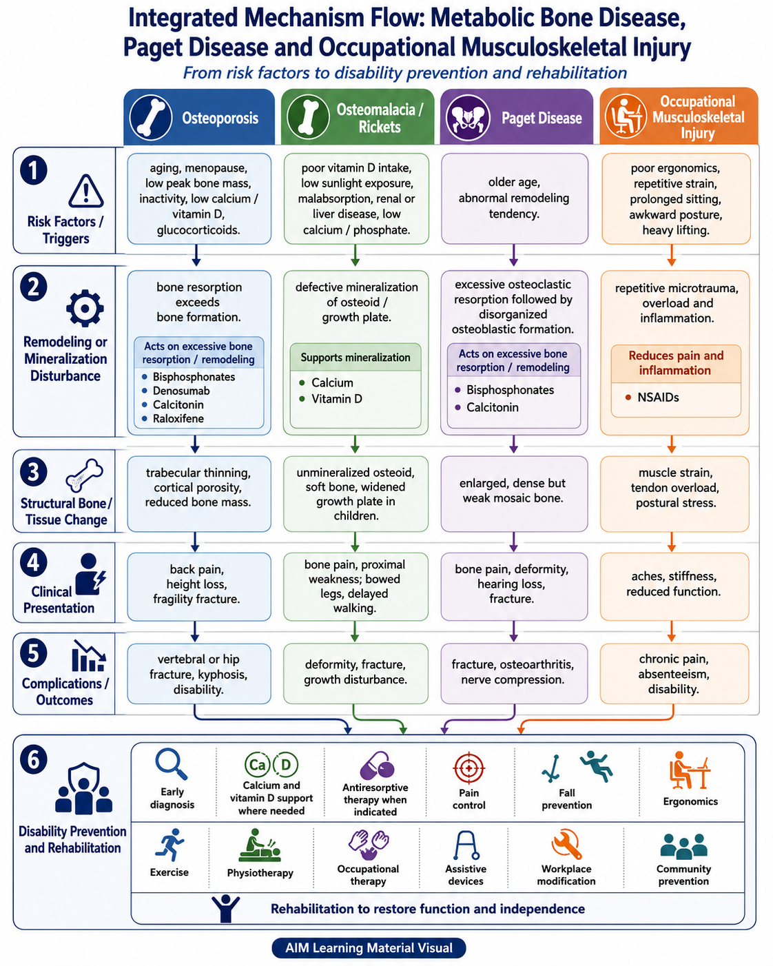
Integrated Mechanism Flow
AIM High-Yield Review
“`
Video Explanation
Watch this concise explanation to reinforce the differences between osteoporosis, osteomalacia and Paget disease.
This video reviews the major pathological differences, risk factors, clinical features, diagnostic findings and basic management principles of the three core metabolic bone disorders covered in this topic.
External educational video hosted on YouTube. It opens in a new browser tab.
“`