
📖 AIM Learning Material
Topic Introduction
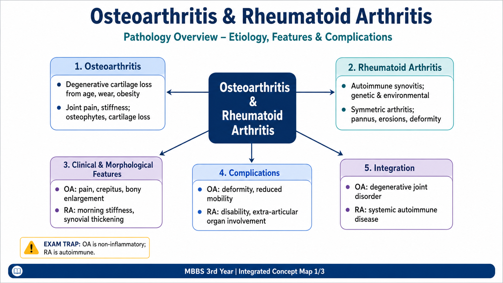
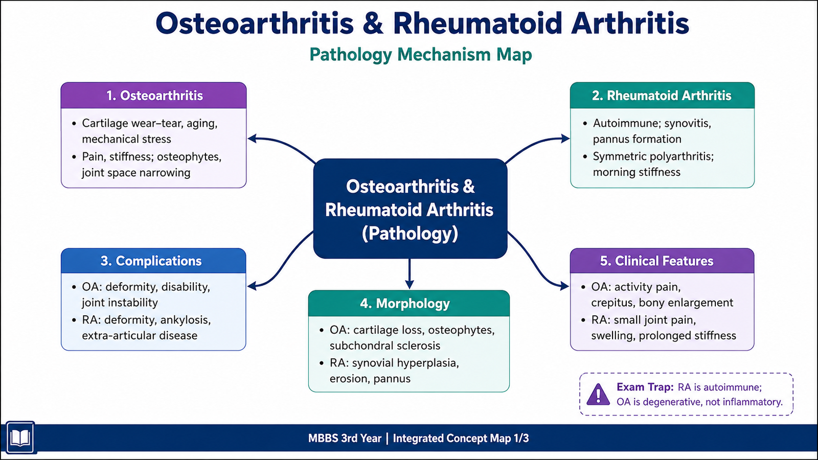
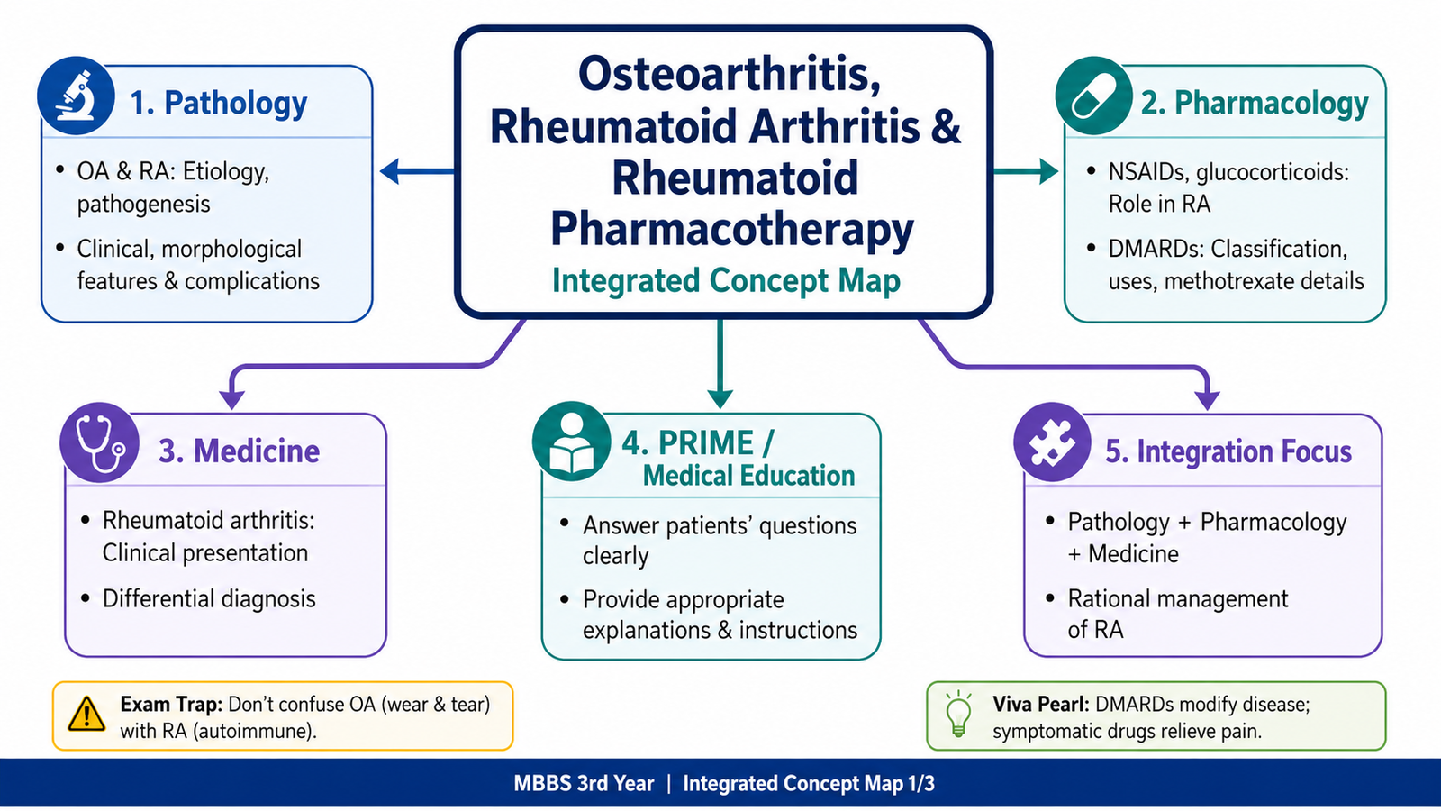
Osteoarthritis and rheumatoid arthritis are common causes of chronic joint pain, but they develop through different processes. Osteoarthritis mainly results from failure of articular cartilage repair together with changes in subchondral bone. Rheumatoid arthritis is a systemic autoimmune disease in which persistent synovial inflammation forms pannus and damages cartilage and bone. Understanding this difference makes the clinical patterns, morphology and treatment easier to remember. This chapter explains the causes, mechanisms, structural changes, clinical features and complications of both disorders. It also classifies the drugs used in rheumatoid arthritis, with special attention to methotrexate, other disease-modifying antirheumatic drugs and safe communication with patients.
Core Learning Material
A. Osteoarthritis: Etiology and Pathogenesis
Definition and Core Concept
Osteoarthritis is a chronic degenerative disease of synovial joints characterized by progressive loss of articular cartilage, remodeling of subchondral bone and variable synovial inflammation. It is not simply “wear and tear.” Mechanical stress interacts with cellular and biochemical responses within cartilage, bone and synovium.
Etiological Classification
- Primary osteoarthritis: develops without a single identifiable initiating disease; age, inherited susceptibility, obesity and cumulative mechanical loading contribute.
- Secondary osteoarthritis: follows a recognizable abnormality such as trauma, congenital or developmental joint deformity, metabolic disease, neuropathic arthropathy or previous inflammatory joint damage.
Important Risk Factors
Pathogenesis
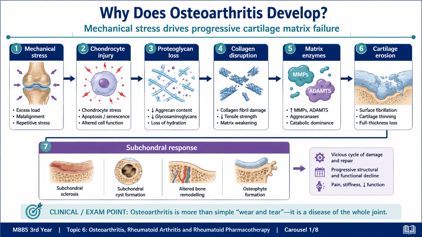
Excessive or abnormal mechanical loading injures chondrocytes and the extracellular matrix. Initially, chondrocytes increase synthesis of matrix components in an attempted repair response. They also produce inflammatory mediators and matrix-degrading enzymes, including metalloproteinases and aggrecan-degrading enzymes. Degradation eventually exceeds repair.
Mechanical stress or cartilage vulnerability
Chondrocyte injury and abnormal activation
Loss of proteoglycans and disruption of type II collagen
Cartilage softening, fibrillation and progressive loss
Exposed subchondral bone, sclerosis, cyst formation and osteophytes
Pain, crepitus, restricted movement and deformity
Mild secondary synovitis may occur when cartilage fragments and matrix products enter the joint cavity, but inflammation is not the primary destructive driver as it is in rheumatoid arthritis.
B. Osteoarthritis: Morphology, Clinical Features and Complications
Gross Morphology
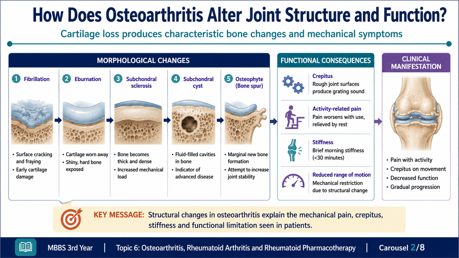
- Early cartilage becomes softened, granular and less resilient.
- Surface fibrillation produces vertical splits and irregularity.
- Progressive erosion exposes polished subchondral bone, producing an ivory-like appearance called eburnation.
- Subchondral bone becomes thickened and sclerotic.
- Small fractures permit synovial fluid to enter bone, producing subchondral cysts.
- New bone forms at joint margins as osteophytes.
- Loose fragments of cartilage or bone may form joint mice.
Microscopic Morphology
- Chondrocyte proliferation in clusters during the early repair response.
- Loss of proteoglycans from the matrix.
- Disorganization and cleavage of collagen framework.
- Fibrillation and fissuring of cartilage.
- Chondrocyte loss and complete denudation of cartilage in advanced disease.
- Reactive subchondral bone formation and mild chronic synovitis.
Clinical Features Explained by Pathology
- Pain worsened by activity: mechanical loading stresses subchondral bone, capsule and periarticular tissues.
- Brief morning stiffness: inactivity causes temporary stiffness, usually improving soon after movement begins.
- Crepitus: irregular cartilage and exposed surfaces move against one another.
- Reduced range of movement: pain, osteophytes, capsular thickening and altered joint contour limit motion.
- Bony enlargement: osteophyte formation and remodeling enlarge joint margins.
- Heberden nodes: osteophyte-related enlargement of distal interphalangeal joints.
- Bouchard nodes: involvement of proximal interphalangeal joints.
Complications
- Progressive pain and functional disability.
- Joint deformity and instability.
- Reduced mobility with muscle wasting and loss of independence.
- Loose bodies and intermittent locking.
- Secondary synovitis and joint effusion.
- Marked impairment of quality of life, especially when weight-bearing joints are involved.
C. Rheumatoid Arthritis: Etiology and Pathogenesis
Definition and Core Concept
Rheumatoid arthritis is a chronic systemic autoimmune inflammatory disease that primarily affects synovial joints. Persistent synovitis produces pannus, cartilage destruction, bone erosion and eventual deformity. Because the immune process is systemic, organs outside the joints may also be affected.
Etiology and Predisposing Factors
Rheumatoid arthritis develops through interaction between genetic susceptibility and environmental triggers. No single cause explains every case.
- Genetic susceptibility: certain HLA class II alleles favor presentation of arthritogenic peptides to CD4-positive T cells.
- Environmental influences: cigarette smoke and mucosal inflammation may increase protein citrullination.
- Loss of immune tolerance: modified self-proteins are recognized as antigens.
- Autoantibody formation: rheumatoid factor and anti-citrullinated protein antibodies develop before or during clinical disease.
- Female predominance and hormonal influences: indicate biological modulation of immune susceptibility.
Integrated Immunopathogenesis
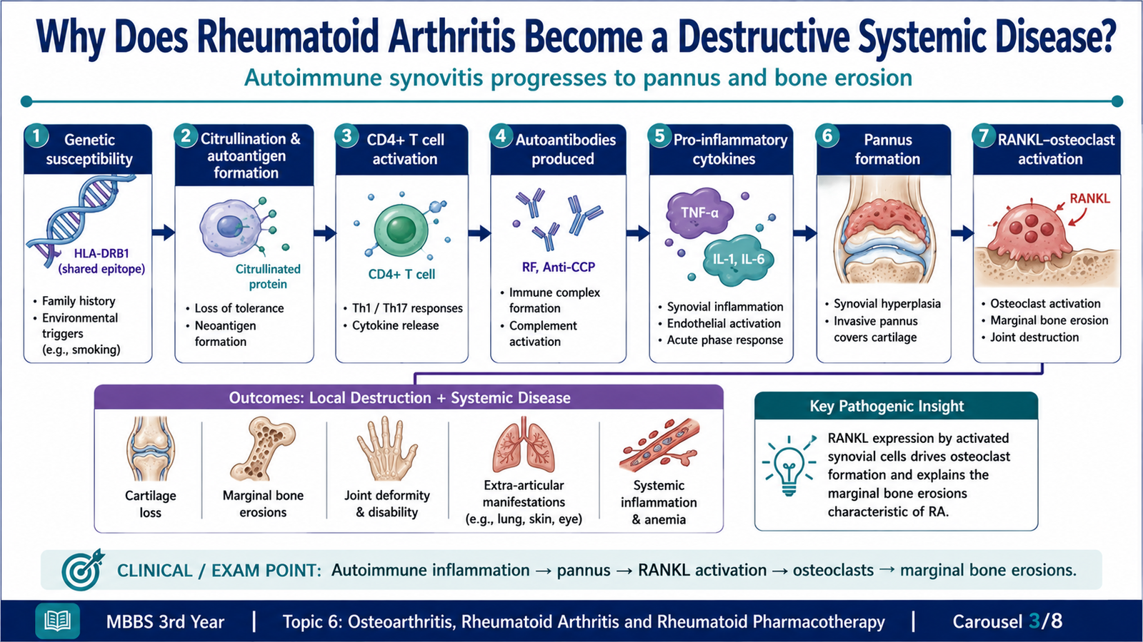
Citrullination converts arginine residues in proteins into citrulline, creating altered self-antigens. Antigen-presenting cells activate CD4-positive T cells, which stimulate macrophages, B cells and synovial fibroblasts. B cells produce autoantibodies, while activated macrophages and fibroblast-like synoviocytes release TNF, IL-1, IL-6 and other mediators.
Genetic susceptibility plus environmental trigger
Citrullinated self-proteins and loss of immune tolerance
CD4-positive T-cell and B-cell activation
Autoantibodies, immune complexes and cytokine release
Chronic proliferative synovitis and pannus formation
Cartilage degradation, osteoclast activation and marginal erosions
Deformity, ankylosis, disability and extra-articular disease
TNF and IL-1 promote endothelial activation, leukocyte recruitment and synovial proliferation. RANKL expressed by activated T cells and synovial stromal cells stimulates osteoclast differentiation, explaining marginal bone erosions. Proteases released by inflammatory cells and synoviocytes degrade cartilage matrix.
D. Rheumatoid Arthritis: Morphology, Clinical Presentation and Complications
Synovial and Joint Morphology
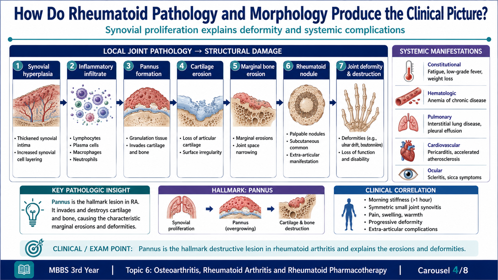
The earliest major lesion is chronic synovitis. The synovium becomes edematous, hyperplastic and heavily infiltrated by inflammatory cells. Proliferating synovial tissue grows over the articular surface as pannus and directly damages cartilage and bone.
Gross Morphology
- Thickened, edematous and congested synovium.
- Villous synovial hypertrophy projecting into the joint cavity.
- Increased turbid synovial fluid containing inflammatory cells.
- Granulation tissue spreading across the articular cartilage as pannus.
- Progressive cartilage destruction and marginal bone erosion.
- Fibrous ankylosis, which may later become bony ankylosis in advanced disease.
Microscopic Morphology
- Synovial lining-cell hyperplasia.
- Dense infiltrates of CD4-positive T cells, B cells, plasma cells and macrophages.
- Lymphoid aggregates that may resemble germinal centers.
- Increased vascularity due to angiogenesis.
- Fibrin deposition on the synovial surface and within the joint.
- Activated synovial fibroblasts and inflammatory cells invading cartilage and bone.
Rheumatoid Nodules
Rheumatoid nodules are firm subcutaneous lesions usually found over pressure points such as the elbows. Microscopically, they show central fibrinoid necrosis surrounded by palisading macrophages and an outer zone of chronic inflammatory cells and granulation tissue.
Clinical Presentation
- Insidious onset of fatigue, malaise and musculoskeletal discomfort.
- Symmetrical inflammatory polyarthritis.
- Common involvement of wrists, metacarpophalangeal and proximal interphalangeal joints.
- Relative sparing of distal interphalangeal joints.
- Prolonged morning stiffness that improves with movement.
- Warmth, tenderness, soft swelling and restricted joint movement.
- Later deformities caused by tendon, ligament and joint destruction.
| Deformity | Underlying Structural Change | Clinical Result |
|---|---|---|
| Ulnar deviation | MCP joint and soft-tissue destruction | Fingers deviate toward ulna |
| Swan-neck deformity | PIP hyperextension with DIP flexion | Impaired grip and fine movement |
| Boutonnière deformity | PIP flexion with DIP hyperextension | Reduced finger extension |
| Z-thumb | MCP flexion with interphalangeal hyperextension | Weak pinch function |
Extra-Articular Manifestations and Complications
- Rheumatoid nodules.
- Anemia of chronic inflammation.
- Vasculitis with skin, nerve or organ ischemia.
- Pleuritis, interstitial lung disease or pulmonary nodules.
- Pericardial involvement.
- Ocular inflammation and dry-eye symptoms.
- Peripheral neuropathy and carpal tunnel syndrome.
- Secondary amyloidosis in prolonged active disease.
- Increased susceptibility to infection due to disease and immunosuppressive treatment.
- Cervical spine instability, particularly at the atlantoaxial joint.
E. Clinical Diagnosis and Differential Diagnosis of Rheumatoid Arthritis
Diagnostic Reasoning in Rheumatoid Arthritis
Rheumatoid arthritis is suspected from the pattern of persistent inflammatory synovitis rather than from a single laboratory result. Symmetry, prolonged morning stiffness, characteristic small-joint involvement and objective swelling are major clues.
Relevant Investigations
- Inflammatory markers: ESR and C-reactive protein may reflect inflammatory activity but are not disease-specific.
- Rheumatoid factor: supports the diagnosis but may occur in other autoimmune, infectious or chronic conditions and in some healthy individuals.
- Anti-citrullinated protein antibodies: are more specific and may be associated with erosive disease.
- Complete blood count: may show anemia and provides a baseline before treatment.
- Radiography: may show periarticular osteopenia, uniform joint-space loss and marginal erosions.
- Ultrasound or other imaging: may detect synovitis and erosions when clinically indicated.
- Synovial fluid examination: is important when infection or crystal arthritis must be excluded.
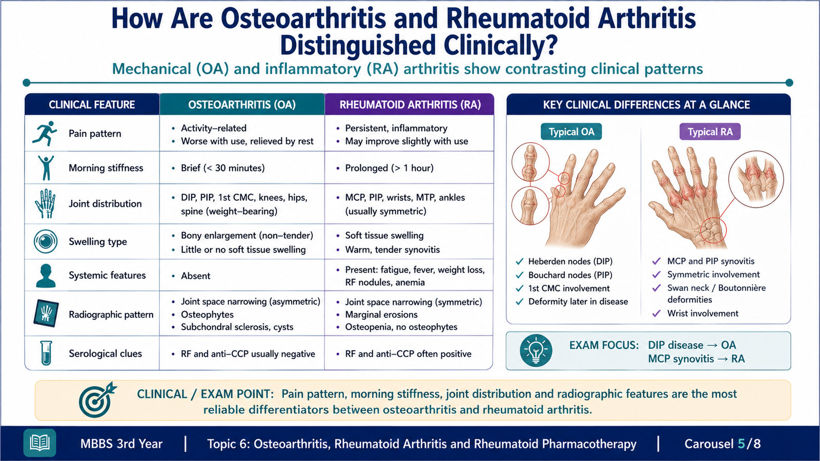
| Feature | Osteoarthritis | Rheumatoid Arthritis |
|---|---|---|
| Primary process | Degenerative cartilage and bone remodeling | Systemic autoimmune synovitis |
| Typical pain | Worse with use, relieved by rest | Prominent after rest; improves with movement |
| Morning stiffness | Usually brief | Usually prolonged |
| Joint pattern | Often knees, hips, spine, DIP and PIP joints | Symmetrical wrists, MCP and PIP joints; DIP usually spared |
| Swelling | Hard and bony | Soft, warm and inflammatory |
| Systemic features | Absent | Fatigue, anemia and extra-articular disease may occur |
| Radiographic pattern | Non-uniform joint-space narrowing, osteophytes, sclerosis, cysts | Uniform narrowing, periarticular osteopenia, marginal erosions |
Important Differential Diagnoses
- Osteoarthritis: mechanical pain, brief stiffness, bony enlargement and osteophytes.
- Systemic lupus erythematosus: inflammatory polyarthritis with systemic autoimmune features but usually non-erosive joint disease.
- Psoriatic arthritis: psoriasis, nail changes, possible DIP involvement and asymmetrical patterns.
- Crystal arthritis: acute episodic attacks; crystal identification is diagnostic.
- Septic arthritis: acute severe pain, systemic illness and urgent need for synovial fluid evaluation.
- Viral polyarthritis: temporal relation to infection and often self-limited course.

F. Classification and Roles of Drugs Used in Rheumatoid Arthritis
Therapeutic Goals
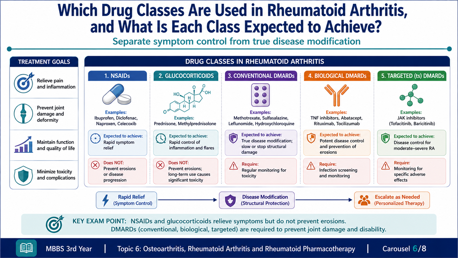
Treatment aims to suppress inflammation, reduce pain and stiffness, prevent cartilage and bone destruction, preserve function and limit systemic complications. Symptom relief and disease modification are different therapeutic objectives.
Classification of Drugs Used in Rheumatoid Arthritis
| Therapeutic Group | Examples | Main Role | Key Limitation |
|---|---|---|---|
| NSAIDs | Ibuprofen, naproxen, diclofenac, selective COX-2 inhibitors | Rapid relief of pain and stiffness | Do not prevent structural joint damage |
| Glucocorticoids | Prednisolone and intra-articular preparations | Rapid anti-inflammatory control or bridging therapy | Cumulative systemic toxicity |
| Conventional synthetic DMARDs | Methotrexate, hydroxychloroquine, sulfasalazine, leflunomide | Modify disease and reduce progression | Delayed onset and monitoring requirements |
| Biological DMARDs | TNF inhibitors, IL-6 pathway inhibitors, abatacept, rituximab, IL-1 antagonist | Target specific immune pathways | Infection risk, cost and parenteral use for many agents |
| Targeted synthetic DMARDs | Janus kinase inhibitors | Interrupt intracellular cytokine signaling | Serious infection and other important safety concerns |
Role of NSAIDs
NSAIDs are useful for rapid symptomatic relief while disease-modifying therapy begins to work. They do not adequately suppress the autoimmune process and do not prevent erosions or deformity.
- Important adverse effects include dyspepsia, peptic ulceration, gastrointestinal bleeding, renal impairment, fluid retention, hypertension and hypersensitivity reactions.
- Selective COX-2 inhibition may reduce some gastrointestinal toxicity but does not remove renal or cardiovascular concerns.
- Caution is required in patients with peptic ulcer disease, renal impairment, heart failure, uncontrolled hypertension or increased bleeding risk.
Role of Glucocorticoids
Glucocorticoids may be used for rapid control of severe inflammation, as temporary bridging therapy while a DMARD takes effect, or by local intra-articular administration for selected joints. Their use should be limited to the lowest effective exposure because toxicity increases with dose and duration.
- Hyperglycemia and worsening of diabetes.
- Hypertension, fluid retention and weight gain.
- Osteoporosis and fracture risk.
- Infection risk and impaired wound healing.
- Skin thinning, muscle weakness and mood disturbance.
- Adrenal suppression with prolonged systemic use.
- Cataract and glaucoma.
G. Methotrexate: Prototype Conventional DMARD
Definition of a DMARD
A disease-modifying antirheumatic drug suppresses the underlying inflammatory disease process and can reduce or delay structural joint damage. This distinguishes DMARDs from medicines that provide only analgesic or short-term anti-inflammatory benefit.
Methotrexate: Mechanism of Action
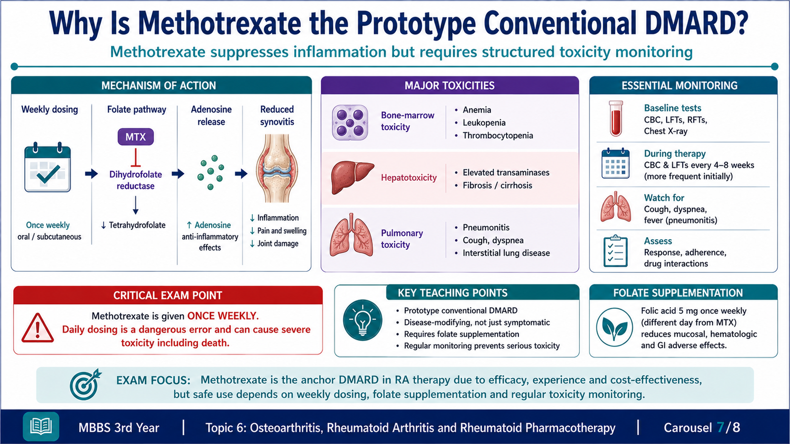
Methotrexate is a folate antagonist. At the lower intermittent exposure used in rheumatoid arthritis, its anti-inflammatory effect is not explained solely by inhibition of cell proliferation. It inhibits folate-dependent enzymes and promotes extracellular accumulation of adenosine, which suppresses inflammatory cell activity and cytokine production.
Methotrexate
Folate-pathway enzyme inhibition and increased anti-inflammatory adenosine signaling
Reduced lymphocyte and inflammatory-cell activity
Reduced cytokine-driven synovitis
Reduced symptoms and slower structural joint damage
Pharmacokinetic Relevance
- May be administered orally or by a parenteral route when clinically appropriate.
- Rheumatoid arthritis treatment uses intermittent weekly administration rather than daily administration.
- It undergoes intracellular polyglutamation, which contributes to persistence of pharmacological activity.
- Renal elimination is clinically important; impaired renal function can increase toxicity.
- Drug interactions that reduce renal clearance or increase antifolate effects may increase adverse reactions.
Clinical Uses
- Prototype and commonly preferred conventional DMARD for active rheumatoid arthritis.
- May be used alone or with other conventional, biological or targeted agents.
- Also has established uses in selected inflammatory disorders and malignant diseases, although indications and treatment schedules differ.
Adverse Effects
- Gastrointestinal: nausea, mucosal irritation and stomatitis.
- Hematological: bone-marrow suppression causing cytopenias.
- Hepatic: elevation of liver enzymes and chronic hepatotoxicity.
- Pulmonary: hypersensitivity pneumonitis or interstitial lung injury.
- Infectious: increased susceptibility due to immunosuppression.
- Reproductive: teratogenicity and fetal toxicity.
- Dermatological: rash and alopecia may occur.
Contraindications and Important Cautions
- Pregnancy and planned conception because of teratogenicity.
- Significant liver disease or persistent heavy alcohol exposure.
- Severe renal impairment because clearance is reduced.
- Pre-existing major cytopenia or bone-marrow dysfunction.
- Serious active infection.
- Pre-existing significant pulmonary disease requires careful evaluation if respiratory symptoms arise.
Monitoring and Interaction Logic
Baseline and periodic assessment of blood-cell counts, liver function and renal function is necessary because toxicity may initially be clinically silent. New cough, breathlessness, fever, mouth ulcers, bruising or severe gastrointestinal symptoms require prompt review.
- Folic acid supplementation reduces several mucosal and hematological adverse effects without removing the need for monitoring.
- Other antifolate medicines may increase marrow and mucosal toxicity.
- Medicines that impair renal elimination may increase methotrexate exposure.
- Combined immunosuppressive therapy increases infection risk.
- Alcohol increases concern regarding hepatotoxicity.
H. Other DMARDs, Safety and Patient Communication
Non-Biological DMARDs
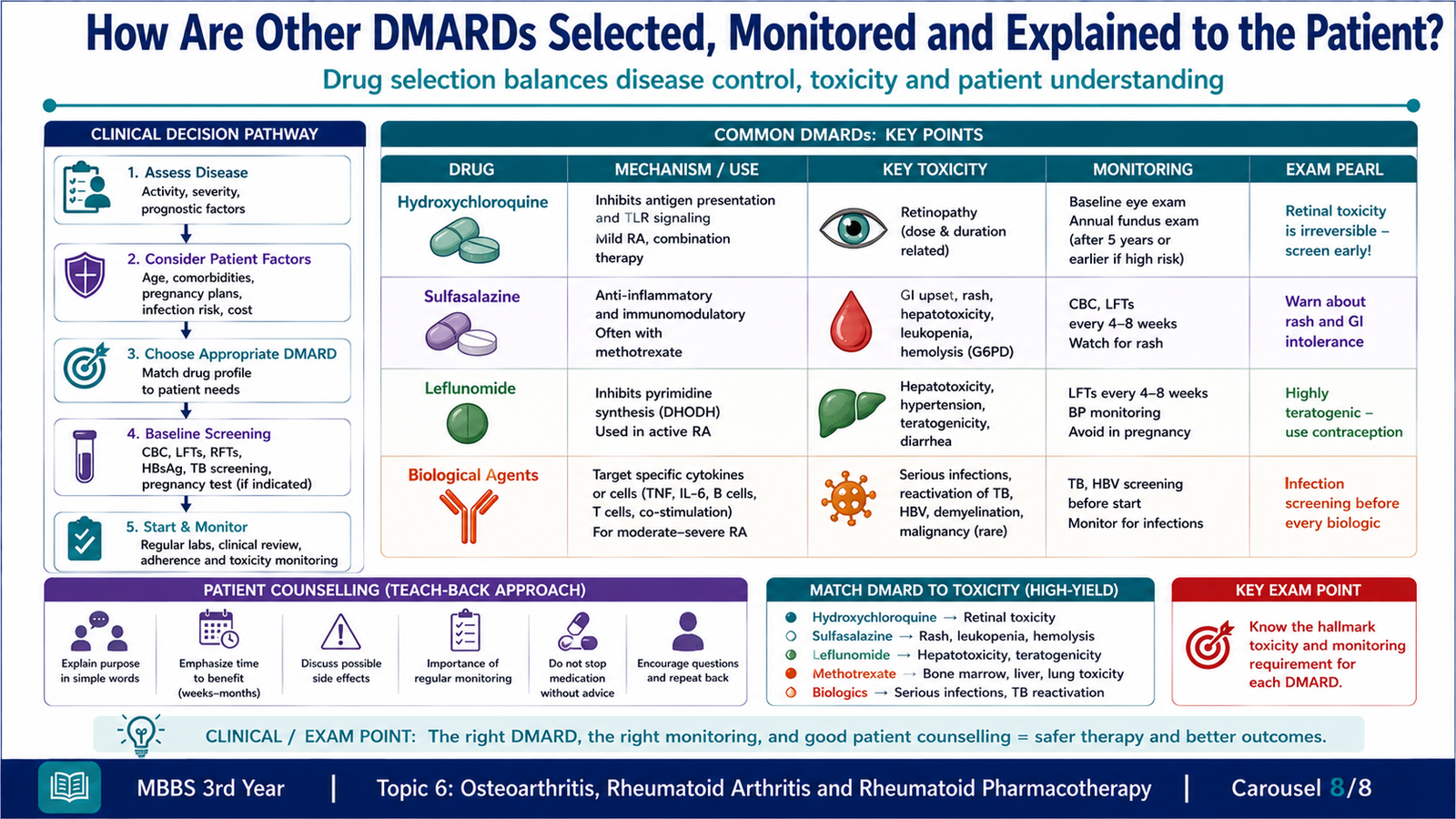
| Drug | Core Action | Therapeutic Role | Important Adverse Effects or Cautions |
|---|---|---|---|
| Hydroxychloroquine | Modifies antigen processing and innate immune signaling | Milder disease or combination therapy | Retinal toxicity; visual monitoring is important |
| Sulfasalazine | Immunomodulatory anti-inflammatory actions | Monotherapy or combination conventional DMARD | Gastrointestinal upset, rash, cytopenia, hepatotoxicity and reversible oligospermia |
| Leflunomide | Inhibits pyrimidine synthesis and lymphocyte proliferation | Alternative to methotrexate or combination therapy | Hepatotoxicity, diarrhea, hypertension and teratogenicity |
| Azathioprine | Interferes with purine synthesis | Selected refractory or systemic cases | Marrow suppression, hepatotoxicity and infection |
| Cyclosporine | Calcineurin inhibition reduces T-cell cytokine transcription | Selected resistant disease | Nephrotoxicity, hypertension and interactions |
Biological DMARDs
- TNF inhibitors: infliximab, adalimumab, etanercept, golimumab and certolizumab reduce TNF-mediated inflammation.
- IL-6 pathway inhibitors: suppress IL-6-mediated systemic and articular inflammation.
- Abatacept: interferes with T-cell co-stimulation.
- Rituximab: targets CD20-positive B cells.
- IL-1 antagonist: reduces selected IL-1-mediated inflammatory effects.
Biological agents may be considered when conventional therapy is inadequate or unsuitable. They target specific immune pathways and can markedly reduce disease activity, but immune suppression creates important infection risks.
Targeted Synthetic DMARDs
Janus kinase inhibitors reduce intracellular signaling from several cytokine receptors. They are orally active but require careful assessment because serious infections, hematological changes and other clinically important risks may occur.
Common Safety Principles
- Assess for active infection before initiating major immunosuppressive therapy.
- Evaluate for latent infections such as tuberculosis and relevant viral infections before selected biological or targeted therapy.
- Avoid combining biological DMARDs routinely because severe immunosuppression and infection risk may increase.
- Monitor blood counts, hepatic function, renal function or other organ-specific parameters according to the medicine used.
- Consider reproductive safety because several DMARDs are teratogenic.
- Review vaccination status before substantial immunosuppression when clinically appropriate.
- Teach the patient to report fever, persistent cough, unexplained bruising, jaundice, breathlessness or other warning symptoms.
Drug-Selection Logic
Drug choice depends on disease activity, prognostic features, comorbidity, pregnancy potential, infection risk, previous response and medicine toxicity. Methotrexate is commonly used as the conventional anchor drug unless contraindicated. Hydroxychloroquine may suit milder disease; sulfasalazine or leflunomide may serve as alternatives or combination partners. Biological or targeted therapy is considered when disease remains active despite adequate conventional treatment or when conventional agents cannot be used.
PRIME: Answering the Patient’s Questions
Patients are more likely to use treatment safely when explanations are specific, respectful and linked to their concerns. The clinician should not simply state that methotrexate is “strong medicine.” The explanation should distinguish its disease-modifying role from ordinary pain relief.
“The anti-inflammatory pain medicine can reduce discomfort, but it does not reliably prevent joint damage. Methotrexate acts on the immune process causing the arthritis, so its purpose is to control inflammation and protect the joints over time.”
“Regular blood tests are needed because the medicine can sometimes affect blood cells, the liver or kidney handling of the drug before you notice symptoms. Monitoring allows us to detect problems early and use the treatment more safely.”
Essential Communication Steps
- Invite the patient to state concerns and expectations.
- Explain the diagnosis in plain language.
- Differentiate symptom control from disease modification.
- Explain expected benefits and delayed onset of DMARD action.
- Discuss major risks without overwhelming the patient.
- Give clear instructions about weekly methotrexate use.
- Explain monitoring and warning symptoms.
- Check understanding by asking the patient to repeat key instructions in their own words.
- Document important counselling and provide written instructions when possible.
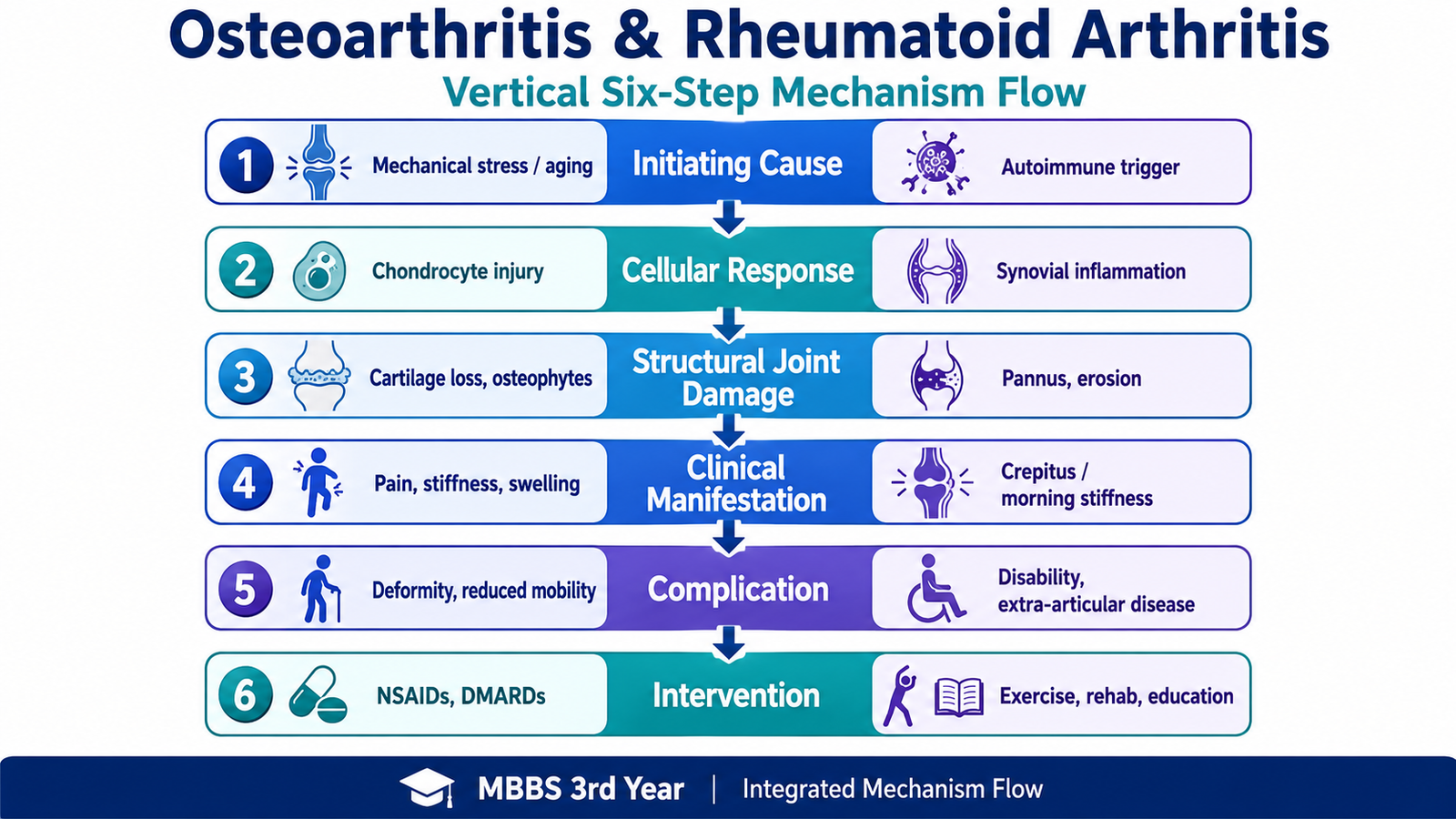
Integrated Mechanism Flow
AIM High-Yield Review
- Osteoarthritis: failed cartilage repair and abnormal mechanical stress produce cartilage loss, subchondral sclerosis, cysts and osteophytes.
- Mechanical pattern: osteoarthritis pain worsens with use, morning stiffness is brief, and crepitus reflects irregular joint surfaces.
- Rheumatoid arthritis: chronic autoimmune synovitis produces pannus, cartilage destruction and marginal bone erosions.
- Key inflammatory mechanism: TNF, IL-1, IL-6 and RANKL promote synovial inflammation and osteoclast-mediated bone loss.
- Typical rheumatoid pattern: symmetrical involvement of wrists, MCP and PIP joints with prolonged morning stiffness and relative sparing of DIP joints.
- Hallmark morphology: proliferative synovitis with pannus formation is central to rheumatoid joint destruction.
- Diagnostic clue: anti-citrullinated protein antibodies are more specific than rheumatoid factor, but diagnosis still depends on the clinical pattern.
- Imaging distinction: osteophytes and subchondral sclerosis suggest osteoarthritis; marginal erosions and periarticular osteopenia suggest rheumatoid arthritis.
- Important red flag: neck pain or neurological symptoms in rheumatoid arthritis may indicate atlantoaxial instability.
- Symptom control: NSAIDs and glucocorticoids reduce pain and inflammation but do not replace disease-modifying therapy.
- Methotrexate: a prototype conventional DMARD that is usually given weekly and requires blood count, liver and renal monitoring.
- Serious methotrexate toxicity: marrow suppression, hepatotoxicity, pulmonary toxicity and fetal harm require clear counselling and follow-up.
- Other DMARDs: conventional, biological and targeted synthetic agents are selected according to disease activity, previous response, comorbidity and safety.
- Patient communication: explain the treatment goal, delayed DMARD benefit, major warning symptoms and monitoring plan, then use teach-back to confirm understanding.
“`
Rheumatoid Arthritis: Pathogenesis, Clinical Features and Treatment
Watch this video to connect the autoimmune pathogenesis of rheumatoid arthritis with synovial inflammation, pannus formation, joint damage, clinical presentation, diagnosis and treatment.
“`