
📖 AIM Learning Material
Topic Introduction
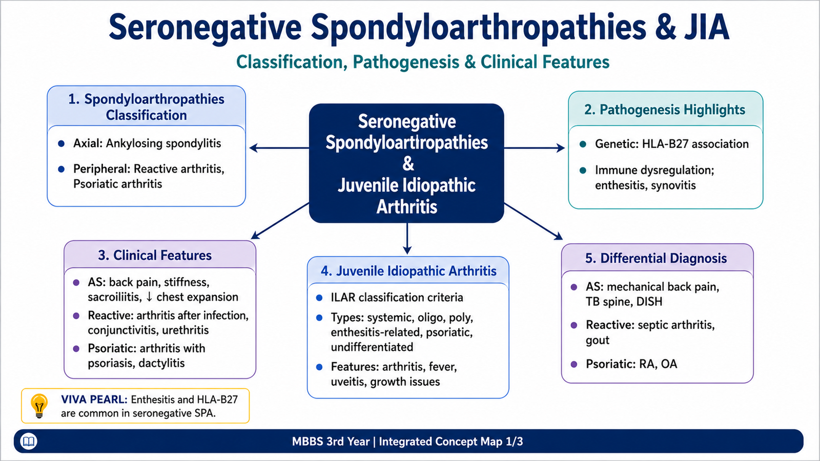
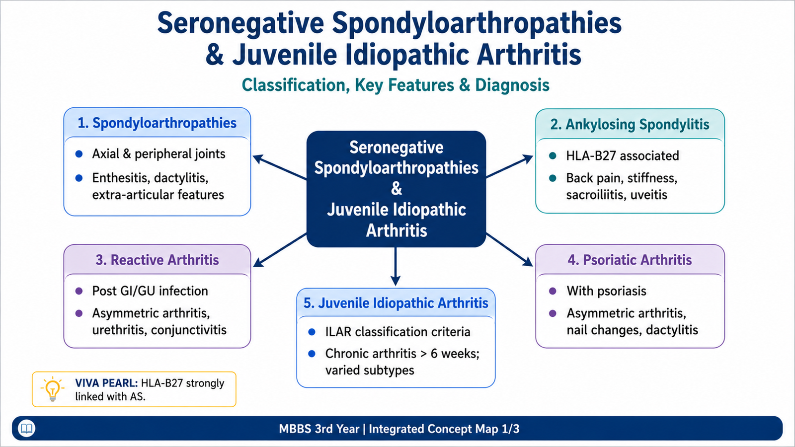
Seronegative spondyloarthropathies are inflammatory disorders that commonly affect the sacroiliac joints, spine, entheses and selected peripheral joints. They are called seronegative because rheumatoid factor is usually absent, but their diagnosis depends on the full clinical pattern rather than one blood test. Ankylosing spondylitis mainly produces inflammatory axial disease, reactive arthritis follows an infection elsewhere in the body, and psoriatic arthritis is linked with psoriasis and distinctive joint, nail and digit changes. Juvenile idiopathic arthritis is a separate group of persistent childhood arthritides classified by joint pattern and associated features. This chapter explains how these conditions develop, how they present and how they are distinguished.
A. Seronegative Spondyloarthropathies: Classification and Shared Pattern
Definition and Core Concept
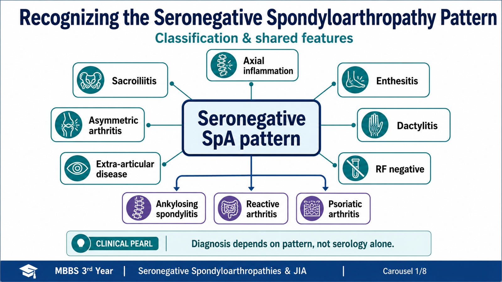
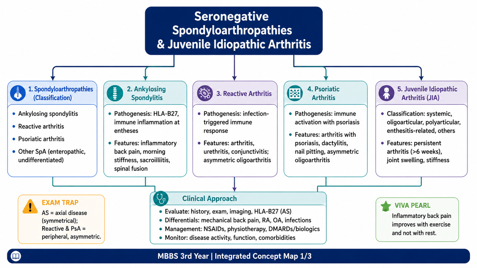
Seronegative spondyloarthropathies are a related group of chronic inflammatory disorders that preferentially involve the sacroiliac joints, spine, entheses and selected peripheral joints. They share overlapping genetic, pathological and clinical characteristics and usually lack rheumatoid factor.
Examinable Classification
- Ankylosing spondylitis
- Reactive arthritis
- Psoriatic arthritis
- Arthritis associated with inflammatory bowel disease
- Undifferentiated forms that do not yet meet the pattern of one defined disorder
Shared Clinical and Pathological Features
- Axial inflammation, especially sacroiliitis
- Enthesitis, commonly affecting the heel or plantar fascia
- Asymmetric peripheral arthritis, often involving lower limbs
- Dactylitis caused by inflammation of joints, tendons and surrounding tissues of a digit
- Extra-articular disease involving the eye, skin, bowel or genitourinary tract
- Association with HLA-B27, particularly in axial disease
- Absence of the typical rheumatoid-factor pattern of rheumatoid arthritis
B. Ankylosing Spondylitis: Pathogenesis and Structural Changes
Definition
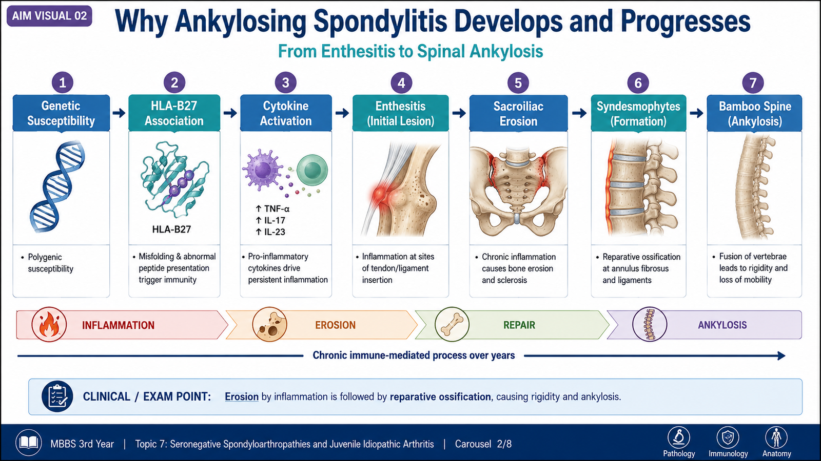
Ankylosing spondylitis is a chronic inflammatory spondyloarthropathy in which inflammation begins predominantly in the sacroiliac joints and entheses and may progressively extend through the axial skeleton, producing stiffness and eventual bony fusion.
Etiological Framework
No single cause explains the disease. It arises from interaction between genetic susceptibility and immune responses influenced by mucosal or environmental triggers. HLA-B27 is strongly associated, but its presence is neither necessary nor sufficient for disease.
Pathogenesis
Morphology and Structural Progression
- Sacroiliac joints: Synovitis, erosion, subchondral sclerosis and later joint-space obliteration.
- Vertebral entheses: Inflammation and erosion at outer annular-fiber insertions.
- Spine: Vertical ossification bridges adjacent vertebral bodies as marginal syndesmophytes.
- Advanced disease: Multiple bridging syndesmophytes create the radiographic “bamboo spine” appearance.
- Other entheses: Involvement may produce Achilles tendon or plantar-fascial pain.
C. Ankylosing Spondylitis: Clinical Presentation and Differential Diagnosis
Typical Clinical Presentation
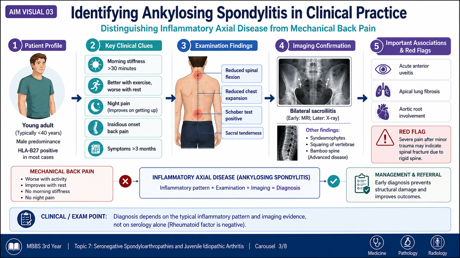
Ankylosing spondylitis typically begins in adolescence or early adulthood. Its central clinical feature is inflammatory back pain rather than mechanical pain.
- Insidious onset of low-back or deep buttock pain
- Morning stiffness and worsening after prolonged rest
- Improvement with exercise or movement
- Night pain, particularly during the latter part of the night
- Alternating buttock pain due to sacroiliitis
- Reduced lumbar flexion and decreased chest expansion
- Progressive loss of normal spinal posture in advanced disease
- Enthesitis, especially at the Achilles tendon or plantar fascia
- Hip or shoulder arthritis in some patients
Extra-Articular Features
- Acute anterior uveitis: Painful red eye, photophobia and blurred vision.
- Cardiovascular involvement: Inflammation around the aortic root may produce aortic regurgitation; conduction abnormalities may occur.
- Respiratory consequence: Reduced chest-wall movement may cause a restrictive ventilatory pattern.
- Neurological risk: A rigid osteoporotic spine is susceptible to fracture and possible spinal-cord injury.
- Renal complication: Chronic inflammation may rarely lead to secondary amyloid deposition.
Investigations and Diagnostic Clues
- Inflammatory markers may be elevated but can be normal.
- Rheumatoid factor is usually absent.
- HLA-B27 supports the diagnosis in an appropriate clinical setting but is not a diagnostic test by itself.
- Pelvic imaging may show bilateral sacroiliitis, erosions, sclerosis and progressive fusion.
- Spinal imaging in established disease may show vertebral squaring and marginal syndesmophytes.
- Early inflammatory lesions may be detected by magnetic resonance imaging before definite radiographic fusion develops.
Differential Diagnosis
| Condition | Pain Pattern | Key Distinguishing Clue |
|---|---|---|
| Ankylosing spondylitis | Improves with exercise; worsens with rest | Sacroiliitis, reduced spinal mobility, possible uveitis |
| Mechanical back pain | Worsens with activity; improves with rest | No inflammatory sacroiliitis or typical extra-articular features |
| Rheumatoid arthritis | Peripheral inflammatory joint pain | Symmetric small-joint polyarthritis; cervical rather than lumbar axial emphasis |
| Diffuse idiopathic skeletal hyperostosis | Stiffness, usually in older adults | Flowing ligamentous ossification without inflammatory sacroiliac erosion |
| Infective sacroiliitis | Acute severe pain | Fever, systemic illness and often unilateral involvement |
D. Reactive Arthritis
Definition
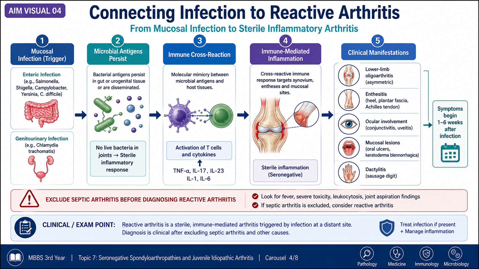
Reactive arthritis is a sterile inflammatory arthritis that develops after an infection elsewhere in the body, commonly involving the gastrointestinal or genitourinary tract. Viable organisms are not ordinarily present within the affected joint.
Preceding Triggers
The disease may follow enteric infection or genitourinary infection. The important concept is not memorization of every organism but recognition that a mucosal infection precedes an immune-mediated joint syndrome.
Pathogenesis
Persistence of microbial antigens or altered immune signaling
Cross-reactive inflammation in genetically susceptible host
Sterile synovitis, enthesitis and extra-articular inflammation
Clinical Features
- Acute asymmetric oligoarthritis, usually affecting lower-limb joints
- Enthesitis, particularly heel pain
- Dactylitis in some patients
- Inflammatory back pain or sacroiliitis in selected cases
- Urethritis or cervicitis related to the preceding infection
- Conjunctivitis or acute anterior uveitis
- Oral ulcers that may be painless
- Keratoderma blennorrhagicum, a hyperkeratotic skin eruption
- Circinate balanitis in affected males
The classic teaching triad of arthritis, urethritis and conjunctivitis is memorable but is not present in every patient. Diagnosis therefore depends on the temporal relationship between infection and the characteristic musculoskeletal pattern.
Diagnostic Reasoning and Differentials
- Ask specifically about recent diarrhea, dysuria, urethral discharge or sexual exposure.
- Joint aspiration may be needed when septic arthritis is possible.
- Synovial inflammation is sterile, but the triggering infection may still be demonstrable at its original site.
- Gout, septic arthritis, psoriatic arthritis and other spondyloarthropathies may produce overlapping peripheral patterns.
E. Psoriatic Arthritis
Definition
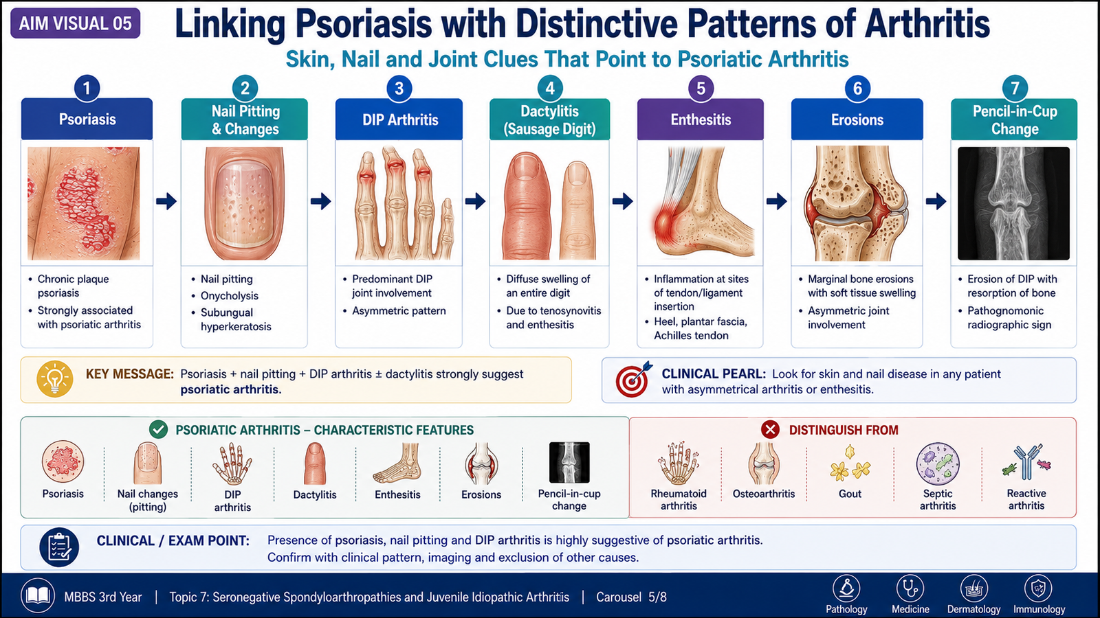
Psoriatic arthritis is an inflammatory arthritis associated with psoriasis. It may affect peripheral joints, entheses, digits, sacroiliac joints or the spine and can produce both destructive erosions and abnormal new-bone formation.
Pathogenesis
Genetic predisposition, dysregulated T-cell responses and inflammatory cytokines promote inflammation at the synovium, entheses, skin and nail apparatus. Because inflammation affects both bone-resorbing and bone-forming pathways, erosions may occur beside irregular periosteal new bone.
Skin, nail, synovial and entheseal inflammation
Erosion plus irregular new-bone formation
Peripheral arthritis, dactylitis, axial disease or deformity
Clinical Patterns
- Asymmetric oligoarthritis: A common presentation involving a few joints.
- Symmetric polyarthritis: May resemble rheumatoid arthritis.
- Distal interphalangeal-predominant disease: Strongly associated with nail changes.
- Arthritis mutilans: Severe destructive disease causing marked shortening and deformity of digits.
- Axial disease: Sacroiliitis or spondylitis, which may be asymmetric.
High-Value Clinical Clues
- Current or previous psoriasis, including hidden lesions on the scalp, umbilicus, natal cleft or behind the ears
- Nail pitting, onycholysis or hyperkeratosis
- Dactylitis producing a “sausage digit”
- Enthesitis
- Distal interphalangeal-joint involvement
- Negative rheumatoid factor in the typical case
Morphology and Imaging Correlation
- Marginal erosions with adjacent reactive bone formation
- Joint-space narrowing and ankylosis in established disease
- Severe erosion with tapering of one bone end and expansion of the adjacent bone may produce a “pencil-in-cup” appearance
- Axial disease may show coarse, non-marginal or asymmetric syndesmophyte formation
F. Important Comparison of Major Spondyloarthropathies
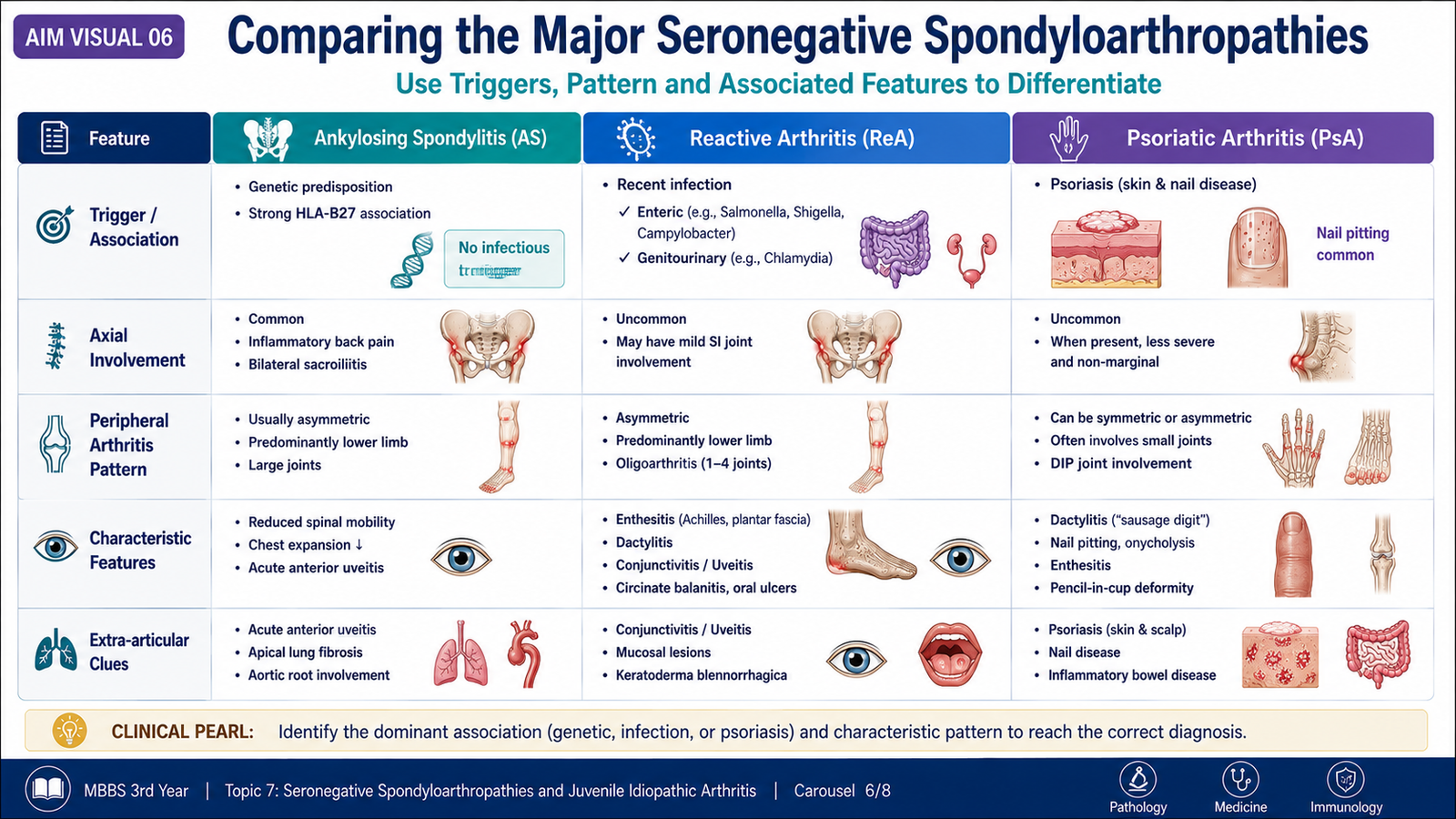
These disorders overlap, so a single manifestation such as uveitis, enthesitis or sacroiliitis cannot establish the diagnosis. The most useful approach is to identify the dominant disease pattern and its associated trigger or tissue manifestation.
| Feature | Ankylosing Spondylitis | Reactive Arthritis | Psoriatic Arthritis |
|---|---|---|---|
| Dominant clue | Inflammatory axial disease | Arthritis after mucosal infection | Psoriasis or nail disease |
| Joint pattern | Sacroiliac and spinal; possible hip or shoulder disease | Acute asymmetric lower-limb oligoarthritis | Variable; distal joints, oligoarthritis, polyarthritis or axial disease |
| Enthesitis | Common | Common | Common |
| Dactylitis | Less characteristic | May occur | Highly characteristic |
| Eye disease | Acute anterior uveitis | Conjunctivitis or uveitis | Uveitis may occur |
| Key associated history | Young onset and inflammatory back pain | Recent diarrhea or genitourinary infection | Skin or nail psoriasis |
| Characteristic imaging clue | Bilateral sacroiliitis and marginal syndesmophytes | Usually nonspecific early; sacroiliitis may develop | Erosion with new bone; pencil-in-cup deformity |
G. Juvenile Idiopathic Arthritis: Definition and Classification
Definition and Classification Principle
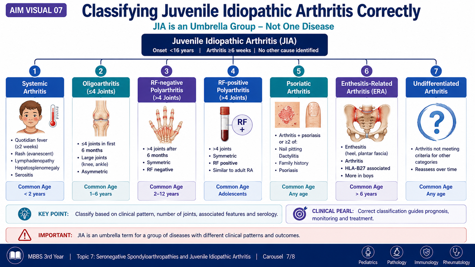
Juvenile idiopathic arthritis is persistent arthritis beginning before the age of 16 years, lasting for at least six weeks and remaining unexplained after other recognized causes have been excluded. It is an umbrella term rather than one uniform disease.
Classification is based mainly on the clinical pattern during the early course of illness, including the number of joints involved, systemic features, psoriasis, enthesitis, rheumatoid-factor status and features that exclude placement in a single category.
Major Classification Categories
| Category | Defining Pattern | Important Clue |
|---|---|---|
| Systemic arthritis | Arthritis with characteristic systemic inflammatory features | Quotidian fever and evanescent rash |
| Oligoarthritis | Four or fewer joints involved during the initial disease period | Young child, large joints and risk of silent uveitis |
| RF-negative polyarthritis | Five or more joints with absent rheumatoid-factor pattern | Variable onset and joint distribution |
| RF-positive polyarthritis | Polyarthritis with persistent rheumatoid-factor positivity | Resembles adult rheumatoid arthritis |
| Psoriatic arthritis | Arthritis associated with psoriasis or characteristic psoriatic features | Dactylitis, nail pitting or family history |
| Enthesitis-related arthritis | Arthritis with enthesitis or a characteristic spondyloarthritis pattern | Older boy, lower-limb disease or sacroiliac symptoms |
| Undifferentiated arthritis | Does not fit one category or fits more than one category | Overlapping or incomplete features |
Why Classification Matters
The category helps predict the likely pattern of joint damage, uveitis risk, systemic complications and need for specialist monitoring. However, classification is a framework for communication and prognosis; the individual child must still be assessed comprehensively.
H. Juvenile Idiopathic Arthritis: Clinical Features and Differential Diagnosis
Clinical Features
Children may not describe pain clearly. Persistent swelling, morning stiffness, reduced play, limping or avoidance of using one limb may therefore be more informative than a verbal pain history.
- Persistent joint swelling, warmth and restricted movement
- Morning stiffness or stiffness after inactivity
- Limping, reduced activity or difficulty with daily tasks
- Large-joint oligoarthritis, commonly affecting knees or ankles
- Symmetric or asymmetric polyarthritis depending on subtype
- Enthesitis and inflammatory axial symptoms in enthesitis-related disease
- Dactylitis or nail changes in juvenile psoriatic disease
- Growth disturbance, muscle wasting or limb-length discrepancy in chronic disease
Systemic Juvenile Idiopathic Arthritis
Systemic disease may present with high spiking fever, an evanescent salmon-colored rash, lymph-node enlargement, hepatosplenomegaly or serosal inflammation. Arthritis may be present at onset or become more obvious during follow-up.
Ocular Disease
Chronic anterior uveitis associated with some juvenile idiopathic arthritis patterns may be initially asymptomatic. Absence of eye pain or redness does not exclude ocular inflammation, making scheduled ophthalmic screening essential in children at risk.
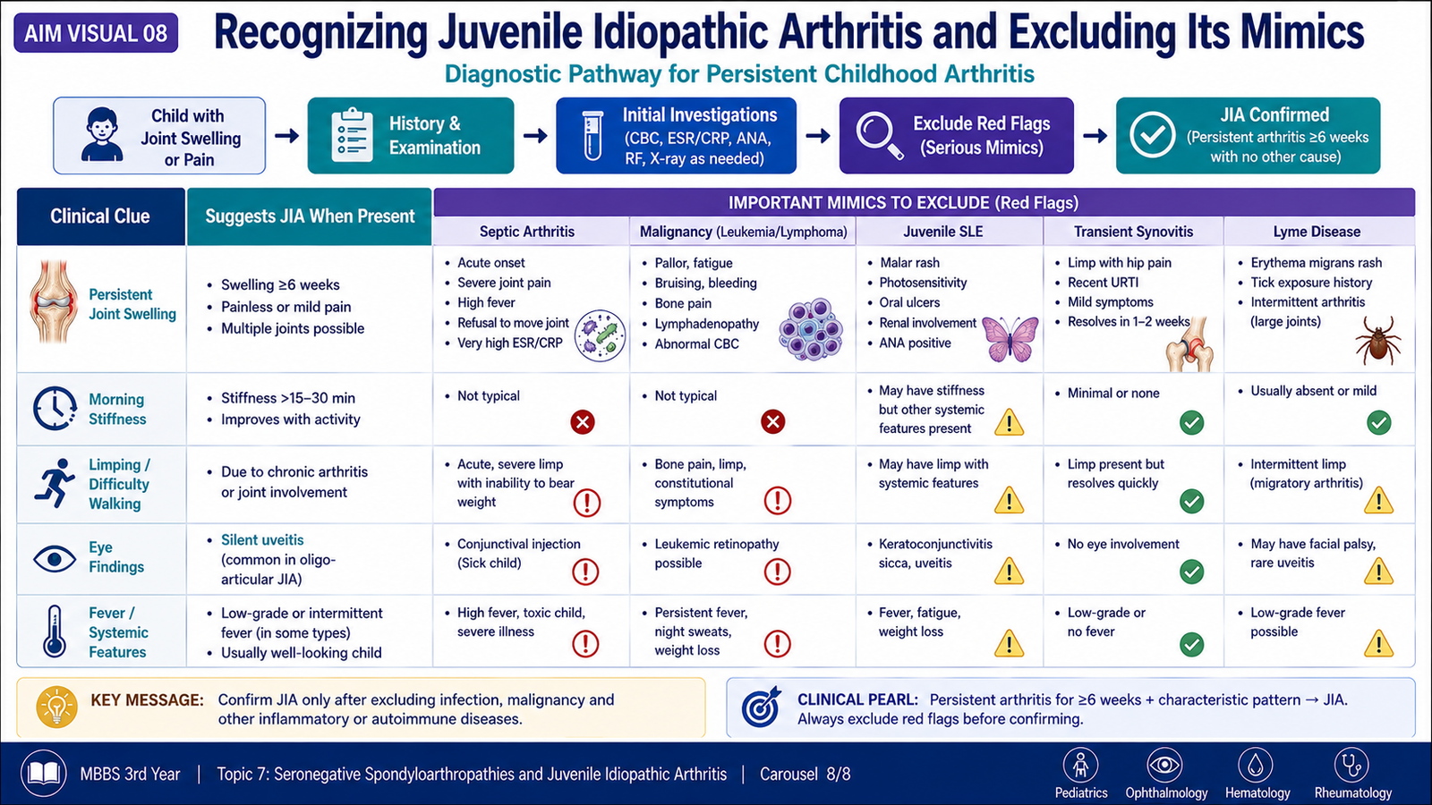
Differential Diagnosis
| Competing Condition | Clue Suggesting the Mimic | Reason It Matters |
|---|---|---|
| Septic arthritis | Acute severe monoarthritis, fever, toxicity or inability to bear weight | Requires urgent diagnosis and antimicrobial treatment |
| Acute rheumatic fever | Migratory large-joint arthritis after streptococcal infection with other characteristic manifestations | Cardiac involvement changes management and prognosis |
| Systemic lupus erythematosus | Multisystem disease with mucocutaneous, renal or hematological findings | Requires a different immunological evaluation |
| Leukemia or malignancy | Bone pain, pallor, bruising, organomegaly, night pain or abnormal blood counts | Immunosuppression must not be started before exclusion |
| Trauma or orthopedic disease | Clear injury, mechanical limitation or localized structural abnormality | May not produce persistent inflammatory stiffness |
| Viral or post-infectious arthritis | Short duration and close association with an acute infection | Often resolves without a chronic arthritic course |
Basic Diagnostic Approach
- Confirm objective arthritis rather than pain alone.
- Establish duration and number of joints involved.
- Search for fever, rash, psoriasis, enthesitis, dactylitis and ocular symptoms.
- Assess growth, gait, muscle bulk and functional limitation.
- Use blood tests to support inflammation and investigate mimics; no single test confirms all forms of juvenile idiopathic arthritis.
- Perform joint aspiration when infection or another intra-articular process is suspected.
- Arrange ophthalmic assessment according to the child’s risk pattern.
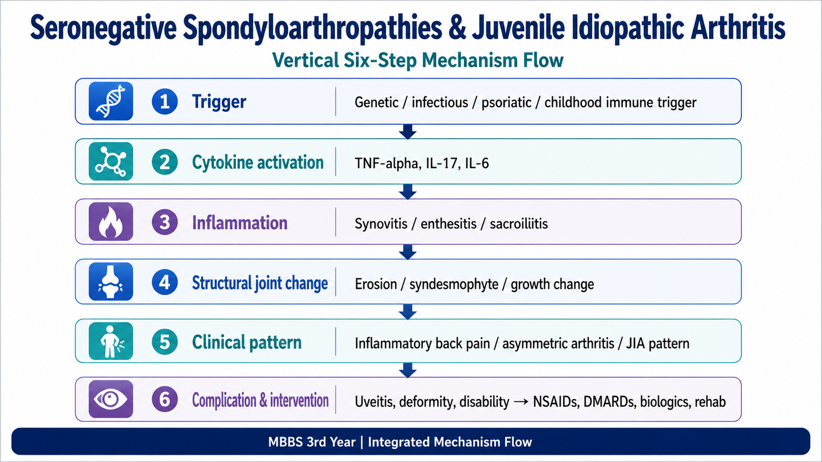
Integrated Mechanism Flow
AIM High-Yield Review
Seronegative Spondyloarthropathies and Juvenile Idiopathic Arthritis
Watch these videos after reading the learning material to reinforce the major disease patterns, mechanisms, clinical features and pediatric classification.
Video 1 — Seronegative Spondyloarthropathies
Focus on ankylosing spondylitis, reactive arthritis, psoriatic arthritis, sacroiliitis, enthesitis and the shared clinical pattern.
Video 2 — Juvenile Idiopathic Arthritis
Focus on the definition, childhood presentation, pathological basis, major clinical features and classification patterns of juvenile idiopathic arthritis.