
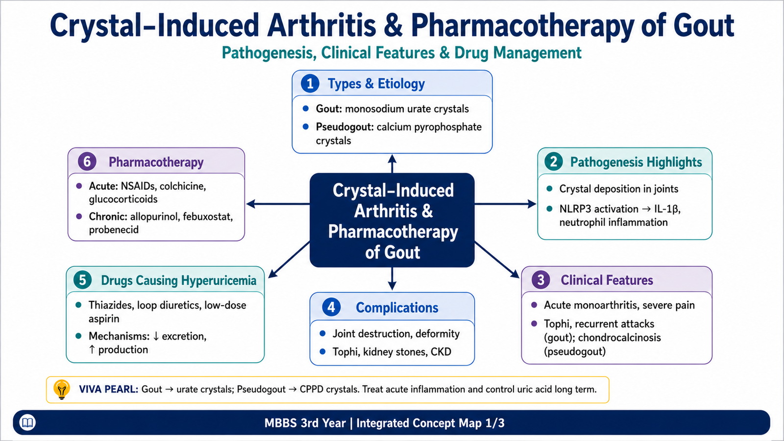
Crystal-induced arthritis develops when microscopic crystals deposit in a joint or nearby tissue and trigger inflammation. The two main disorders in this chapter are gout, caused by monosodium urate crystals, and calcium pyrophosphate deposition disease, commonly called pseudogout when it produces an acute attack.
This chapter explains why these crystals form, how they produce acute and chronic joint disease, how their morphology and clinical findings differ, and how medicines are selected. Acute treatment controls inflammation, whereas long-term treatment reduces urate production or increases urate excretion. Important adverse effects, drug interactions and medicines that cause hyperuricemia are also covered.
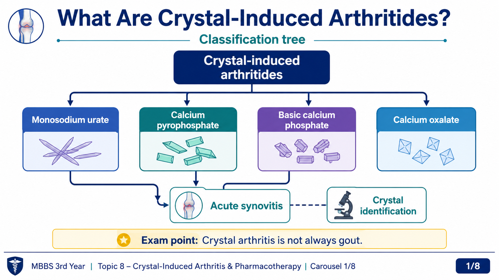
Crystal-induced arthritis refers to joint and periarticular inflammation caused by deposition of microscopic crystals. These crystals behave as endogenous danger signals. Once present in synovial tissue or fluid, they may activate macrophages, inflammatory mediators and neutrophils, producing episodes that can clinically resemble infection.
- Monosodium urate crystal disease: gout.
- Calcium pyrophosphate dihydrate deposition: CPPD disease or pseudogout.
- Basic calcium phosphate deposition: may contribute to calcific periarthritis and destructive arthropathy.
- Calcium oxalate deposition: uncommon and usually associated with severe renal dysfunction or oxalate excess.
Gout and CPPD disease are the principal undergraduate disorders. Both may cause acute synovitis, but they differ in crystal composition, typical joint distribution, associated conditions and radiological findings.
Gout is a disorder caused by tissue deposition of monosodium urate crystals, usually in the setting of persistent hyperuricemia. Hyperuricemia increases risk but is not by itself synonymous with gout because some hyperuricemic individuals never develop crystal deposition or arthritis.
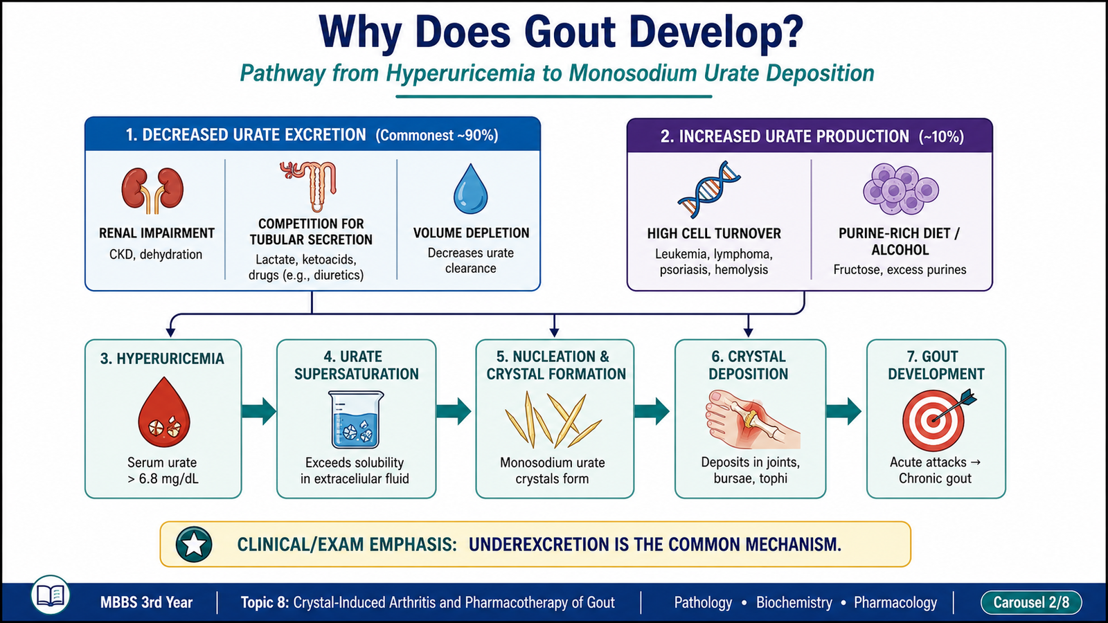
- Primary gout: usually reflects genetically influenced underexcretion of urate, sometimes combined with increased production.
- Secondary hyperuricemia: results from another disease, high cellular turnover, renal impairment or medication.
- Renal impairment and reduced urate clearance.
- Alcohol use, especially when associated with increased lactate production and reduced urate excretion.
- High cell turnover, such as myeloproliferative disorders, malignancy treatment or extensive tissue breakdown.
- Inherited enzyme abnormalities that increase purine synthesis or impair purine salvage.
- Obesity and metabolic syndrome-associated hyperuricemia.
- Drugs that reduce urate excretion.
Monosodium urate is less soluble in cooler peripheral tissues. The first metatarsophalangeal joint is also exposed to repeated minor mechanical stress, helping explain the classic presentation of podagra.
Monosodium urate crystal formation and deposition
Crystal phagocytosis by macrophages
NLRP3 inflammasome activation and interleukin-1 release
Neutrophil recruitment, crystal ingestion and mediator release
Intense acute synovitis with pain, warmth, erythema and swelling
The attack may resolve spontaneously because crystals become coated by proteins, inflammatory mediators are degraded, neutrophils undergo apoptosis and anti-inflammatory mechanisms gradually dominate. However, persistent hyperuricemia allows further crystal deposition and recurrent attacks.
- Asymptomatic hyperuricemia: elevated serum urate without clinically apparent arthritis.
- Acute gouty arthritis: abrupt, severe monoarthritis, classically involving the first metatarsophalangeal joint.
- Intercritical gout: symptom-free interval between attacks, although crystals may remain in tissues.
- Chronic tophaceous gout: persistent deposits with chronic synovitis, erosive joint damage and visible or palpable tophi.
- Acute synovial congestion and edema.
- Dense neutrophilic inflammatory infiltrate.
- Needle-shaped urate crystals within the synovial fluid and inflammatory cells.
- Crystals may dissolve during routine formalin processing, leaving pale, elongated spaces.
A tophus is a mass of deposited urate crystals surrounded by a chronic inflammatory reaction. It may occur in articular cartilage, synovium, tendons, bursae, soft tissue, pinna of the ear and periarticular tissues.
- Grossly: firm, chalky-white or yellow-white nodules.
- Microscopically: aggregates of urate crystals or crystal-shaped spaces surrounded by macrophages, foreign-body giant cells, lymphocytes and fibrosis.
- Chronic deforming and erosive arthritis.
- Tophi in periarticular tissues, tendons and ear pinna.
- Uric-acid nephrolithiasis.
- Urate-associated renal injury, particularly when marked hyperuricemia or pre-existing renal disease is present.
- Reduced mobility and impaired quality of life due to recurrent attacks.

Calcium pyrophosphate deposition disease results from deposition of calcium pyrophosphate dihydrate crystals in articular and periarticular tissues. When it presents as acute inflammatory arthritis resembling gout, the episode is termed pseudogout.
- Increasing age and degenerative cartilage changes.
- Previous joint damage or trauma.
- Familial forms with altered pyrophosphate metabolism.
- Metabolic associations such as hyperparathyroidism, hemochromatosis, hypomagnesemia and hypothyroidism.
Increased extracellular inorganic pyrophosphate within cartilage → combination with calcium → CPPD crystal formation → deposition in cartilage, fibrocartilage and synovium → crystal shedding into the joint → macrophage and neutrophil activation → acute inflammatory arthritis.
- Crystals deposit within hyaline cartilage and fibrocartilage.
- Calcification may be visible radiologically as chondrocalcinosis.
- The knee is commonly affected, although wrists, shoulders, elbows and ankles may also be involved.
- Clinical patterns include asymptomatic chondrocalcinosis, acute pseudogout and chronic degenerative arthropathy.
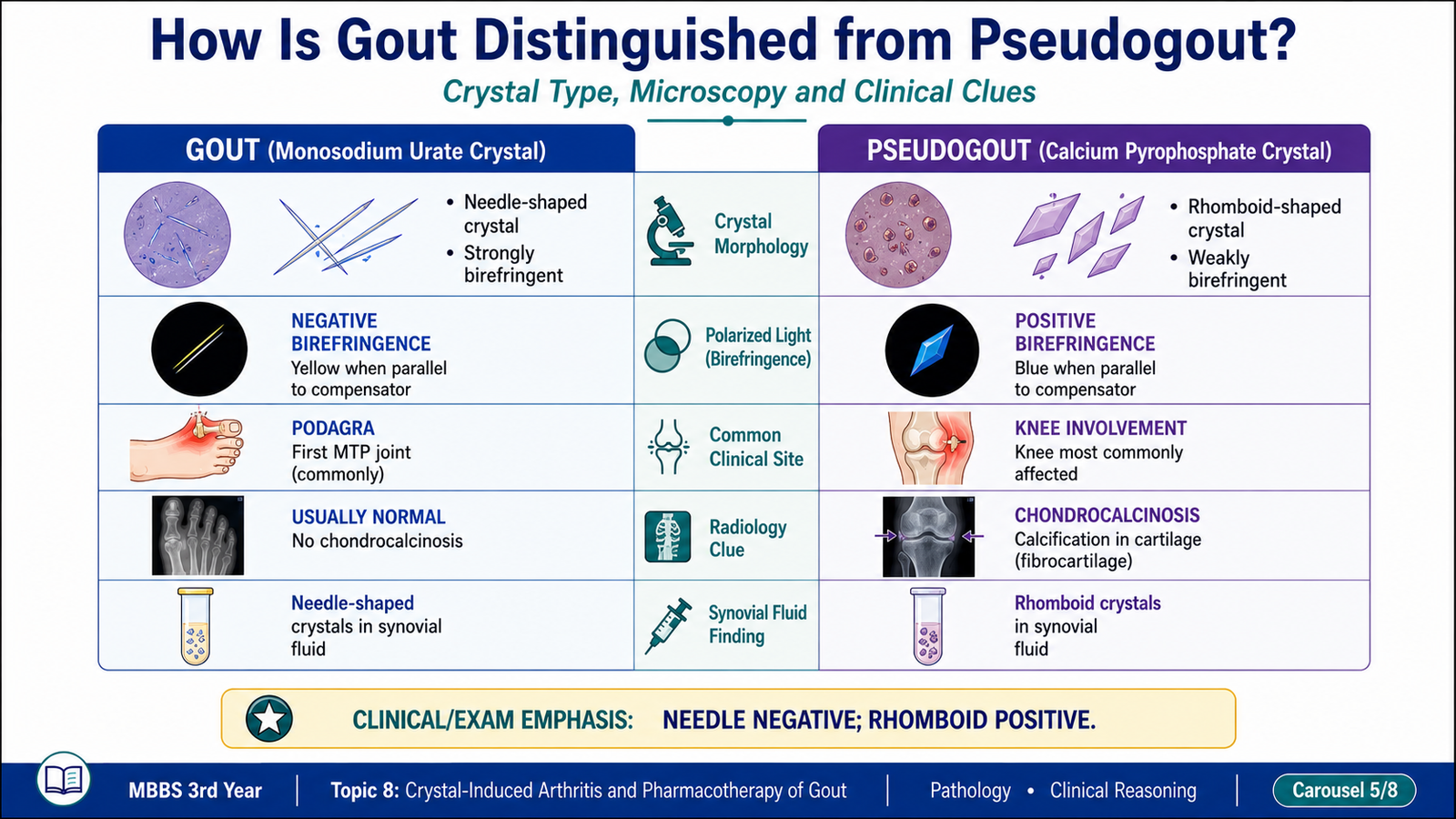
| Feature | Gout | CPPD / Pseudogout |
|---|---|---|
| Crystal | Monosodium urate | Calcium pyrophosphate dihydrate |
| Shape | Needle-shaped | Rhomboid or short rod-shaped |
| Birefringence | Strongly negative | Weakly positive |
| Typical joint | First metatarsophalangeal joint | Knee or wrist |
| Imaging clue | Erosions in chronic disease | Chondrocalcinosis |
| Chronic deposit | Tophus | Cartilage calcification |
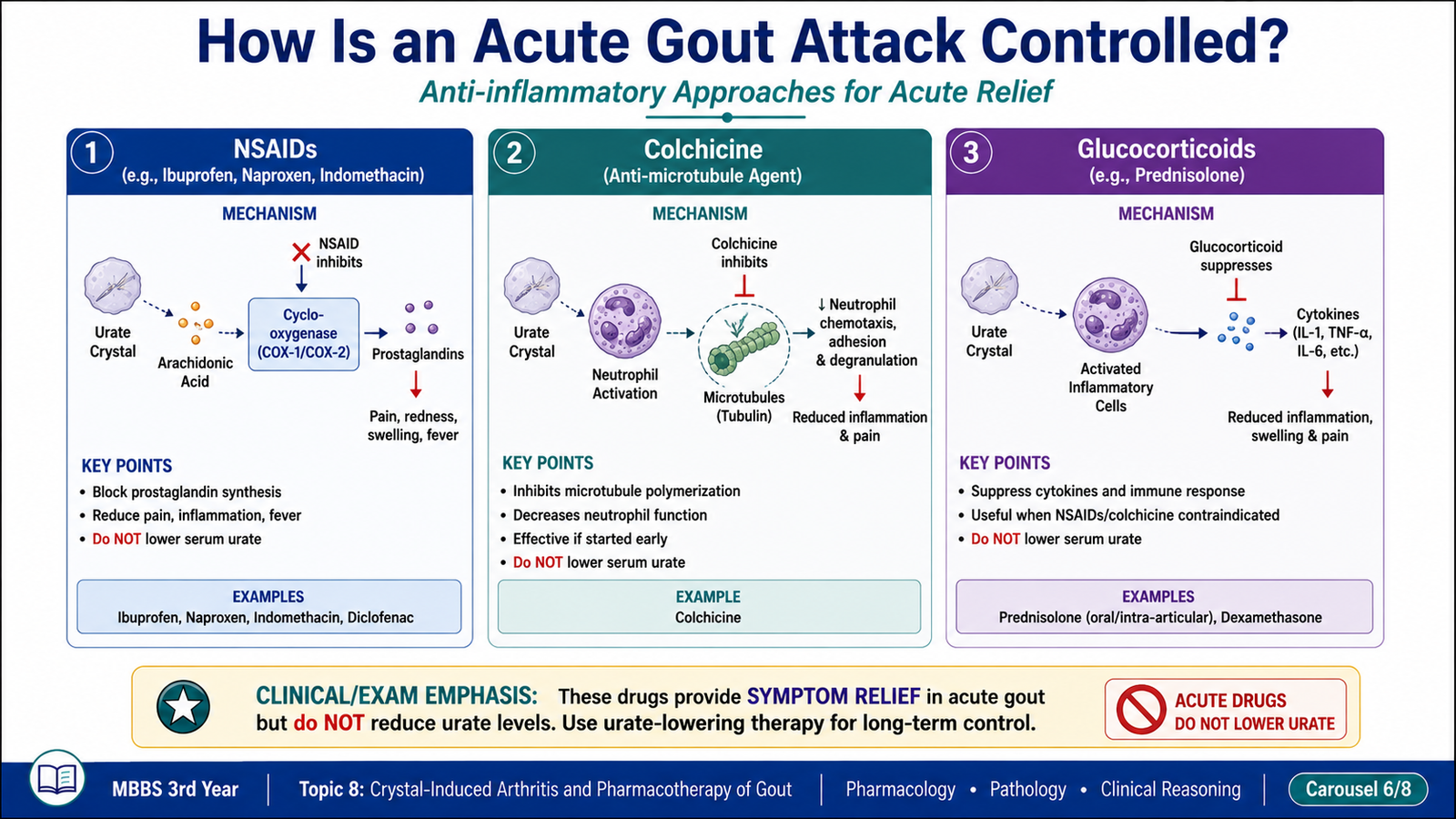
Acute gout treatment suppresses the crystal-triggered inflammatory response. It does not rapidly remove existing urate deposits. Choice among an NSAID, colchicine and a glucocorticoid depends on comorbidities, contraindications, severity and tolerability.
NSAID → cyclo-oxygenase inhibition → reduced prostaglandin synthesis → reduced vasodilation, edema and pain → rapid improvement in acute gouty inflammation.
- Useful for acute attacks when started promptly.
- They do not reduce serum urate or prevent crystal formation.
- Important adverse effects include gastric irritation, peptic ulceration, gastrointestinal bleeding, sodium retention, renal impairment and hypersensitivity.
- Use cautiously or avoid in significant renal disease, active ulcer disease, high bleeding risk or relevant cardiovascular decompensation.
- Aspirin is not used as routine anti-inflammatory treatment for gout because salicylates can produce dose-dependent effects on renal urate handling.
Colchicine → binds tubulin → inhibits microtubule polymerization → impairs neutrophil migration, chemotaxis, degranulation and crystal phagocytosis → suppresses crystal-induced inflammation.
- Used for acute gout and for prophylaxis against flares when urate-lowering therapy is initiated.
- It is anti-inflammatory in crystal disease but is not a general analgesic and does not lower serum urate.
- Common adverse effects: nausea, abdominal pain, vomiting and diarrhea.
- Serious toxicity: bone-marrow suppression, neuromuscular toxicity, peripheral neuropathy and multiorgan toxicity.
- Toxicity risk increases when drug clearance is impaired or when interacting medicines raise colchicine exposure.
Glucocorticoid → intracellular glucocorticoid receptor activation → altered gene transcription → reduced cytokines, eicosanoids, leukocyte recruitment and vascular inflammation → suppression of acute synovitis.
- Useful when NSAIDs or colchicine are contraindicated, poorly tolerated or ineffective.
- May be administered systemically or into an affected joint when clinically appropriate and infection has been excluded.
- Adverse effects of systemic treatment include hyperglycemia, fluid retention, mood changes, increased infection risk and gastrointestinal irritation.
- Repeated or prolonged exposure increases risks such as osteoporosis, muscle weakness and adrenal suppression.
| Therapeutic purpose | Drug class | Examples | Main action |
|---|---|---|---|
| Acute inflammation | Anti-inflammatory agents | NSAIDs, colchicine, glucocorticoids | Suppress crystal-induced inflammation |
| Reduced urate production | Xanthine-oxidase inhibitors | Allopurinol, febuxostat | Decrease uric-acid synthesis |
| Increased urate excretion | Uricosuric agents | Probenecid | Reduce proximal tubular urate reabsorption |
| Enzymatic urate degradation | Uricase preparations | Specialist-use uricase agents | Convert urate into a more soluble metabolite |
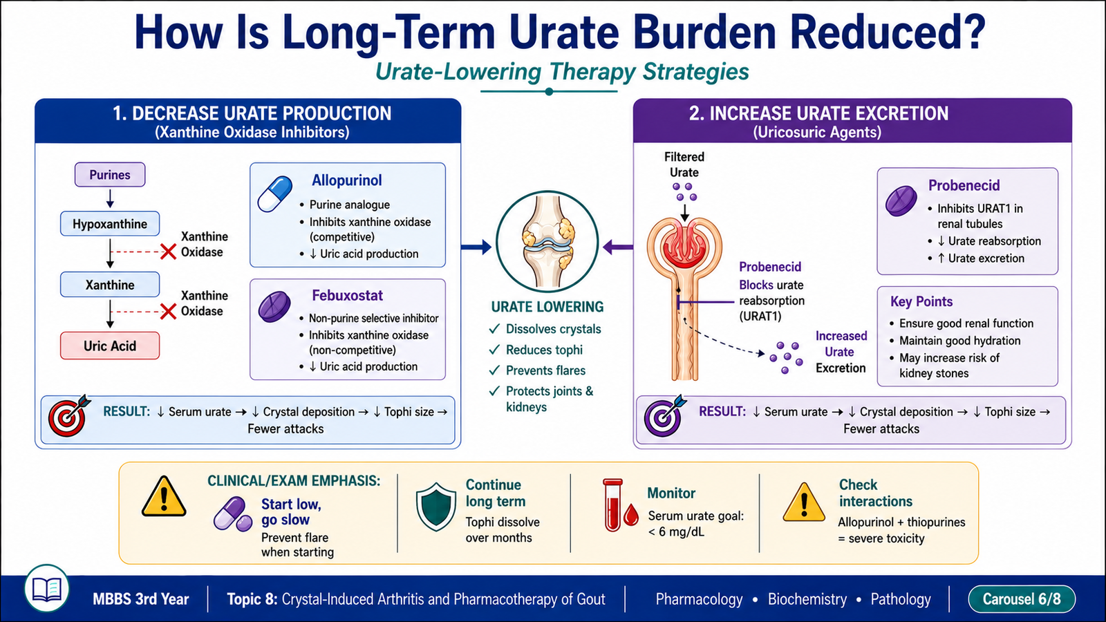
Allopurinol is converted to the active metabolite oxypurinol. Allopurinol and oxypurinol → inhibit xanthine oxidase → reduce conversion of hypoxanthine to xanthine and xanthine to uric acid → lower serum and tissue urate → prevent new crystal deposition and promote gradual dissolution of existing deposits.
- Used for long-term control of recurrent gout, tophaceous disease and clinically important urate overproduction.
- It is not an analgesic and does not directly terminate acute inflammatory pain.
- Initiation or major alteration of urate-lowering therapy may mobilize urate stores and precipitate a flare; anti-inflammatory prophylaxis may therefore be required.
- Adverse effects: gastrointestinal upset, rash, hepatotoxicity, leukopenia and acute gout flares during early therapy.
- Serious adverse effect: allopurinol hypersensitivity syndrome with severe cutaneous reaction, systemic involvement and organ dysfunction.
Febuxostat → selectively inhibits xanthine oxidase → decreases uric-acid production → lowers serum urate and reduces long-term crystal burden.
- Provides an alternative xanthine-oxidase inhibitor when allopurinol is unsuitable or insufficient.
- Adverse effects: nausea, rash, abnormal liver-function tests and early gout flares.
- Cardiovascular history and overall patient risk require careful consideration when selecting long-term therapy.
- Like allopurinol, it can interact dangerously with medicines dependent on xanthine oxidase for metabolism, including azathioprine and 6-mercaptopurine.
Probenecid → inhibits proximal tubular urate reabsorption transport → increases urinary urate excretion → lowers serum urate → reduces future crystal deposition.
- Most useful when hyperuricemia is related to urate underexcretion and renal function is adequate.
- Less suitable in significant renal impairment.
- Increased urinary urate can promote uric-acid stone formation.
- Adverse effects: gastrointestinal irritation, rash, hypersensitivity and nephrolithiasis.
- Adequate hydration is important to reduce urinary crystal concentration.
Probenecid inhibits renal tubular secretion of several organic acids. It may therefore delay renal elimination and increase plasma concentrations of drugs such as penicillins and some cephalosporins. This interaction has historically been used therapeutically, but it can also increase adverse effects. Salicylates can antagonize the uricosuric action of probenecid by altering renal urate transport. Probenecid may also alter the renal elimination of other medicines handled by organic-anion transport systems.
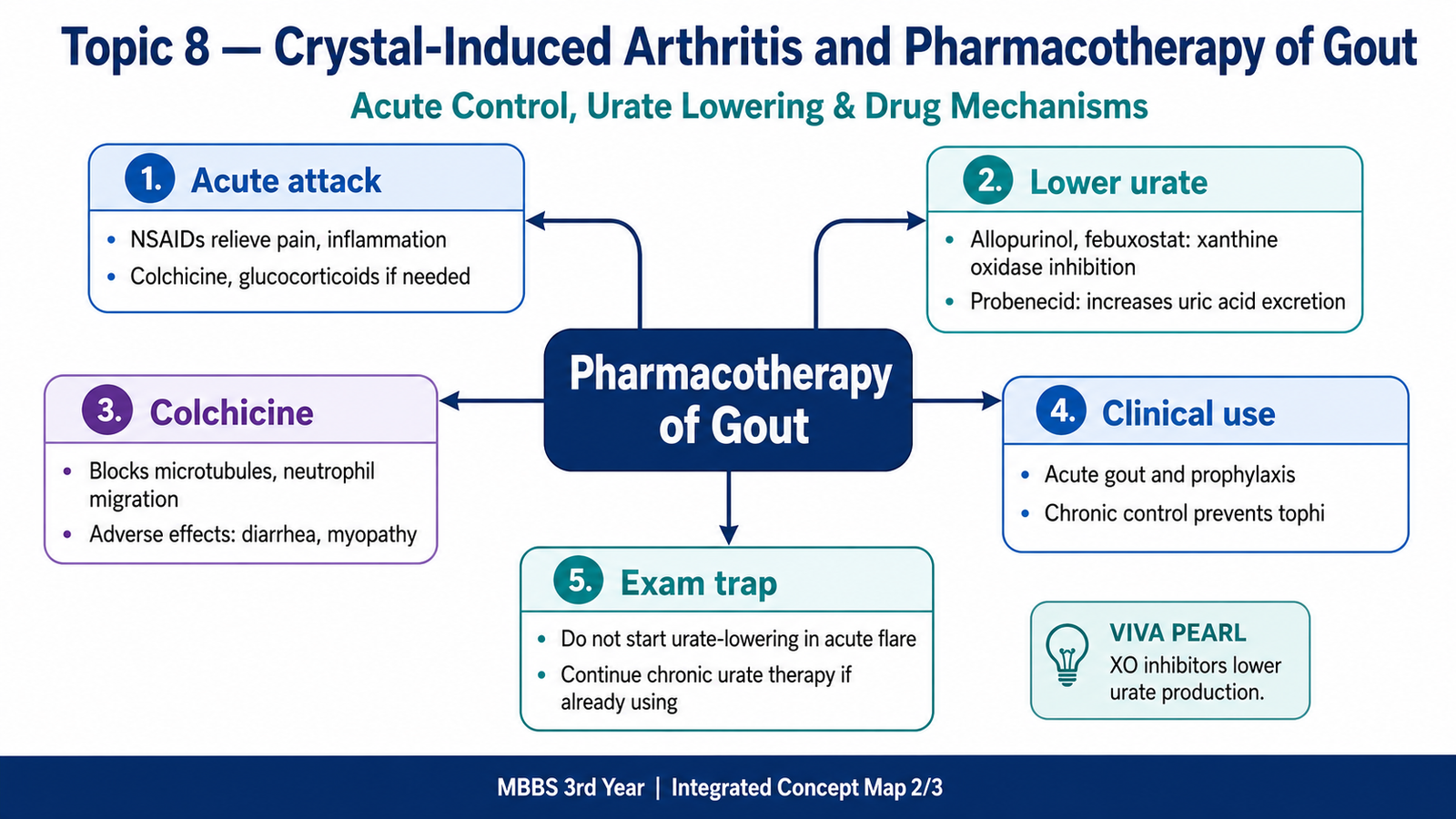
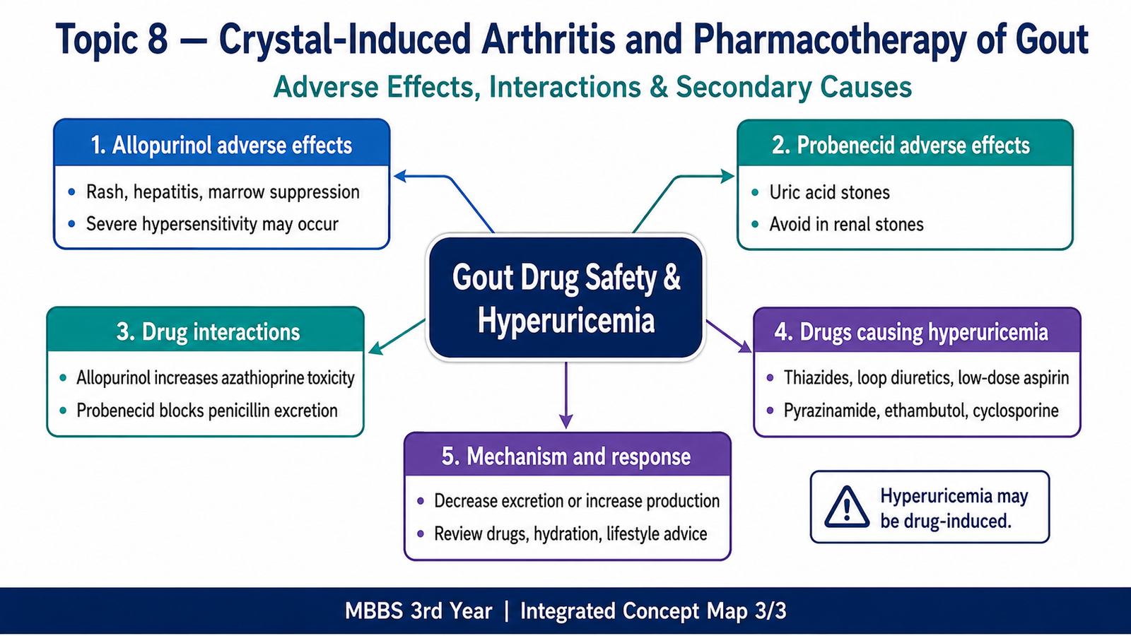
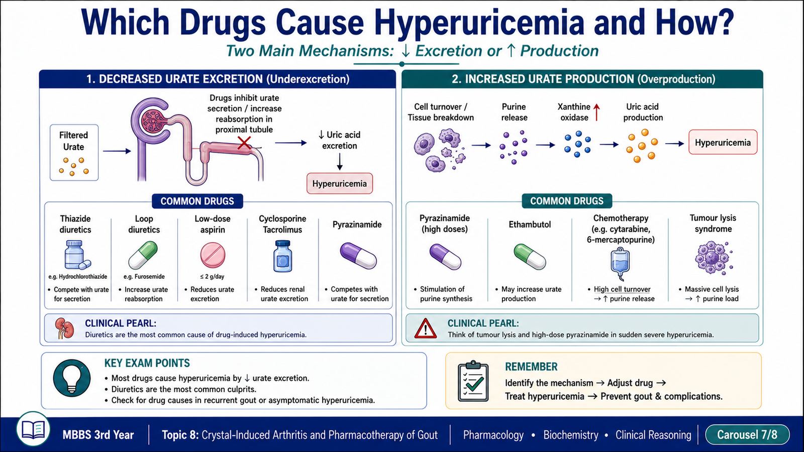
Drug-induced hyperuricemia usually results from reduced renal urate excretion, although increased purine breakdown or altered metabolism can also contribute. Medication review is therefore an essential part of evaluating recurrent gout.
| Drug or class | Mechanism of hyperuricemia | Clinical implication |
|---|---|---|
| Thiazide diuretics | Volume contraction and competition for proximal tubular organic-acid transport reduce urate excretion. | May precipitate gout in susceptible patients. |
| Loop diuretics | Reduced renal urate clearance related to volume contraction and tubular transport effects. | Important in patients with cardiac or renal comorbidity. |
| Low-dose aspirin | Reduces renal urate secretion at low doses. | May modestly increase urate; essential antiplatelet therapy should not be stopped without appropriate clinical reasoning. |
| Pyrazinamide | Metabolites compete with urate for renal tubular secretion. | Commonly tested cause of reduced urate excretion. |
| Ethambutol | Reduces renal urate excretion. | Consider during antituberculous treatment. |
| Cyclosporine and tacrolimus | Renal vasoconstrictive and tubular effects reduce urate clearance. | May cause hyperuricemia in transplant or immunosuppressed patients. |
| Niacin | Reduces renal urate excretion. | May worsen established gout. |
| Cytotoxic chemotherapy | Rapid cell destruction increases purine breakdown and uric-acid production. | Can produce acute urate nephropathy in tumour lysis. |
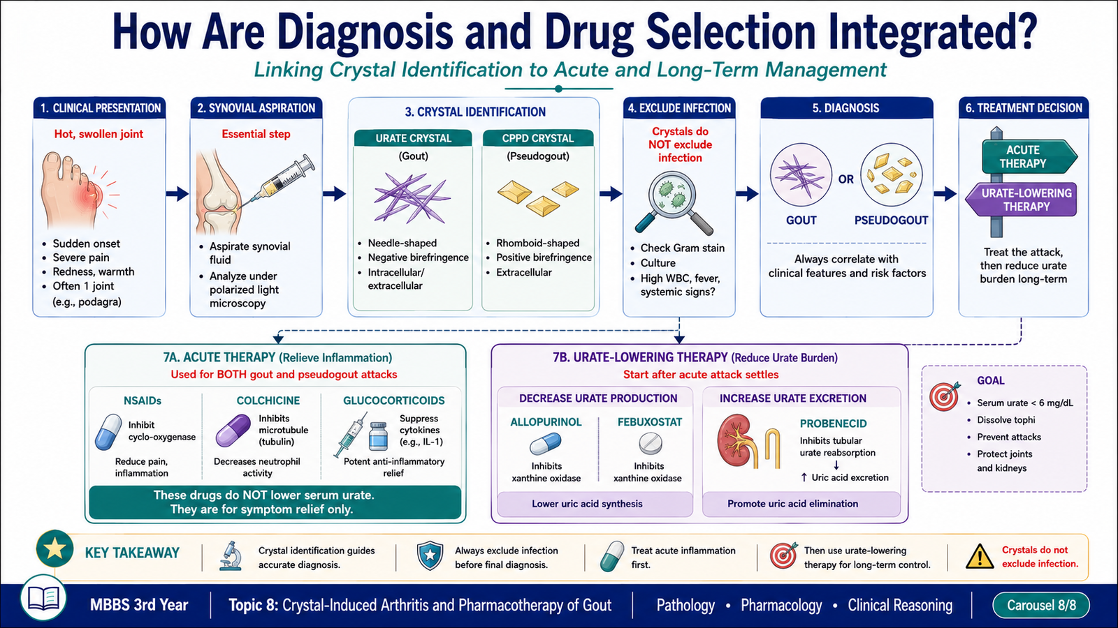
Acute gout should be suspected when a patient develops abrupt severe monoarthritis, especially podagra, but the diagnosis should be confirmed whenever uncertainty exists. Synovial-fluid examination provides the most direct evidence and helps distinguish crystal arthritis from septic arthritis.
- Gout: needle-shaped, strongly negatively birefringent monosodium urate crystals.
- CPPD disease: rhomboid or short rod-shaped, weakly positively birefringent crystals.
- Septic arthritis: organisms may be demonstrated by appropriate microbiological examination; crystals do not exclude coexisting infection.
- Serum urate: supports assessment of urate burden but may be normal during an acute attack and cannot independently confirm or exclude gout.
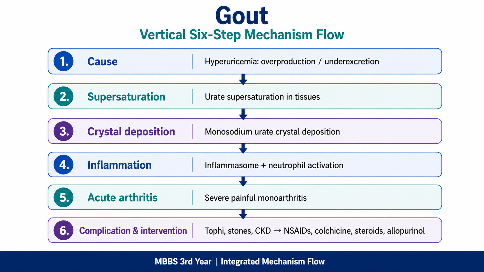
Watch this video to reinforce the pathogenesis, clinical features, diagnosis and pharmacological management of gout.